
Muellerius capillaris in Sheep and Goats: Protostrongylid Lungworm Diagnosis and Control
Introduction
Muellerius capillaris is a small ruminant lungworm belonging to the family Protostrongylidae, subfamily Muelleriinae [1]. It is a highly prevalent nematode parasite of sheep and goats across diverse geographic regions, including Europe, Africa, Asia, the Americas, and the Middle East [2, 3, 4, 5, 39, 47, 50, 53, 54]. Unlike the large lungworm Dictyocaulus filaria, which resides in the trachea and bronchi, M. capillaris inhabits the lung parenchyma, bronchioles, and alveoli, where it induces chronic, often subclinical, verminous pneumonia [6, 7, 45]. The parasite is transmitted via terrestrial gastropod intermediate hosts, and its epidemiology is tightly linked to snail population dynamics, climatic conditions, and grazing management [8, 9, 10, 11, 55]. This article provides a detailed, evidence-based review of the taxonomy, life cycle, epidemiology, pathogenesis, clinical presentation, diagnostic methods, and control strategies for M. capillaris infection in sheep and goats.
Taxonomy and Morphology
Muellerius capillaris (Mueller, 1889) Cameron, 1927, is a slender, filiform nematode [1]. Adult males measure approximately 12 to 23 mm in length, while females are larger, ranging from 18 to 30 mm. The caudal bursa of the male is well developed, with characteristic ray patterns used for species identification [1]. First-stage larvae (L1) are distinctive: they measure 280 to 320 micrometers in length, possess a kinked or wavy tail with a dorsal spine, and are morphologically distinguishable from other protostrongylid larvae such as Protostrongylus rufescens and Cystocaulus ocreatus [12, 54]. The L1 stage is the diagnostic stage recovered from feces.
Life Cycle
The life cycle of M. capillaris is indirect, requiring a terrestrial gastropod intermediate host [11, 1]. Adult worms reside in the lung parenchyma of the definitive host (sheep or goat), where females deposit embryonated eggs [7]. These eggs hatch in the alveoli or bronchioles, and first-stage larvae (L1) migrate up the respiratory tract, are coughed up, swallowed, and then passed in the feces [13, 7].
Once in the external environment, L1 larvae are ingested by a suitable gastropod intermediate host, which includes species from genera such as Trichia, Cernuella, Helicella, and others [8, 10, 11, 55]. Within the snail, the L1 molts to the second stage (L2) and then to the third, infective stage (L3) over a period of 2 to 4 weeks, depending on ambient temperature and humidity [9, 11]. The L3 larvae accumulate in the foot and mantle of the snail [11].
Definitive hosts become infected by accidentally ingesting infected snails while grazing [14, 57]. After ingestion, the L3 larvae are liberated in the gastrointestinal tract, penetrate the intestinal wall, and migrate via the lymphatic system and bloodstream to the lungs [15, 16, 13]. The prepatent period is approximately 5 to 8 weeks [17, 13]. The parasite can also be transmitted experimentally to laboratory animals such as mice, guinea pigs, and rabbits, although these are not natural hosts [16, 71].
Epidemiology
Muellerius capillaris is one of the most prevalent lungworm species in small ruminants worldwide [18, 2, 3, 47, 50, 53]. Prevalence rates vary widely depending on geographic location, management practices, and diagnostic methods used. In a study from northwestern Spain, protostrongylid infection prevalence in meat sheep was 42.5%, with M. capillaris being the dominant species [2]. In northeastern Ethiopia, a prevalence of 27.3% was reported in sheep [3]. In the Republic of Belarus, infection rates in sheep and goats reached 35.1% and 44.3%, respectively. In Iceland, a prevalence of 35.1% was found in lambs. In Algeria, M. capillaris accounted for 43% of lungworm infections in sheep. In Turkey, the prevalence was 5.6% in sheep and 34.4% in goats. In Argentina, M. capillaris was reported for the first time in goat flocks in the northwestern region.
Several risk factors influence the prevalence and intensity of infection. Climatic conditions, particularly temperature and humidity, directly affect the survival of L1 larvae on pasture and the development of L3 larvae within snail intermediate hosts [19, 9]. L1 larvae are resistant to desiccation and can survive for extended periods under cool, moist conditions [20, 21]. Grazing management is a critical factor; continuous grazing on permanent pastures with abundant snail populations increases exposure risk [10, 22, 57]. Mixed grazing of sheep and goats can facilitate transmission between species [14, 53]. Breed susceptibility has been documented, with Romanov sheep showing higher resistance compared to Lacaune sheep [23]. Age is also a factor, with older animals often harboring higher worm burdens due to cumulative exposure [2, 63].
Pathogenesis and Pathology
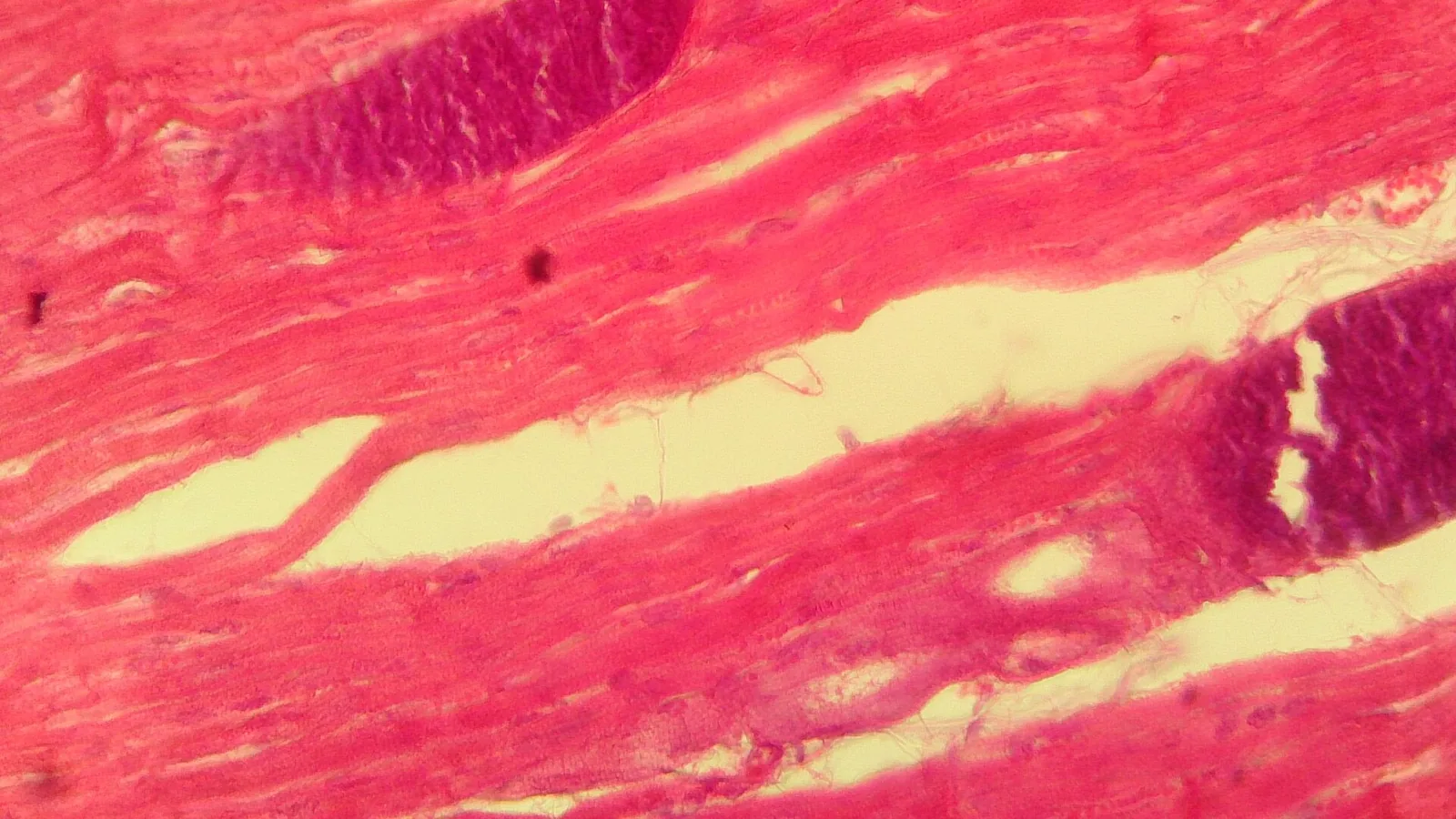
The pathological changes induced by M. capillaris are primarily confined to the lung parenchyma [6, 7, 45]. Adult worms and eggs elicit a chronic inflammatory response characterized by the formation of small, firm, greyish-white nodules (1 to 5 mm in diameter) scattered throughout the lung tissue, particularly in the diaphragmatic lobes [6, 39, 50]. Histologically, these nodules consist of a central core of adult worms, eggs, and L1 larvae surrounded by a zone of inflammatory cells, including macrophages, eosinophils, lymphocytes, and giant cells [6, 45, 50]. The surrounding alveolar tissue often shows emphysema and atelectasis [6, 39]. In heavy infections, coalescing nodules can lead to extensive verminous pneumonia and fibrosis [6, 45]. The eosinophilic response is a hallmark of infection, with peripheral eosinophilia observed in infected animals.
The pathological impact is generally considered subclinical in well-nourished animals, but heavy burdens can impair pulmonary function, leading to reduced exercise tolerance, chronic cough, and secondary bacterial pneumonia [24, 45]. In a study from Mexico, naturally infected sheep showed significant pathological and immunological changes, including increased serum globulins and eosinophil counts.
Clinical Signs
Clinical signs of mulleriosis are often subtle and non-specific, particularly in low to moderate infections [24, 45]. In heavy infections, affected animals may exhibit a chronic, intermittent cough, nasal discharge, tachypnea, and dyspnea, especially after exercise [24, 40]. Reduced weight gain, decreased milk production, and poor overall condition are common production losses [43, 59, 60]. In lambs and kids, severe infections can lead to unthriftiness and increased susceptibility to other respiratory pathogens [40, 59]. Concurrent infections with other respiratory pathogens, such as Mannheimia haemolytica or Mycoplasma ovipneumoniae, can exacerbate clinical disease [40, 45].
Diagnosis
Accurate diagnosis of M. capillaris infection relies on a combination of clinical, parasitological, and pathological methods.
Fecal Examination: The Baermann Technique
The gold standard for antemortem diagnosis is the recovery of first-stage larvae (L1) from feces using the Baermann technique [44, 53, 63]. This method exploits the active migration of larvae from fecal material into warm water. A modified Baermann apparatus, consisting of a funnel, rubber tubing, and a clamp, is used. Feces (5 to 10 grams) are wrapped in cheesecloth or placed on a sieve and suspended in warm water (25 to 30 degrees Celsius) for 12 to 24 hours. Larvae migrate through the mesh and sediment at the bottom of the funnel. The sediment is then collected and examined microscopically for the characteristic L1 larvae of M. capillaris [12, 44]. The larvamigration (pipette) method has been described as a more efficient alternative for detecting M. capillaris larvae.
Larval Identification
First-stage larvae of M. capillaris must be differentiated from other protostrongylid larvae. Key morphological features include the presence of a dorsal spine on the tail and a kinked or wavy tail tip [12, 54]. The total length (280 to 320 micrometers) and the shape of the buccal cavity are also used for species identification [12, 54]. Protostrongylus rufescens larvae have a straight tail with a small spine, while Cystocaulus ocreatus larvae have a characteristic S-shaped tail [12].
Postmortem Examination
At necropsy, the lungs should be carefully examined for the presence of small, firm nodules on the pleural surface and within the lung parenchyma [6, 39, 50]. The Baermann technique can also be applied to lung tissue to recover larvae. Adult worms can be recovered by dissecting the nodules under a stereomicroscope [6, 39].
Molecular Diagnostics
While less commonly used in routine field diagnosis, molecular methods such as conventional PCR and real-time PCR have been developed for the detection and differentiation of protostrongylid larvae in fecal samples. These assays target ribosomal DNA (ITS-2) or mitochondrial DNA (cox1) sequences and offer high sensitivity and specificity, particularly for mixed infections. However, these methods are not yet widely adopted in commercial diagnostic laboratories.
Serology
Serological tests, such as enzyme-linked immunosorbent assays (ELISAs), have been developed for experimental purposes but are not routinely used for clinical diagnosis. They may be useful for herd-level surveillance in research settings.
Control and Treatment
Control of M. capillaris is challenging due to the indirect life cycle involving snail intermediate hosts and the limited efficacy of some anthelmintics against the adult worms.
Anthelmintic Therapy
Several anthelmintic classes have been evaluated for efficacy against M. capillaris. Macrocyclic lactones (MLs) are the most commonly used drugs.
Ivermectin: Ivermectin administered subcutaneously at the standard dose (0.2 mg/kg) has shown variable efficacy against M. capillaris. A controlled-release capsule formulation of ivermectin demonstrated high efficacy (greater than 99%) against adult worms in sheep [25]. However, injectable formulations may have lower efficacy, particularly against larval stages. In a study from Algeria, twelve different registered ivermectin preparations showed variable efficacy against M. capillaris.
Moxidectin: Moxidectin (0.2 mg/kg subcutaneously) has demonstrated high efficacy (greater than 95%) against M. capillaris in sheep [26]. It is often preferred due to its longer persistence in tissues.
Doramectin: Doramectin (0.2 mg/kg intramuscularly) has shown 100% efficacy against M. capillaris larvae in naturally infected sheep. Another study confirmed its efficacy against lungworms in pregnant sheep.
Benzimidazoles: Fenbendazole and albendazole have limited efficacy against M. capillaris at standard doses. Flubendazole has been used in mouflon with some success [27]. Luxabendazole has shown activity against lungworms in sheep [28].
Other Drugs: Tetramisole and levamisole have been used historically but are associated with toxicity in heavily infected animals [29]. Doramectin has also been studied for its efficacy against mixed nematode infections.
Pasture Management
Reducing exposure to infected snails is a key component of control. Strategies include:
- Rotational grazing: Moving animals to clean pastures every 2 to 3 weeks can reduce the ingestion of infected snails [22, 57].
- Avoiding overgrazing: Maintaining adequate sward height reduces snail habitat.
- Mixed grazing with cattle: Cattle are not hosts for M. capillaris and can help break the life cycle by reducing snail populations through trampling.
- Snail control: Molluscicides are generally not practical or cost-effective for large-scale use.
Integrated Control
An integrated approach combining strategic anthelmintic treatment with pasture management is recommended [18, 30]. Treatment should be targeted at high-risk periods, such as late autumn or early spring, when snail activity is high [19, 10, 61, 63]. Regular monitoring of fecal larval counts using the Baermann technique can help guide treatment decisions and assess the effectiveness of control programs.
Differential Diagnosis
Respiratory disease in sheep and goats can be caused by a variety of infectious and non-infectious agents. The differential diagnosis for M. capillaris infection includes:
- Other lungworms: Dictyocaulus filaria (large lungworm), Protostrongylus rufescens, Cystocaulus ocreatus, and Neostrongylus linearis [2, 50, 53, 54].
- Bacterial pneumonia: Mannheimia haemolytica, Pasteurella multocida, Mycoplasma ovipneumoniae, and Trueperella pyogenes.
- Viral infections: Respiratory syncytial virus (RSV), parainfluenza-3 virus (PI-3), and adenoviruses.
- Parasitic conditions: Nasal bots (Oestrus ovis) and liver fluke (Fasciola hepatica) can cause secondary respiratory signs.
Diagnostic Workflow
The following Mermaid diagram illustrates a recommended diagnostic workflow for suspected M. capillaris infection in a sheep or goat flock.
flowchart TD
 A["Clinical Signs: Chronic cough, poor growth, dyspnea"] --> B{Individual or Flock Investigation?}
 B -->|Individual| C[Fecal Baermann Examination]
 B -->|Flock| D[Pooled Fecal Baermann from 5-10 animals]
 C --> E{Larvae Detected?}
 D --> E
 E -->|No| F["Consider other respiratory pathogens: Bacterial culture, PCR for viruses/mycoplasma"]
 E -->|Yes| G[Morphological Identification of L1 Larvae]
 G --> H{Species Confirmed as M. capillaris?}
 H -->|No| I[Identify other protostrongylid or dictyocaulid species]
 H -->|Yes| J[Assess larval count per gram of feces]
 J --> K{High larval count?}
 K -->|Yes| L["Implement anthelmintic treatment: Moxidectin or Doramectin"]
 K -->|No| M["Monitor; consider pasture management"]
 L --> N["'Post-treatment Baermann (14-21 days later')"]
 N --> O{Efficacy >90% reduction?}
 O -->|Yes| P["Continue integrated control: Rotational grazing, snail habitat reduction"]
 O -->|No| Q["Investigate anthelmintic resistance; consider alternative drug class"]
Conclusion
Muellerius capillaris is a highly prevalent and economically important lungworm of sheep and goats worldwide. Its indirect life cycle, involving terrestrial gastropod intermediate hosts, complicates control efforts. Diagnosis relies on the Baermann technique for fecal larval recovery, with morphological identification of the characteristic L1 larvae. Macrocyclic lactones, particularly moxidectin and doramectin, are the most effective anthelmintics. An integrated control strategy combining strategic treatment with pasture management is essential for reducing the impact of this parasite on small ruminant health and productivity.
References
[1] Morgan D. On the Morphology and Biology of a Larval Stage of Muellerius capillaris (Mueller, 1889) Cameron, 1927; a Lungworm of Sheep and Goats. J Helminthol. 1929. URL: https://www.semanticscholar.org/paper/ca734087e5a6bca2b75a8b397bfbc2dcf7377f5d
[2] López CM, Fernández G, Viña M, et al. Protostrongylid infection in meat sheep from Northwestern Spain: prevalence and risk factors. Vet Parasitol. 2011. URL: https://pubmed.ncbi.nlm.nih.gov/21269772/
[3] Alemu S, Leykun EG, Ayelet G, et al. Study on small ruminant lungworms in northeastern Ethiopia. Vet Parasitol. 2006. URL: https://pubmed.ncbi.nlm.nih.gov/16919877/
[4] Berrag B, Urquhart GM. Epidemiological aspects of lungworm infections of goats in Morocco. Vet Parasitol. 1996. URL: https://pubmed.ncbi.nlm.nih.gov/8750686/
[5] Cabaret J, Chartier C. Muellerius capillaris in north-east Zaire: prevalence in sheep and goats and determination of intermediate hosts. J Helminthol. 1989. URL: https://pubmed.ncbi.nlm.nih.gov/2600412/
[6] Panayotova-Pencheva MS, Alexandrov MT. Some pathological features of lungs from domestic and wild ruminants with single and mixed protostrongylid infections. Vet Med Int. 2010. URL: https://pubmed.ncbi.nlm.nih.gov/20445790/
[7] Rose J. Site of development of the lungworm Muellerius capillaris in experimentally infected lambs. J Comp Pathol. 1958. URL: https://pubmed.ncbi.nlm.nih.gov/13563687/
[8] Reguera-Feo A, Castañón-Ordóñez L, Cordero-del-Campillo M. Population variability of Cernuella arigonis (Haas, 1929) (Mollusca: Helicidae) experimentally infected with Muellerius capillaris (Mueller, 1889) (Nematoda: Protostrongylidae). Vet Parasitol. 1994. URL: https://pubmed.ncbi.nlm.nih.gov/8091607/
[9] Cabaret J, Riseani SR, Baeza E. Survival of sheep and goat first stage protostrongylid larvae in experimental conditions: influence of humidity and temperature. J Helminthol. 1991. URL: https://pubmed.ncbi.nlm.nih.gov/1940250/
[10] Lahmar S, Cabaret J, Cheniti T. Land snails and periods at high risk for protostrongylid infection on a sheep-grazed pasture of northeast Tunisia. Vet Parasitol. 1990. URL: https://pubmed.ncbi.nlm.nih.gov/2382378/
[11] Beresford-Jones WP. Observations on Muellerius capillaris (Muller, 1889), Cameron, 1927. I. The bionomics and development in Trichia hispida (Linnaeus) of larvae obtained from sheep grazed on permanent pasture. Res Vet Sci. 1966. URL: https://pubmed.ncbi.nlm.nih.gov/5950125/
[12] Reguera-Feo A, Castañón-Ordóñez L, del Campillo MC. Ecological relations among first-stage larvae of four species of Protostrongylidae (Nematoda) within their ovine host. Appl Parasitol. 1996. URL: https://pubmed.ncbi.nlm.nih.gov/8574249/
[13] Rose JH. Experimental infection of lambs with Muellerius capillaris. J Comp Pathol. 1959. URL: https://pubmed.ncbi.nlm.nih.gov/14438839/
[14] Foreyt WJ, Jenkins EJ, Appleyard GD. Transmission of lungworms (Muellerius capillaris) from domestic goats to bighorn sheep on common pasture. J Wildl Dis. 2009. URL: https://pubmed.ncbi.nlm.nih.gov/19395736/
[15] Beresford-Jones WP. Observations on Muellerius capillaris (Müller, 1889) Cameron, 1927. 3. Experimental infection of sheep. Res Vet Sci. 1967. URL: https://pubmed.ncbi.nlm.nih.gov/5634078/
[16] Beresford-Jones WP. Observations on Muellerius capillaris (Müller, 1889), Cameron, 1927. II. Experimental infection of mice, guinea-pigs and rabbits with third stage larvae. Res Vet Sci. 1966. URL: https://pubmed.ncbi.nlm.nih.gov/6008019/
[17] Sauerländer R. Experimental infection of sheep and goats with Muellerius capillaris (Protostrongylidae, Nematoda). Zentralbl Veterinarmed B. 1988. URL: https://pubmed.ncbi.nlm.nih.gov/3188728/
[18] Bentounsi B, Cabaret J. Small-Lungworm (Protostrongylidae) Infections in Relation to Meat Sheep Breeds, Mediterranean Climates, and Anthelmintic Regimens. Vet Sci. 2025. URL: https://pubmed.ncbi.nlm.nih.gov/40431564/
[19] Díez-Baños P, Morrondo-Pelayo P, Feijoo-Penela A, et al. Relationship between the excretion of protostrongylid larvae in sheep in north-west Spain and climatic conditions. J Helminthol. 1994. URL: https://pubmed.ncbi.nlm.nih.gov/7829839/
[20] Reguera-Feo A, Rojo-Vazquez FA, Cordero-Del-Campillo M. On first-stage larval resistance under controlled conditions of three species of nematodes of the family Protostrongylidae Leiper, 1926. Ann Parasitol Hum Comp. 1986. URL: https://pubmed.ncbi.nlm.nih.gov/3813415/
[21] Observations on the Bionomics of the Free-living First Stage Larvae of the Sheep Lungworm, Muellerius capillaris. J Helminthol. 1957. URL: https://www.semanticscholar.org/paper/f6c13b3d50fdee25994a77af52a2a85e8405957f
[22] Cabaret J, Pandey VS. The use of tracer lambs for monitoring protostrongylid infection on extension pastures of Morroco. Ann Rech Vet. 1986. URL: https://pubmed.ncbi.nlm.nih.gov/3777804/
[23] Gruner L, Cabaret J, Sauve C, et al. Comparative susceptibility of Romanov and Lacaune sheep to gastrointestinal nematodes and small lungworms. Vet Parasitol. 1986. URL: https://pubmed.ncbi.nlm.nih.gov/3962166/
[24] López CM, Cienfuegos S, Dacal V, et al. Efficacy of anthelminthic control programs against natural Muellerius capillaris infection in sheep in the north-west of Spain. Effect on blood gases and pH in venous blood samples. Parasite. 2010. URL: https://pubmed.ncbi.nlm.nih.gov/20597445/
[25] Rehbein S, Visser M. Efficacy of ivermectin delivered via a controlled-release capsule against small lungworms (Protostrongylidae) in sheep. J Vet Med B Infect Dis Vet Public Health. 2002. URL: https://pubmed.ncbi.nlm.nih.gov/12420864/
[26] Papadopoulos E, Sotiraki S, Himonas C, et al. Treatment of small lungworm infestation in sheep by using moxidectin. Vet Parasitol. 2004. URL: https://pubmed.ncbi.nlm.nih.gov/15135874/
[27] Lamka J, Vondrejc M, Klecá ková J. [Effect of flubendazole on Muellerius capillaris in mouflon]. Vet Med (Praha). 1996. URL: https://pubmed.ncbi.nlm.nih.gov/9036620/
[28] Kassai T, Takáts C, Fok E, et al. Activity of luxabendazole against liver flukes, gastrointestinal roundworms, and lungworms in naturally infected sheep. Parasitol Res. 1988. URL: https://pubmed.ncbi.nlm.nih.gov/2974592/
[29] Halhead WA. Tetramisole toxicity in sheep infected with Muellerius capillaris. 1968. URL: https://www.semanticscholar.org/paper/edd8b01c91980ccd2c9b3d416257e32e2da110be
[30] Vaivarinya G, Keidan P, Krūklīte A. Epidemiology of Dictyocaulus filaria and protostrongylid (Muellerius capillaris) infections in sheep and their control in Latvia. 1980. URL: https://www.semanticscholar.org/paper/8e68edd0bd3b9236b44856b7889a37eada4e2a01
[31] Ezenwa VO, Hines AM, Archie EA, et al. Muellerius capillaris dominates the lungworm community of bighorn sheep at the National Bison Range, Montana. J Wildl Dis. 2010. URL: https://pubmed.ncbi.nlm.nih.gov/20688711/
[32] Polley L. Quantitative observations on intrapulmonary populations of Muellerius capillaris (Mueller, 1889) Cameron, 1927, in sheep. J Parasitol. 1987. URL: https://pubmed.ncbi.nlm.nih.gov/3572660/
[33] Pybus MJ, Shave H. Muellerius capillaris (Mueller, 1889) (Nematoda: Protostrongylidae): an unusual finding in Rocky Mountain bighorn sheep (Ovis canadensis canadensis Shaw) in South Dakota. J Wildl Dis. 1984. URL: https://pubmed.ncbi.nlm.nih.gov/6530714/
[34] Bwangamoi O, Aruo SK, Nkwizire GS. Muellerius capillaris in goats and sheep in Uganda. Bull Anim Health Prod Afr. 1976. URL: https://pubmed.ncbi.nlm.nih.gov/16300133/
[35] Moges N, Bogale B, Chanie M. Dictyocaulus Filaria and Muellerius capillaris are Important Lungworm Parasites of Sheep in Wogera District, Northern Ethiopia. 2011. URL: https://www.semanticscholar.org/paper/dcc29154c9dbaffedfd63ddd536b1d4753da1df8
[36] Shumakovich EE. Treatment of lung disease in sheep caused by Muellerius capillaris. 1948. URL: https://www.semanticscholar.org/paper/1d06d6a44b87a03cc5caa5ac4110b925f045b804
Disclaimer: This article is for educational and informational purposes only. It is not intended to substitute for professional veterinary advice, diagnosis, treatment, or regulatory guidance. Always consult a licensed veterinarian or qualified specialist regarding animal health, disease diagnosis, and therapeutic decisions.