Haemonchus contortus (Barber's Pole Worm)
By Dr. Zubair Khalid, DVM, MS, PhD ·
Key Takeaways
- Haemonchus contortus is a highly pathogenic, blood-feeding nematode of the abomasum in small ruminants, causing anemia, hypoproteinemia, and significant economic losses globally, particularly in warm, moist climates.
- The parasite's direct life cycle involves ingestion of infective third-stage larvae (L3) from pasture, with development to adult worms in the abomasum and eggs passed in feces; L4 larvae can undergo hypobiosis for survival.
- Diagnosis relies on clinical signs of anemia (e.g., pale mucous membranes assessed by the FAMACHA system), fecal egg counts (EPG), larval culture for species identification, and postmortem worm counts.
- Anthelmintic resistance is a critical challenge, with resistance documented across all major drug classes; management necessitates integrated strategies including targeted selective treatment (TST), pasture rotation, and breeding for host resistance.
- Control strategies focus on reducing pasture contamination through rotational grazing, mixed grazing, and strategic anthelmintic use, alongside host management such as nutritional support and genetic selection for resilience.
- While not zoonotic, H. contortus poses significant indirect public health implications through its economic impact on livestock production and food security.
Introduction
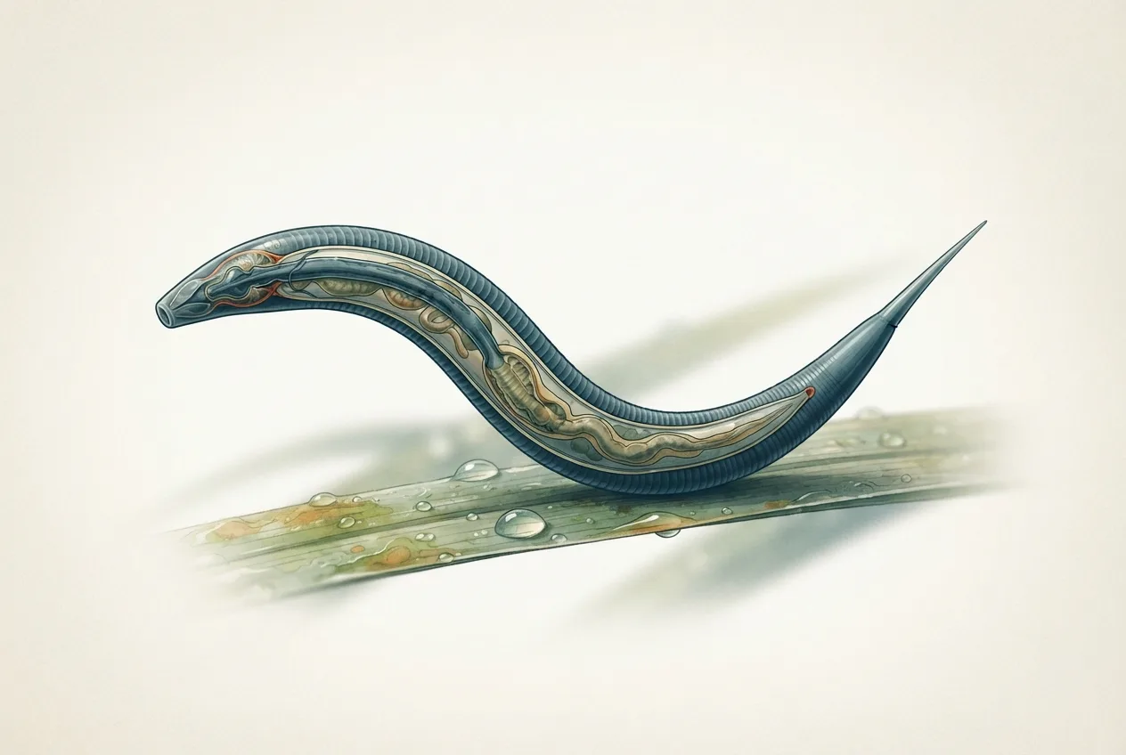
Haemonchus contortus, commonly known as the barber's pole worm, is a highly pathogenic blood-feeding nematode parasite of the abomasum in small ruminants, particularly sheep and goats. This parasite is a member of the family Trichostrongylidae and is responsible for significant economic losses in the livestock industry worldwide. The common name "barber's pole worm" derives from the characteristic spiral appearance of the female reproductive tract (white) wrapped around the blood-filled intestine (red), creating a striped pattern. Haemonchosis, the disease caused by this parasite, is characterized by anemia, hypoproteinemia, weight loss, and reduced productivity. The global distribution of H. contortus is extensive, with particularly high prevalence in tropical, subtropical, and warm temperate regions where conditions favor the development and survival of free-living larval stages on pasture. The parasite's high fecundity, short generation time, and ability to undergo hypobiosis (arrested larval development) contribute to its epidemiological success and the challenges faced in its control.
Taxonomy and Morphology
Haemonchus contortus belongs to the phylum Nematoda, order Strongylida, and family Trichostrongylidae. The genus Haemonchus is characterized by a lancet-like tooth within the buccal capsule, which is used to lacerate the abomasal mucosa and feed on host blood. Adult worms are slender and measure 10 to 30 mm in length. Females are larger than males, reaching up to 30 mm, and are easily identified by the visible barber's pole pattern. Males are smaller, measuring 10 to 20 mm, and possess a well-developed copulatory bursa with characteristic spicules and a gubernaculum. The eggs are oval, thin-shelled, and measure approximately 70 to 90 micrometers by 40 to 50 micrometers. They are passed in the feces and contain a morula of 16 to 32 cells when freshly deposited. The first-stage larvae (L1) hatch from the eggs and develop through two molts to the third-stage infective larvae (L3), which are ensheathed and resistant to environmental conditions.
Life Cycle
The life cycle of H. contortus is direct, meaning it does not require an intermediate host. Adult female worms in the abomasum produce large numbers of eggs, which are passed in the feces of the infected host. Under favorable conditions of moisture and temperature (optimal range 18 to 26 degrees Celsius), the eggs hatch, releasing first-stage larvae (L1). These larvae feed on bacteria in the feces and molt to second-stage larvae (L2), which then molt to the third-stage infective larvae (L3). The L3 retain the cuticle of the L2 as a protective sheath. This development from egg to L3 typically takes 7 to 14 days under optimal conditions. The L3 migrate from the fecal pellet onto herbage, where they await ingestion by a grazing host. After ingestion, the L3 exsheath in the rumen and migrate to the abomasum. Within the abomasum, they penetrate the mucosa and undergo two more molts to become fourth-stage larvae (L4) and then young adults. The prepatent period, from ingestion of L3 to the appearance of eggs in the feces, is approximately 18 to 21 days. A key feature of the H. contortus life cycle is the ability of the L4 to undergo hypobiosis, a state of arrested development within the abomasal mucosa. This occurs in response to adverse environmental conditions, such as cold winters or dry summers, and allows the parasite to survive periods when transmission is not possible. Hypobiotic larvae resume development when conditions become favorable again, often synchronizing with the spring or rainy season.
graph TD
    A["Adult worms in abomasum"] --> B["Eggs passed in feces"]
    B --> C["L1 larvae hatch in feces"]
    C --> D["L2 larvae develop in feces"]
    D --> E["L3 infective larvae on pasture"]
    E --> F["Ingestion by grazing host"]
    F --> G["L3 exsheath in rumen"]
    G --> H["L3 migrate to abomasum"]
    H --> I["L4 larvae penetrate mucosa"]
    I --> J["Young adults in abomasum"]
    J --> A
    I -.-> K["Hypobiosis (arrested L4)"]
    K -.-> I
Pathogenesis and Clinical Signs
The primary pathogenic mechanism of H. contortus is blood feeding. Each adult worm can consume approximately 0.05 mL of blood per day. In heavy infections, with thousands of worms present, this can result in a daily blood loss of 50 to 100 mL or more. The blood loss leads to a severe, often fatal, anemia. The pathogenesis is further compounded by the loss of plasma proteins, resulting in hypoproteinemia and edema. The clinical signs of haemonchosis are directly related to the degree of anemia and are often categorized as peracute, acute, or chronic. Peracute disease is seen in young lambs with a sudden, massive intake of L3. It is characterized by sudden death due to severe blood loss before anemia is clinically apparent. Acute disease is the most common form and presents with progressive anemia, pale mucous membranes, weakness, lethargy, and submandibular edema (bottle jaw). Affected animals may also show weight loss, reduced wool growth, and diarrhea. Chronic disease is seen in older animals with lower, persistent worm burdens. It is characterized by a more gradual onset of anemia, poor body condition, and reduced productivity. The FAMACHA system, a clinical scoring method based on the color of the ocular mucous membranes, is a widely used tool for assessing the degree of anemia in individual animals and guiding treatment decisions. This system is detailed in the article Haemonchus contortus in Sheep: Anthelmintic Resistance and FAMACHA-Based Control.
Diagnosis
The diagnosis of haemonchosis is based on a combination of clinical signs, epidemiological data, and laboratory findings. Fecal examination using quantitative techniques, such as the modified McMaster method, is used to determine the number of eggs per gram (EPG) of feces. While EPG counts are useful for estimating the level of infection, they do not always correlate perfectly with worm burden due to factors such as the fecundity of female worms and the consistency of the feces. The eggs of H. contortus are morphologically similar to those of other strongyle-type nematodes, such as Trichostrongylus and Oesophagostomum species. Therefore, differentiation requires larval culture and identification of the infective L3. The L3 of H. contortus are characterized by a prominent, refractile buccal capsule and a long, whip-like tail. Postmortem examination is the definitive diagnostic method, allowing for the direct visualization and counting of adult worms in the abomasum. The characteristic barber's pole appearance of the female worms makes them easily identifiable. Molecular diagnostic techniques, including polymerase chain reaction (PCR) and real-time PCR assays, have been developed for the specific detection and quantification of H. contortus DNA in fecal samples. These methods offer high sensitivity and specificity and can differentiate H. contortus from other trichostrongylid species. Serological tests, such as enzyme-linked immunosorbent assays (ELISAs) for the detection of antibodies against H. contortus antigens, are also available but are more commonly used for research purposes than for routine clinical diagnosis.
Epidemiology
The epidemiology of H. contortus is heavily influenced by environmental factors, particularly temperature and moisture. The free-living stages (eggs, L1, L2, and L3) are highly susceptible to desiccation and extreme temperatures. Optimal conditions for development and survival are warm (18 to 26 degrees Celsius) and moist. In tropical and subtropical regions, transmission can occur year-round, with peaks during the rainy season. In temperate regions, transmission is typically seasonal, with a peak in late spring and early summer. The ability of the L4 to undergo hypobiosis is a critical survival strategy. Hypobiotic larvae accumulate in the abomasal mucosa during the winter or dry season and resume development in the spring, providing a source of infection for new lambs. The periparturient rise in fecal egg counts in ewes is another important epidemiological feature. This phenomenon, which occurs around the time of lambing, is due to a temporary relaxation of immunity and results in a significant increase in pasture contamination. Management practices, such as stocking density, grazing management, and anthelmintic use, also play a major role in the epidemiology of haemonchosis. Overgrazing and continuous use of the same pastures can lead to high levels of pasture contamination. The widespread and often indiscriminate use of anthelmintics has led to the development of resistance in H. contortus populations, which is now a major global concern.
Anthelmintic Resistance
Anthelmintic resistance in H. contortus is a well-documented and increasingly prevalent problem. Resistance has been reported to all major classes of anthelmintics, including benzimidazoles, imidazothiazoles (levamisole), macrocyclic lactones (ivermectin, moxidectin), and, more recently, monepantel and derquantel. The mechanisms of resistance are diverse and include target site mutations (e.g., beta-tubulin gene mutations for benzimidazole resistance), increased drug efflux (e.g., P-glycoprotein overexpression for macrocyclic lactone resistance), and enhanced drug metabolism. The diagnosis of anthelmintic resistance is typically performed using the fecal egg count reduction test (FECRT). This test involves measuring the fecal egg count in a group of animals before and after treatment with a specific anthelmintic. A reduction of less than 95% (or a lower 95% confidence interval below 90%) is indicative of resistance. Molecular tests for detecting resistance-associated alleles, such as PCR-based assays for the benzimidazole resistance-associated mutations in the isotype-1 beta-tubulin gene, are also available. The management of anthelmintic resistance requires an integrated approach that includes the use of targeted selective treatment (TST) strategies, such as the FAMACHA system, to reduce the selection pressure for resistance. Other strategies include the use of combination anthelmintic products, the maintenance of a refugia of susceptible worms, and the implementation of pasture management practices to reduce the reliance on anthelmintics.
Control and Management
The control of H. contortus requires an integrated approach that combines strategic anthelmintic use with pasture management and host management practices. The goal is to reduce the level of pasture contamination and minimize the exposure of susceptible animals to infective larvae. Strategic anthelmintic treatments are timed to coincide with periods of high transmission risk, such as the spring and early summer in temperate regions. Targeted selective treatment (TST) strategies, such as the FAMACHA system, are increasingly recommended to reduce the selection pressure for anthelmintic resistance. Under this system, only animals showing clinical signs of anemia are treated, leaving a proportion of the worm population unexposed to the drug (refugia). Pasture management practices include rotational grazing, mixed grazing with cattle or other species, and the use of hay or silage crops to break the parasite life cycle. Resting pastures for extended periods (e.g., 6 to 12 months) can also reduce the number of infective larvae. Host management practices include breeding for genetic resistance to haemonchosis, as some breeds of sheep (e.g., Red Maasai, Gulf Coast Native) are known to be more resistant or resilient to infection. Nutritional management, particularly ensuring adequate protein intake, can also improve the host's ability to tolerate infection. Biological control methods, such as the use of nematophagous fungi (e.g., Duddingtonia flagrans) to reduce the number of L3 on pasture, are under investigation. The development of a vaccine against H. contortus has been a long-standing research goal. A commercial vaccine based on the gut membrane proteins of the adult worm (Barbervax) is available in some countries and has shown efficacy in reducing worm burdens and egg counts.
Differential Diagnosis
The differential diagnosis for haemonchosis includes other causes of anemia and weight loss in small ruminants. Other gastrointestinal nematodes, such as Teladorsagia circumcincta and Trichostrongylus species, can cause similar clinical signs, although they are typically less pathogenic. Liver fluke (Fasciola hepatica) infection can also cause anemia and hypoproteinemia, particularly in chronic cases. Nutritional deficiencies, such as copper or cobalt deficiency, can lead to poor growth and anemia. Other infectious diseases, such as caseous lymphadenitis and Johne's disease, can cause chronic weight loss. A thorough clinical examination, combined with appropriate diagnostic testing, is essential for an accurate diagnosis. The article Gastrointestinal Parasites in Sheep: Worms and Control provides further information on the differential diagnosis of parasitic infections in sheep.
Public Health Significance
Haemonchus contortus is not considered a zoonotic pathogen. It is a highly host-specific parasite of ruminants and does not infect humans. Therefore, there are no direct public health concerns associated with this parasite. However, the economic impact of haemonchosis on the livestock industry can have indirect effects on food security and the cost of animal products.
Conclusion
Haemonchus contortus remains one of the most economically important parasites of small ruminants globally. Its high pathogenicity, high fecundity, and ability to develop resistance to anthelmintics make it a formidable challenge for livestock producers and veterinarians. Effective control requires a comprehensive, integrated approach that combines strategic anthelmintic use, pasture management, and host management practices. The continued development of novel control strategies, including vaccines and biological control agents, is essential for the sustainable management of this parasite in the face of increasing anthelmintic resistance.
Frequently Asked Questions
What is the barber's pole worm?
Haemonchus contortus, known as the barber's pole worm, is a blood-feeding nematode parasite of the abomasum in sheep and goats, named for the striped appearance of the female worm.
How is haemonchosis transmitted?
Transmission occurs through the ingestion of third-stage infective larvae (L3) on pasture, which develop from eggs passed in the feces of infected animals.
What are the clinical signs of haemonchosis?
The primary clinical sign is anemia, which can range from mild to severe, and is often accompanied by weakness, lethargy, submandibular edema (bottle jaw), and weight loss.
How is haemonchosis diagnosed?
Diagnosis is based on clinical signs, fecal egg counts, larval culture for species identification, and postmortem examination for adult worms.
What is the FAMACHA system?
The FAMACHA system is a clinical tool that uses the color of the ocular mucous membranes to assess the degree of anemia in individual animals, guiding targeted selective treatment decisions.
What is anthelmintic resistance?
Anthelmintic resistance is the heritable ability of a parasite population to survive a dose of an anthelmintic drug that would normally be lethal, and it is a major problem in H. contortus control.
How can anthelmintic resistance be managed?
Management strategies include targeted selective treatment, maintaining a refugia of susceptible worms, using combination anthelmintic products, and implementing pasture management practices.
Is Haemonchus contortus a zoonotic parasite?
No, H. contortus is not zoonotic and does not infect humans.
What is hypobiosis in H. contortus?
Hypobiosis is a state of arrested larval development (L4) within the abomasal mucosa, allowing the parasite to survive adverse environmental conditions.
What are the key control strategies for haemonchosis?
Key strategies include strategic anthelmintic use, pasture rotation, mixed grazing, breeding for resistance, and nutritional management.
Related Clinical & Scientific Guides
- Tick-Borne Diseases in Dogs: Pathogens, Clinical Signs, Diagnosis, and Prevention
- Toxoplasmosis in Cats and the Risk of Brain Infection in Humans
- Dog Heartworm and Tick-Borne Disease Prevention
References
- Merck Veterinary Manual. Haemonchosis. Merck & Co., Inc.
- Taylor, M. A., Coop, R. L., & Wall, R. L. Veterinary Parasitology. 4th ed. Wiley-Blackwell.
- Bowman, D. D. Georgis' Parasitology for Veterinarians. 11th ed. Elsevier.
- Zajac, A. M., & Conboy, G. A. Veterinary Clinical Parasitology. 8th ed. Wiley-Blackwell.
- Besier, R. B., Kahn, L. P., Sargison, N. D., & Van Wyk, J. A. The Pathophysiology, Ecology and Epidemiology of Haemonchus contortus Infection in Small Ruminants. Advances in Parasitology, 93, 95-143.
- Sargison, N. D. Pharmaceutical Control of Endoparasitic Infections in Sheep. Veterinary Clinics of North America: Food Animal Practice, 27(1), 139-156.
- Kaplan, R. M. Drug Resistance in Nematodes of Veterinary Importance: A Status Report. Trends in Parasitology, 20(10), 477-481.
- Van Wyk, J. A., & Bath, G. F. The FAMACHA System for Managing Haemonchosis in Sheep and Goats by Clinically Identifying Individual Animals for Treatment. Veterinary Research, 33(5), 509-529.
- Knox, D. P., & Smith, W. D. Vaccination Against Haemonchus contortus: A Review. Veterinary Parasitology, 98(1-3), 135-150.
- Coles, G. C., Jackson, F., Pomroy, W. E., Prichard, R. K., von Samson-Himmelstjerna, G., Silvestre, A., Taylor, M. A., & Vercruysse, J. The Detection of Anthelmintic Resistance in Nematodes of Veterinary Importance. Veterinary Parasitology, 136(3-4), 167-185.
Disclaimer This article is for educational and informational purposes only. It is not intended to substitute for professional veterinary advice, diagnosis, treatment, or regulatory guidance. Always consult a licensed veterinarian or qualified specialist regarding animal health, disease diagnosis, and therapeutic decisions.