Ichthyophthirius multifiliis (White Spot / Ich): A Comprehensive Reference on the Ciliate Parasite of Freshwater Fish
By Dr. Zubair Khalid, DVM, MS, PhD ·
Key Takeaways
- Ichthyophthirius multifiliis is an obligate parasitic ciliate causing white spot disease in freshwater fish, leading to significant global aquaculture losses exceeding one billion USD annually. The parasite's life cycle involves infective theronts, parasitic trophonts within host epithelium, and free-swimming protomonts and encysted tomonts for reproduction.
- Pathogenesis involves severe histopathological changes in fish gills and skin, including lamellar fusion, hyperplasia, necrosis, and inflammatory infiltration, leading to osmoregulatory and respiratory dysfunction. Immune responses are species-specific, with some fish exhibiting efficient clearance while others show subdued early responses.
- Diagnostic methods include classical microscopic identification of trophonts in scrapings and histopathology, alongside highly sensitive molecular techniques like TaqMan probe-based qPCR targeting the ICP2 gene, capable of detecting as few as 4 theronts per liter of water. AI-driven behavioral monitoring can detect early infection indicators before visible signs appear.
- Treatment and control strategies are diverse, ranging from chemical antiparasitics (e.g., BHTCA, catechol compounds, berberine) and natural plant extracts to nanoparticle-based therapies and dietary immunostimulants. Biological control using cyclopoid copepods that prey on theronts offers a sustainable approach.
- I. multifiliis infection significantly disrupts the fish microbiome, leading to dysbiosis and increased susceptibility to secondary bacterial infections, particularly motile Aeromonas septicemia. The parasite has also been detected in wild amphibian larvae, indicating a broader host range.
- Experimental DNA vaccines targeting antigens like IAG52A and β-tubulin have demonstrated immunogenicity, and functional gene silencing via antisense oligonucleotides (ASOs) targeting essential parasite genes like hsp90 shows therapeutic potential. Cysteine proteases are critical for invasion and represent a key therapeutic target.
Introduction
Ichthyophthirius multifiliis is an obligate parasitic ciliate that causes ichthyophthiriasis, commonly known as white spot disease, in freshwater fish worldwide. The pathogen infects virtually all species of freshwater teleost fish and is responsible for substantial economic losses in global aquaculture, with annual losses exceeding one billion USD [<a href="#ref-1">1</a>]. The disease is characterized by the appearance of small white nodules (trophonts) on the skin, fins, and gills of infected fish, leading to osmoregulatory dysfunction, respiratory distress, and secondary infections [<a href="#ref-2">2</a>, <a href="#ref-3">3</a>, <a href="#ref-4">4</a>]. This article provides an exhaustive review of the biology, pathogenesis, diagnostics, and control of I. multifiliis, with emphasis on recent molecular and ecological advances. Cross-referencing is made to the companion article on Ichthyophthirius multifiliis (White Spot Disease) in Farmed Fish: Advances in Molecular Detection and Treatment for additional context.
Taxonomy and Morphology
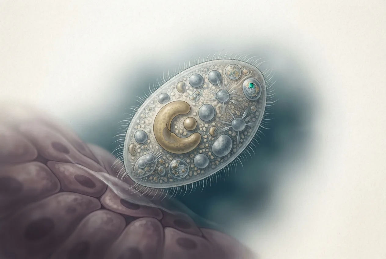
I. multifiliis belongs to the phylum Ciliophora, class Oligohymenophorea, order Ichthyophthiriidae. The parasite possesses two distinct extrusome types: type I mucocysts (crystalline, oval, 0.7-1.4 × 0.6-1.1 μm) and type II toxicysts (rod-shaped, 2.0-3.0 × 0.2-0.3 μm), which are involved in attachment and invasion [<a href="#ref-5">5</a>]. The trophont stage is characterized by a horseshoe-shaped macronucleus and uniform ciliation [<a href="#ref-6">6</a>]. Single-cell transcriptome sequencing has identified stage-specific gene expression, with theronts highly expressing leishmanolysin family proteins, heat shock proteins, transmembrane proteins, and cysteine proteases critical for invasion [<a href="#ref-7">7</a>]. Rab family GTPases (25 genes) govern extrusome exocytosis across the life cycle [<a href="#ref-5">5</a>].
Life Cycle
The life cycle of I. multifiliis consists of four main stages: theront (infective free-swimming stage), trophont (parasitic feeding stage in host epithelium), protomont (short free-swimming stage after exiting host), and tomont (encysted reproductive stage) [<a href="#ref-5">5</a>]. The following Mermaid diagram summarizes the life cycle:
graph TD
 A["Theront: free-swimming, infective"] --> B["Attaches to host skin/gills"]
 B --> C["Trophont: feeding stage in host epithelium"]
 C --> D["Protomont: exits host, short free-swimming"]
 D --> E["Tomont: encysted, divides within cyst"]
 E --> F["Theronts released from tomont"]
 F --> A
Theronts actively seek and invade host epithelial tissues, facilitated by extrusome discharge and cysteine protease activity [<a href="#ref-5">5</a>, <a href="#ref-7">7</a>]. The trophont feeds on host cellular debris, enlarges significantly, and then exits the host as a protomont. Protomonts attach to substrates and form tomonts, which undergo repeated binary fission to produce 200-1000 new theronts, completing the cycle in 3-7 days at optimal water temperatures (20-25°C).
Pathogenesis and Host Immune Response
Infection by I. multifiliis induces profound pathological changes in host tissues. Histopathological examination of infected goldfish (Carassius auratus) gills reveals lamellar fusion, cell hyperplasia, hyperemia, inflammatory infiltration, necrosis, and desquamation [<a href="#ref-3">3</a>]. Similar findings are reported in grass carp (Ctenopharyngodon idella), with epithelial necrosis and inflammatory cell infiltration in gill and fin tissues [<a href="#ref-2">2</a>]. In rainbow trout (Oncorhynchus mykiss), systemic pathological disorders include decreased erythrocyte counts, increased leukocyte counts with neutrophilia, elevated serum AST (750.9 ± 147.7 U/L), ALT (39.4 ± 6.7 U/L), and alkaline phosphatase (467.8 ± 72.6 U/L), together with histological damage to gill epithelium, renal tubules, hepatocytes, and splenic reticuloendothelial cells [<a href="#ref-4">4</a>].
The ocular mucosa of rainbow trout serves as an additional invasion site for I. multifiliis, triggering a strong mucosal immune response with upregulation of immune-related genes. Transcriptomic analyses of infected rainbow trout and zebrafish reveal species-specific early immune responses: zebrafish clear the infection efficiently, whereas rainbow trout exhibit a subdued early response [<a href="#ref-8">8</a>]. Comparative immune gene expression profiling across five fish pathogens demonstrates that I. multifiliis induces a distinct Th1/Th2/Th17 and innate immune gene expression cluster in rainbow trout.
Mucosal immunity is central to host defense. The mucosal immune gene IgT and pro-inflammatory cytokine TNF-α are upregulated in grass carp skin, gill, and gut following infection [<a href="#ref-2">2</a>]. In Takifugu fasciatus, RT-qPCR analysis shows immune responses in spleen, liver, gill, skin, and gut, with RNA-seq revealing enrichment of Th1 and Th2 cell differentiation pathways in spleen and ECM-receptor interaction, calcium signaling, and PI3K-Akt signaling pathways in skin [<a href="#ref-9">9</a>]. Transcriptomic and metabolomic analyses of Rhinogobio ventralis skin identify 388 differentially expressed genes and 135 differentially expressed metabolites at 7 days post-infection (dpi), involving ribosome, protein processing, antigen processing, and glycerophospholipid metabolism pathways [<a href="#ref-10">10</a>]. Co-expression network analysis in grass carp has identified genes associated with resistance to I. multifiliis.
DNA vaccination studies have progressed. A genetic vaccine containing the IAG52A antigen from I. multifiliis, adjuvanted with IL-8 from Astyanax lacustris, induced IL-1β mRNA expression and leukocyte migration at the injection site [<a href="#ref-11">11</a>]. A β-tubulin DNA vaccine also elicited immune responses in grass carp.
Diagnostic Methods
Classical and Histological Diagnosis
Presumptive diagnosis is based on clinical signs (white spots, flashing, lethargy) and microscopic identification of trophonts in skin or gill scrapings [<a href="#ref-12">12</a>]. Histopathology using hematoxylin and eosin staining reveals the characteristic ciliated trophonts with macronucleus within epithelial lesions [<a href="#ref-3">3</a>, <a href="#ref-6">6</a>, <a href="#ref-10">10</a>]. Transmission electron microscopy (TEM) further identifies extrusome types and cellular damage [<a href="#ref-5">5</a>, <a href="#ref-10">10</a>].
Molecular Detection
A TaqMan probe-based quantitative PCR (qPCR) assay targeting the cathepsin L cysteine protease (ICP2) gene has been developed, with a detection limit of 4 theronts per liter of water [<a href="#ref-13">13</a>]. The method demonstrates high sensitivity, specificity, and reproducibility, and can quantify parasite loads in water, sediment, and fish tissues [<a href="#ref-13">13</a>]. The linear model Ct = -3.312 lg(SQ) + 34.47 (R² = 0.9636) allows estimation of theront numbers from Ct values [<a href="#ref-13">13</a>].
An environmental DNA/RNA (eDNA/eRNA) approach has also been implemented for non-invasive detection of I. multifiliis in aquaculture systems [<a href="#ref-14">14</a>]. This method detects the parasite in water samples without requiring fish handling, enabling early warning.
AI-Driven Behavioral Monitoring
Artificial intelligence-based video monitoring systems can detect early behavioral changes in infected rainbow trout, such as movement towards higher water currents at 4-5 dpi, before visible white spots appear [<a href="#ref-12">12</a>]. This approach uses object detection, classification, and tracking to quantify fish positioning and movement patterns, improving animal welfare and enabling earlier intervention [<a href="#ref-12">12</a>].
Host-Microbiota-Parasite Interactions
I. multifiliis infection induces significant dysbiosis in the fish microbiota, which may predispose hosts to secondary bacterial infections. In grass carp, 16S rRNA sequencing reveals reduced skin microbiota diversity, with proliferation of opportunistic pathogens (Aeromonas, Vibrio) in the intestine and increases in Flavobacterium and Candidatus Megaira in skin and gills [<a href="#ref-2">2</a>]. Correlation analyses show positive associations between Aeromonas/Vibrio abundance and host pathology [<a href="#ref-2">2</a>].
In goldfish, I. multifiliis infection decreases alpha and beta diversity of gill microbiota, with Candidatus Megaira becoming dominant [<a href="#ref-3">3</a>]. Gut microbiota show no significant diversity changes but exhibit increased abundances of Aeromonas and Achromobacter [<a href="#ref-3">3</a>]. In Takifugu fasciatus, infection reduces gut microbiota alpha diversity, with a decrease in commensal Chryseobacterium and an increase in opportunistic Elizabethkingia [<a href="#ref-9">9</a>].
The naturally resistant fish species Percocypris pingi possesses a unique skin metabolome characterized by high glutathione levels and a distinct, more diverse microbial community lacking certain parasitic bacteria, compared to susceptible crucian carp and yellow catfish [<a href="#ref-15">15</a>]. This suggests that both antioxidant capacity and microbiome composition contribute to resistance.
Treatment and Control
Chemical and Natural Antiparasitic Compounds
Formaldehyde and malachite green have historically been used but are now restricted due to environmental and food safety concerns [<a href="#ref-1">1</a>]. Numerous candidate compounds have been evaluated. The synthetic isoquinoline derivative BHTCA shows stage-selective efficacy: theront EC50 = 0.10 mg/L at 4 h, tomont EC50 = 0.40 mg/L at 24 h, and reduces parasite loads by 78.1% and host mortality by 66.7% in grass carp at 0.6 mg/L, with a therapeutic index >167 [<a href="#ref-1">1</a>]. Catechol compounds (quercetin, luteolin, caffeic acid, chlorogenic acid) exhibit potent antiparasitic effects in vitro and in vivo, targeting dipeptidyl peptidase (DPP) and promoting macrophage M2 polarization via STAT1 inhibition [<a href="#ref-16">16</a>]. Magnolol-based compounds also demonstrate strong activity [<a href="#ref-17">17</a>].
Plant extracts and essential oils offer eco-friendly alternatives. Sophoraflavanone G from Sophora flavescens kills theronts at 0.5 mg/L and tomonts at 2 mg/L in vitro, with a 96-h LC50 of 46.6 mg/L in grass carp. Berberine kills 99.3% of theronts at 15 mg/L and reduces theront release from tomonts at 5 mg/L, with a safety margin of 67-fold (LC50 528.44 mg/L in goldfish). Essential oils from sage (Salvia officinalis), lavender (Lavandula officinalis), and oregano (Origanum onites) achieve 100% mortality of trophonts at 0.50 mL/L (sage, lavender) and 0.10-0.50 mL/L (oregano) within 60 minutes. Menthe, onion, and garlic oils yield 92-94% mortality. Extracts from Nelumbo nucifera, Glycines testa, and Agrimonia eupatoria are also effective against multiple life stages [<a href="#ref-18">18</a>].
Turmeric oil supplementation at 10 ppm enhances survival against co-infection with I. multifiliis and Aeromonas hydrophila in Pangasianodon hypophthalmus, modulating stress, antioxidant, and immune responses (increased SOD, CAT, IL-1β, transferrin, C3, HSP70, HSP90, IgM) [<a href="#ref-19">19</a>].
Nanoparticle-Based Therapies
Chlorella vulgaris extract conjugated to magnetic iron oxide nanoparticles (ChVE@Mag iron NPs) fed to Nile tilapia improved growth, antioxidant status, and immune parameters, and reduced I. multifiliis infection (62% decrease in infection, 84% decrease in mortality, 92% fewer white spots) [<a href="#ref-20">20</a>]. The treatment also mitigated inflammatory gene expression (IL-1β, TNFα, COX-2, iNOS) and enhanced survival during subsequent A. hydrophila challenge (2% mortality vs 43% in controls) [<a href="#ref-20">20</a>].
Dietary Immunostimulants
Concentrated mannan oligosaccharide (cMOS) supplementation in goldfish increased gill lamella length, dermal thickness, mucin cell counts, lysozyme and alkaline phosphatase activity, and Muc-2 expression, leading to lower mortality and fewer white spots after I. multifiliis challenge.
Vaccination and Gene Silencing
DNA vaccines encoding I. multifiliis antigens (IAG52A, β-tubulin) have shown immunogenicity, with measurable mRNA expression and leukocyte infiltration at the injection site [<a href="#ref-11">11</a>]. Functional gene silencing via antisense oligonucleotides (ASOs) targeting heat shock protein 90 (hsp90) in I. multifiliis reduces theront motility, growth, and infectivity; common carp exposed to ASO-treated theronts had 0% mortality vs 100% in controls. This identifies hsp90 as a promising therapeutic target. The cysteine protease inhibitor E-64 kills theronts and protomonts in vitro and inhibits tomont division and theront invasion, underscoring the essential role of cysteine proteases [<a href="#ref-7">7</a>].
Biological Control
Cyclopoid copepods (Cyclops vicinus, Thermocyclops taihokuensis, Mesocyclops spp., Macrocyclops sp., Paracyclopina sp.) prey on free-swimming theronts, as demonstrated by fluorescence labeling and qPCR [<a href="#ref-21">21</a>]. In co-culture, the presence of these copepods significantly reduces I. multifiliis infection in goldfish. The freshwater clam Corbicula fluminea has also been assessed for controlling infections [<a href="#ref-22">22</a>]. This biological control strategy offers a sustainable complement to chemical treatments.
Co-Infections with Bacteria
I. multifiliis infection predisposes fish to secondary bacterial infections, particularly motile Aeromonas septicemia caused by Aeromonas hydrophila. Natural outbreaks in Pangasianodon hypophthalmus confirmed co-infection with I. multifiliis and A. hydrophila via 16S rRNA sequencing [<a href="#ref-19">19</a>]. In rainbow trout, a mixed Gram-negative bacterial flora (Enterobacter sp., Citrobacter freundii, Acinetobacter calcoaceticus) was isolated from the liver of infected fish [<a href="#ref-4">4</a>]. The bacterial load of A. hydrophila in co-infected fish is higher than in fish infected with bacteria alone, indicating synergistic pathogenesis [<a href="#ref-19">19</a>, <a href="#ref-20">20</a>].
Amphibian Infections
I. multifiliis has been detected in wild amphibian larvae (Salamandra salamandra and Rana temporaria) in Catalonia, based on histological identification of trophonts in gill tissue [<a href="#ref-6">6</a>]. These findings confirm that the parasite is not strictly fish-specific and highlight the need to include I. multifiliis in amphibian disease monitoring programs [<a href="#ref-6">6</a>].
FAQ
What is the causative agent of white spot disease in freshwater fish?
The causative agent is the ciliate protozoan Ichthyophthirius multifiliis, an obligate parasite that infects the skin, gills, and fins of freshwater teleosts.
How does Ichthyophthirius multifiliis infect fish?
The infective theront stage actively penetrates host epithelium using specialized extrusomes (toxicysts) and cysteine proteases, then feeds and matures as a trophont within the host tissue [<a href="#ref-5">5</a>, <a href="#ref-7">7</a>].
What are the typical clinical signs of ichthyophthiriasis?
Clinical signs include visible white nodules (trophonts) on the body, flashing (rubbing against objects), lethargy, anorexia, equilibrium disturbances, and respiratory distress [<a href="#ref-4">4</a>, <a href="#ref-12">12</a>].
Which molecular diagnostic methods are available for I. multifiliis?
A TaqMan probe-based qPCR targeting the ICP2 gene is highly sensitive (4 theronts/L water) and specific [<a href="#ref-13">13</a>]. Environmental DNA/RNA approaches also enable non-invasive detection [<a href="#ref-14">14</a>].
Can I. multifiliis infect amphibians?
Yes, I. multifiliis has been confirmed in wild salamander and frog larvae, indicating it can infect non-fish hosts [<a href="#ref-6">6</a>].
What treatments are effective against I. multifiliis?
Several compounds show efficacy: BHTCA, catechol compounds, sophoraflavanone G, berberine, magnolol derivatives, and essential oils from sage, lavender, and oregano [<a href="#ref-1">1</a>, <a href="#ref-16">16</a>, <a href="#ref-17">17</a>, <a href="#ref-23">23</a>]. Dietary immunostimulants and nanoparticle conjugates also reduce infection [<a href="#ref-20">20</a>].
Are there biological control methods for white spot disease?
Cyclopoid copepods prey on theronts and can reduce infection levels in aquaculture systems [<a href="#ref-21">21</a>].
How does I. multifiliis infection affect the fish microbiome?
Infection causes dysbiosis, with decreased microbial diversity and increased abundance of opportunistic pathogens such as Aeromonas and Vibrio in the gut and skin [<a href="#ref-2">2</a>, <a href="#ref-3">3</a>, <a href="#ref-9">9</a>].
Is there a vaccine for ichthyophthiriasis?
Experimental DNA vaccines encoding IAG52A or β-tubulin have induced immune responses, but no commercial vaccine is yet available [<a href="#ref-11">11</a>].
What is the role of cysteine proteases in I. multifiliis?
Cysteine proteases are highly expressed in theronts and are essential for host invasion. The inhibitor E-64 blocks parasite development and infectivity [<a href="#ref-7">7</a>].
Related Articles
- Argulus (Fish Louse)
- Gyrodactylus salaris: Taxonomy, Pathogenesis, Epidemiology, and Control of the Salmonid Monogenean
- Lernaea (Anchor Worm)
Related Clinical & Scientific Guides
- Tick-Borne Diseases in Dogs: Pathogens, Clinical Signs, Diagnosis, and Prevention
- Toxoplasmosis in Cats and the Risk of Brain Infection in Humans
- Dog Heartworm and Tick-Borne Disease Prevention