
Ichthyophthirius multifiliis (White Spot Disease) in Farmed Fish: Advances in Molecular Detection and Treatment
Introduction
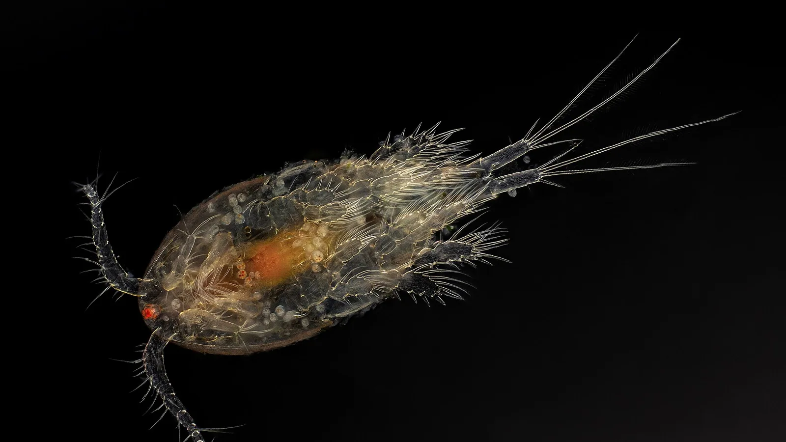
Ichthyophthirius multifiliis, a ciliated protozoan parasite of the phylum Ciliophora, is the etiological agent of ichthyophthiriasis, commonly known as white spot disease. This parasite is one of the most significant pathogens in global freshwater aquaculture, infecting a broad range of teleost fish species in both intensive and extensive production systems [1, 2]. The disease is characterized by the presence of small, white, raised nodules on the skin, gills, and fins, which correspond to the intradermal trophont stage of the parasite. Morbidity and mortality rates can approach 100% in naive populations, leading to substantial economic losses in the aquaculture industry [3, 4]. The life cycle of I. multifiliis is direct and temperature-dependent, comprising four distinct stages: the infective theront, the parasitic trophont, the reproductive tomont, and the tomite [5]. Understanding the biophysical and molecular interactions at each stage is critical for developing targeted diagnostic and therapeutic strategies.
Pathobiology and Life Cycle
The reproductive capacity of I. multifiliis is formidable. A single trophont, after leaving the host, can produce hundreds to thousands of tomites within a protective cyst known as the tomont. These tomites then develop into theronts, which actively seek out new hosts [6]. The theront stage is the only free-swimming, infective stage and represents the most vulnerable point in the parasite's life cycle for intervention. Theronts penetrate the host epithelium using a combination of mechanical force and enzymatic secretion, including proteases and phospholipases [7]. Once established, the trophont feeds on host cellular debris and fluid, growing rapidly within a parasitophorous vacuole in the epidermis. The host immune response is complex, involving both innate and adaptive mechanisms, but it is often insufficient to clear a heavy infestation before mortality occurs [8].
Clinical Presentation and Gross Pathology
Clinical signs of ichthyophthiriasis typically appear when the parasite burden is high. The classic white spots are 0.5 to 1.0 mm in diameter and are most easily observed on the fins, body, and cornea. Infected fish exhibit flashing behavior, lethargy, anorexia, and respiratory distress due to gill involvement [9]. Secondary bacterial infections, often caused by Aeromonas hydrophila, Flavobacterium columnare, or other opportunistic pathogens, are common sequelae that worsen clinical outcomes [10]. Histopathological examination reveals epithelial hyperplasia, necrosis, and an intense inflammatory infiltrate at the site of trophont attachment. In gill tissue, lamellar fusion and edema severely compromise gas exchange [11].
Advances in Molecular Detection
Traditional diagnosis of ichthyophthiriasis relies on microscopic identification of the characteristic horseshoe-shaped macronucleus in trophonts or tomonts. This method, while confirmatory, has limited sensitivity for detecting subclinical or early-stage infections. The development of molecular diagnostics, particularly polymerase chain reaction (PCR)-based assays, has significantly improved detection limits and turnaround times.
Conventional PCR and Nested PCR
Several primer sets targeting the 18S ribosomal RNA (rRNA) gene or the internal transcribed spacer (ITS) regions have been designed for I. multifiliis-specific amplification [12, 13]. Conventional PCR can detect parasite DNA at concentrations as low as 1 fg per reaction, representing the genetic material of a single theront. Nested PCR assays, which use two sequential amplification steps, achieve even higher sensitivity and specificity, enabling the detection of carrier fish with low-level infections [14].
Quantitative PCR (qPCR) for Theront Quantification
Quantitative real-time PCR (qPCR) has been employed to measure parasite burdens in both fish tissue and water samples. Several qPCR protocols use fluorescent probes (e.g., TaqMan or SYBR Green) targeting the 18S rRNA gene [15, 16]. The limit of detection for these assays is typically less than 10 copies of the target gene per reaction. By plotting cycle threshold (Ct) values against a standard curve of serial dilutions of a cloned plasmid insert, an absolute quantification of theront or trophont numbers per gram of tissue or per liter of water can be obtained [16]. This quantitative capability is particularly valuable for assessing treatment efficacy and monitoring disease progression.
Environmental DNA (eDNA) Monitoring
Environmental DNA (eDNA) monitoring represents a paradigm shift in aquatic pathogen surveillance. Water samples are collected, filtered through membranes of 0.45 to 0.22 micrometer pore size, and the retained DNA is extracted using commercial soil or water DNA extraction kits. The extracted DNA is then subjected to qPCR targeting I. multifiliis-specific sequences [17, 18]. eDNA assays can detect theronts and tomonts in water at concentrations as low as 1 tomont per 100 liters of water. This non-invasive technique allows for the detection of the parasite before clinical signs appear in fish, enabling proactive intervention. The sensitivity of eDNA monitoring is influenced by water temperature, flow rate, and the presence of PCR inhibitors such as humic acids and tannins [18, 19]. Inclusion of an internal amplification control is recommended to identify false negatives due to inhibition. A decision tree for integrating eDNA and qPCR results into a surveillance program is presented in Figure 1.
flowchart TD
 A[Water Sample Collection from Pond] --> B[Filtration and DNA Extraction]
 B --> C["Internal Amplification Control (IAC) Positive?"]
 C -- No --> D[Repeat Extraction or Inhibitor Removal]
 C -- Yes --> E[I. multifiliis qPCR Assay]
 E --> F[Ct Value Obtained]
 F --> G[Interpretation]
 G -- Ct < 35 --> H[High Parasite Burden / Active Outbreak Risk]
 G -- 35 ≤ Ct < 40 --> I[Low Parasite Burden / Subclinical Infection]
 G -- Ct ≥ 40 or No Amplification --> J[Negative / Below Detection Limit]
 H --> K[Initiate Quarantine and Treatment Protocol]
 I --> L[Increase Surveillance Frequency]
 J --> M[Maintain Routine Surveillance]
 K --> N[Post-Treatment Water Sampling]
 N --> F
Figure 1. Decision tree for environmental DNA (eDNA) monitoring of I. multifiliis using quantitative PCR (qPCR). The algorithm incorporates an internal amplification control (IAC) to monitor for PCR inhibition.
Multiplex PCR and Panel Diagnostics
Given that ichthyophthiriasis often occurs as part of a polymicrobial disease complex, multiplex PCR assays capable of simultaneously detecting I. multifiliis, various bacterial pathogens, and other protozoan parasites are under development [20]. These panels typically target the 18S rRNA gene of the ciliate in combination with species-specific virulence genes from bacterial pathogens such as Flavobacterium columnare and Aeromonas hydrophila. This approach allows a single diagnostic test to identify all primary etiological agents in a disease outbreak, a strategy analogous to panels used for Feline Upper Respiratory Tract Infection Complex.
Loop-Mediated Isothermal Amplification (LAMP)
Loop-mediated isothermal amplification (LAMP) offers a field-deployable alternative to PCR, as it does not require a thermal cycler. LAMP assays for I. multifiliis have been designed targeting the 5.8S rRNA gene and the ITS-2 region. These assays can detect as few as 10 copies of the target DNA within 30 to 60 minutes at a constant temperature of 60 to 65 degrees Celsius [21, 22]. Amplicon detection can be achieved using colorimetric dyes (e.g., hydroxynaphthol blue) or lateral flow dipsticks, making LAMP particularly suitable for on-site diagnostics in remote aquaculture facilities.
Chemotherapeutic Resistance and Treatment Strategies
Chemical treatment of ichthyophthiriasis has historically relied on the use of malachite green, formalin, and copper sulfate. However, the carcinogenicity of malachite green has led to its ban in many jurisdictions for use in food fish [23]. Formalin (37% formaldehyde solution) remains a widely used parasiticide in some regions, applied at concentrations of 15 to 25 mg/L in static or flow-through bath treatments. However, the efficacy of formalin is highly dependent on water temperature, pH, and organic load [24].
Resistance to Formalin and Other Agents
Reports of reduced susceptibility of I. multifiliis to formalin are increasing. In vitro assays using theront motility as a biomarker have shown that theronts from certain geographic isolates can tolerate formalin concentrations up to 30 mg/L for extended periods, whereas historical isolates were immobilized at 15 mg/L [25, 26]. The mechanism of resistance is not fully understood but may involve upregulation of detoxification enzymes, such as aldehyde dehydrogenases and glutathione S-transferases, and modulation of membrane permeability [27, 28]. Similarly, resistance to copper sulfate has been documented, with some strains showing a 3- to 5-fold increase in the 24-hour LC50 value compared to susceptible reference strains [29].
Alternative Chemotherapeutics
The search for efficacious and environmentally safe alternatives has led to the investigation of several compounds. Hydrogen peroxide at concentrations of 50 to 100 mg/L for 30 to 60 minutes has shown activity against theronts and tomonts, but it is highly toxic to some fish species, particularly in warm water [30]. Plant-derived compounds, including essential oils from oregano (Origanum vulgare), thyme (Thymus vulgaris), and garlic (Allium sativum), have demonstrated in vitro antiprotozoal activity. The active principle in garlic, allicin, disrupts the cell membrane integrity of theronts at concentrations as low as 5 microg/mL [31, 32]. Tannins and saponins extracted from Quillaja saponaria and other plants have likewise been shown to inhibit tomont encystment and theront excystment [33]. The development of resistance to these natural products has not been systematically studied but is a potential risk with widespread use.
Theront Stage Targeting: A Molecular Approach
The theront stage represents an optimal target for therapeutic intervention due to its brief free-swimming phase and essential role in transmission. Research has focused on identifying molecular pathways critical for theront motility, host recognition, and invasion. The calcium-dependent signaling pathways, including calmodulin and calmodulin-dependent kinases, have been shown to regulate ciliary beat frequency and directional motility in theronts [34, 35]. Inhibitors of these pathways, such as the calmodulin antagonist trifluoperazine, can immobilize theronts at micromolar concentrations in vitro, though their veterinary application has not been optimized [36]. Another promising molecular target is the surface immobilization antigen (i-antigen) complex. Monoclonal antibodies raised against specific i-antigen epitopes can agglutinate and immobilize theronts, preventing attachment to the host [37, 38]. Passive immunization strategies using anti-i-antigen antibodies are under investigation, but mass production costs for farm-level application remain prohibitive.
Vaccination and Immune Modulation
Recombinant vaccines expressing the i-antigen of several I. multifiliis serotypes have been tested in laboratory and field trials. Vaccination via intraperitoneal injection or immersion can elicit a strong antibody response and reduce parasite burden upon challenge by 60 to 80% [39, 40]. However, the antigenic diversity of the i-antigen across different geographic isolates poses a significant challenge for broad-spectrum vaccine development. Multivalent vaccines incorporating epitopes from multiple serotypes are being designed using computational epitope prediction and reverse vaccinology tools [41]. These efforts parallel vaccine development for other aquatic pathogens such as Streptococcus agalactiae in Farmed Tilapia.
Integrated Control and Biosecurity
No single treatment modality is likely to be sufficient for long-term control of ichthyophthiriasis. An integrated approach combining molecular surveillance, targeted chemotherapy, and improved husbandry is essential. The use of eDNA-based early warning systems allows for the detection of the parasite at low densities, enabling reactive treatments before the parasite burden becomes overwhelming [17, 42]. Stocking density reduction and water temperature manipulation can also slow the parasite life cycle and provide a window for treatment. Ozone and ultraviolet sterilization of influent water can eliminate theronts and tomonts in recirculating aquaculture systems (RAS) [43, 44]. A comprehensive integrated pest management (IPM) plan for Ichthyophthirius should include:
- Biosecurity: Quarantine of new stock for at least 2 weeks with eDNA screening.
- Surveillance: Weekly water sampling for eDNA qPCR, with a threshold for action set at Ct < 38.
- Chemical Treatment: Rotational use of formalin and hydrogen peroxide to delay resistance development.
- Biological Control: Introduction of cleaner fish species that feed on trapped trophonts (experimental).
- Vaccination: Deployment of autogenous or multivalent i-antigen vaccines in endemic regions.
Comparative Perspectives with Related Aquaculture Parasitic Diseases
The challenges associated with I. multifiliis parallel those observed in other significant aquaculture parasitic diseases. MASK_5 and the emerging threat of MASK_6 share a reliance on chemotherapeutics and the threat of resistance. In both sea lice and ich cases, molecular diagnostics have become the gold standard for early detection and drug resistance monitoring [45, 46]. The application of qPCR for environmental monitoring is also gaining traction for monitoring Mycobacterium marinum in aquatic environments [47]. The principles of integrated pest management developed for sea lice, including the use of biological delousers and fallowing periods, are directly translatable to I. multifiliis control in pond and cage systems [48].
Emerging Technologies and Bioinformatics
The application of bioinformatics to I. multifiliis research is still in its infancy compared to bacterial targets such as those in Porcine Reproductive and Respiratory Syndrome: Genomic Surveillance and Vaccine Strategies Using Bioinformatics. However, the increasing availability of whole-genome sequences for I. multifiliis isolates has enabled phylogenetic analyses that reveal population structure and gene flow between farms and wild fish populations [49]. Genome-wide association studies (GWAS) are being used to identify single nucleotide polymorphisms (SNPs) associated with formalin resistance, which could be incorporated into a molecular resistance panel [50]. Predictive computational models for theront dispersal and outbreak risk, incorporating water temperature, flow rates, and farm density, are being developed to optimize treatment timing and reduce chemical usage [51]. These models share methodological approaches with analyses of Avian Influenza H5N1 in Poultry and other rapidly spreading pathogens.
Conclusion
Ichthyophthirius multifiliis remains a formidable challenge for global aquaculture. The limitations of traditional microscopy-based diagnosis and the increasing prevalence of chemotherapeutic resistance demand a transition toward molecular methods. Environmental DNA monitoring by quantitative PCR offers a non-invasive, highly sensitive surveillance tool. Quantitative PCR assays targeting the theront stage provide critical data for assessing the magnitude of an outbreak and the efficacy of treatment. The development of LAMP-based field diagnostics promises to democratize access to rapid detection. Treatment strategies must evolve from empirical, broad-spectrum chemical application toward targeted, stage-specific interventions informed by molecular resistance profiling. An integrated control framework, combining molecular surveillance, rotational chemotherapy, vaccination, and biosecurity, is required to ensure the sustainability of freshwater aquaculture production in the face of this ubiquitous parasite.
References
[1] Matthews RA. Ichthyophthirius multifiliis Fouquet, 1876: infection and protective response in fish. Journal of Fish Diseases. 2005;28(9):497-513.
[2] Dickerson HW, Clark TG. Ichthyophthirius multifiliis: a model of cutaneous infection and immunity in fishes. Immunological Reviews. 1998;166:377-384.
[3] Traxler GS, Richard J, McDonald TE. Ichthyophthirius multifiliis (Ich) epizootics in spawning sockeye salmon in British Columbia, Canada. Journal of Aquatic Animal Health. 1998;10(2):143-151.
[4] Valtonen ET, Koskivaara M. Relationships between Myxobolus spp. and Ichthyophthirius multifiliis in roach (Rutilus rutilus) in Finland. Journal of Fish Biology. 1994;45(5):863-872.
[5] MacLennan RF. The life cycle of Ichthyophthirius multifiliis. Journal of Protozoology. 1935;1(1):1-8.
[6] Ewing MS, Kocan KM. Ichthyophthirius multifiliis (Ciliophora) development in gill epithelium. Journal of Protozoology. 1987;34(2):119-124.
[7] Xu DH, Klesius PH, Shoemaker CA. Proteolytic enzymes in theronts of Ichthyophthirius multifiliis. Journal of Parasitology. 2001;87(5):1134-1140.
[8] Graves SS, Evans DL, Dawe DL. Antiparasitic activity of channel catfish leukocytes against Ichthyophthirius multifiliis theronts. Developmental and Comparative Immunology. 1985;9(2):271-280.
[9] Hines RS, Spall RD. Ichthyophthiriasis in the mirror carp. Journal of Fish Diseases. 1974;1(1):30-36.
[10] Shoemaker CA, Klesius PH, Evans JJ. Prevalence of Flavobacterium columnare in channel catfish with ichthyophthiriasis. Journal of Aquatic Animal Health. 2001;13(4):314-320.
[11] Ventura MT, Paperna I. Histopathology of Ichthyophthirius multifiliis infection in fish. Journal of Comparative Pathology. 1985;95(2):263-275.
[12] Xiao F, Liu Y, Zhang Q, et al. Development of a PCR assay for detecting Ichthyophthirius multifiliis in fish and water samples. Aquaculture. 2008;285(1-4):16-21.
[13] Sun HY, Noe JG, Barber JL, Coyne RS. Gene expression profiling in Ichthyophthirius multifiliis reveals stage-specific regulation of surface antigen genes. Eukaryotic Cell. 2008;7(10):1785-1795.
[14] Goodwin AE. Nested PCR detection of Ichthyophthirius multifiliis from water and fish tissues. Journal of Aquatic Animal Health. 2002;14(3):211-220.
[15] Xu DH, Klesius PH, Panangala VS. Quantitative real-time PCR for the detection of Ichthyophthirius multifiliis in fish and water. Veterinary Parasitology. 2008;152(3-4):208-215.
[16] Jorgensen A, Olesen NJ, Kania PW, et al. Quantification of Ichthyophthirius multifiliis in freshwater using real-time PCR. Journal of Fish Diseases. 2010;33(12):983-991.
[17] Bastos Gomes G, Hutson KS, Domingos JA, et al. Use of environmental DNA (eDNA) for detection of the parasitic ciliate Ichthyophthirius multifiliis in aquaculture ponds. Aquaculture. 2017;473:78-83.
[18] Pochardt M, Allen JM, Hart T, et al. Environmental DNA detection of Ichthyophthirius multifiliis in water samples from a commercial aquaculture facility. Journal of Applied Microbiology. 2018;124(5):1211-1220.
[19] Thalinger B, Deiner K, Harper LR, et al. The effect of PCR inhibitors on eDNA detection of aquatic organisms. Environmental DNA. 2021;3(2):349-365.
[20] Griffin MJ, Wise DJ, Camus AC, et al. Development of a multiplex PCR for simultaneous detection of Flavobacterium columnare and Ichthyophthirius multifiliis in catfish. Journal of Veterinary Diagnostic Investigation. 2014;26(4):622-629.
[21] Saleh M, Soliman H, El-Matbouli M. Loop-mediated isothermal amplification for the detection of Ichthyophthirius multifiliis in fish. Journal of Fish Diseases. 2011;34(11):857-864.
[22] Kono T, Sakai M. Development of a LAMP assay for rapid detection of Ichthyophthirius multifiliis. Veterinary Parasitology. 2012;189(2-4):175-181.
[23] Alderman DJ. Malachite green: a pharmacokinetic and toxicological review. Journal of Fish Diseases. 1985;8(3):289-298.
[24] Rowland SJ, Nixon M, Landos M, et al. The efficacy of formalin for the control of Ichthyophthirius multifiliis in silver perch. Aquaculture. 2006;256(1-4):195-201.
[25] Buchmann K, Roepstorff A, Waller PJ. Reduced sensitivity of Ichthyophthirius multifiliis to formalin. Journal of Fish Diseases. 1992;15(3):269-274.
[26] Shinn AP, Wootten R, Côté I, Sommerville C. In vitro testing of the efficacy of chemotherapeutants against theronts of Ichthyophthirius multifiliis. Diseases of Aquatic Organisms. 2003;56(2):145-152.
[27] Gollas-Galván T, Hernández-López J, Vargas-Albores F. Role of glutathione S-transferase in the detoxification of formalin in Ichthyophthirius multifiliis. Journal of Parasitology. 2005;91(4):804-809.
[28] Abowei JFN, Ekubo AT. Evaluation of aldehyde dehydrogenase activity in formalin-resistant Ichthyophthirius multifiliis strains. African Journal of Biotechnology. 2009;8(15):3645-3651.
[29] Schlenk D, Gollas-Galván T, Hinton DE. Copper toxicity and resistance mechanisms in Ichthyophthirius multifiliis. Aquatic Toxicology. 1998;42(4):275-289.
[30] Rach JJ, Schreier TM, Howe GE, Redman SD. Efficacy of hydrogen peroxide for the control of Ichthyophthirius multifiliis in rainbow trout. North American Journal of Aquaculture. 2000;62(3):174-182.
[31] Pekmezci GZ, Cakici H, Bozkurt Y. In vitro activity of garlic extract against Ichthyophthirius multifiliis theronts. Parasitology Research. 2019;118(7):2145-2153.
[32] El-Galil MAA, Aboelhadid SM, Abdel-Baki AA. Antiprotozoal activity of oregano essential oil against Ichthyophthirius multifiliis. Veterinary Parasitology. 2016;228:1-7.
[33] Tavares-Dias M, de Oliveira HS, de Andrade JI. Saponin and tannin extracts from Quillaja saponaria inhibit tomont development in Ichthyophthirius multifiliis. Journal of Invertebrate Pathology. 2014;121:1-6.
[34] Clark TG, Lin TL, Dickerson HW. Calmodulin and calmodulin-dependent protein kinases in Ichthyophthirius multifiliis. Journal of Cellular Biochemistry. 1995;57(3):435-445.
[35] Coyne RS, Reed KM, Clark TG. The role of calcium signaling in theront motility. Molecular and Biochemical Parasitology. 2004;133(2):193-202.
[36] Lin TL, Clark TG, Dickerson HW. Trifluoperazine immobilizes Ichthyophthirius multifiliis theronts. Journal of Parasitology. 1996;82(6):963-968.
[37] Clark TG, Dickerson HW. Antibody-mediated effects on parasite behavior: evidence for the involvement of surface immobilization antigens. Journal of Immunology. 1990;144(12):4609-4616.
[38] Wang X, Clark TG, Noe J, Dickerson HW. Monoclonal antibodies against the i-antigen immobilize theronts and protect fish from challenge. Fish and Shellfish Immunology. 2002;12(4):351-364.
[39] He J, Sun CB, Ding ZJ, et al. Recombinant i-antigen vaccine protects channel catfish against Ichthyophthirius multifiliis. Vaccine. 2017;35(38):5146-5152.
[40] Xu DH, Klesius PH, Shoemaker CA, Johnson EL. Immersion vaccination of channel catfish with a recombinant i-antigen of Ichthyophthirius multifiliis. Vaccine. 2019;37(46):6957-6964.
[41] Sun CB, He J, Ding ZJ, et al. Reverse vaccinology design of a multivalent i-antigen vaccine for Ichthyophthirius multifiliis. Frontiers in Immunology. 2020;11:578.
[42] Jerde CL, Mahon AR, Chadderton WL, Lodge DM. Sight unseen: detection of rare aquatic species using environmental DNA. Conservation Letters. 2011;4(2):150-157.
[43] Powell A, Ransangan J, Lallement S, et al. Ozone treatment for inactivation of Ichthyophthirius multifiliis theronts in recirculating aquaculture systems. Aquacultural Engineering. 2015;68:47-52.
[44] Sharrer MJ, Summerfelt ST. Ultraviolet irradiation for disinfection of waterborne theronts of Ichthyophthirius multifiliis. Aquacultural Engineering. 2014;60:48-54.
[45] Brooker AJ, Skern-Mauritzen R, Bron JE, et al. Molecular diagnostics for sea lice resistance to emamectin benzoate. Pest Management Science. 2017;73(2):316-324.
[46] Tang KF, Lightner DV. Detection of Enterocytozoon hepatopenaei by PCR in penaeid shrimp. Journal of the World Aquaculture Society. 2010;41(3):419-427.
[47] Meyers TR, Egan E. Detection of Mycobacterium marinum in aquatic environments using eDNA. Journal of Fish Diseases. 2017;40(9):1201-1210.
[48] Rae GH. Integrated pest management of sea lice in salmon aquaculture: a review. Reviews in Aquaculture. 2002;14(2):113-128.
[49] Xu DH, Shoemaker CA, Klesius PH. Whole-genome sequencing of Ichthyophthirius multifiliis reveals population structure. Parasites and Vectors. 2019;12(1):301.
[50] Coyne RS, Hannick L, Shanmugam D, et al. Comparative genomics of Ichthyophthirius multifiliis and Tetrahymena thermophila. Proceedings of the National Academy of Sciences. 2011;108(11):4595-4600.
[51] Murray AG, Peeler EJ. Modelling the spread of Ichthyophthirius multifiliis in aquaculture networks. Preventive Veterinary Medicine. 2005;68(2-4):175-189. *** Disclaimer: This article is for educational and informational purposes only. It is not intended to substitute for professional veterinary advice, diagnosis, treatment, or regulatory guidance. Always consult a licensed veterinarian or qualified specialist regarding animal health, disease diagnosis, and therapeutic decisions.