Protein Polymerization: Mechanisms and Examples
By Dr. Zubair Khalid, DVM, MS, PhD ·
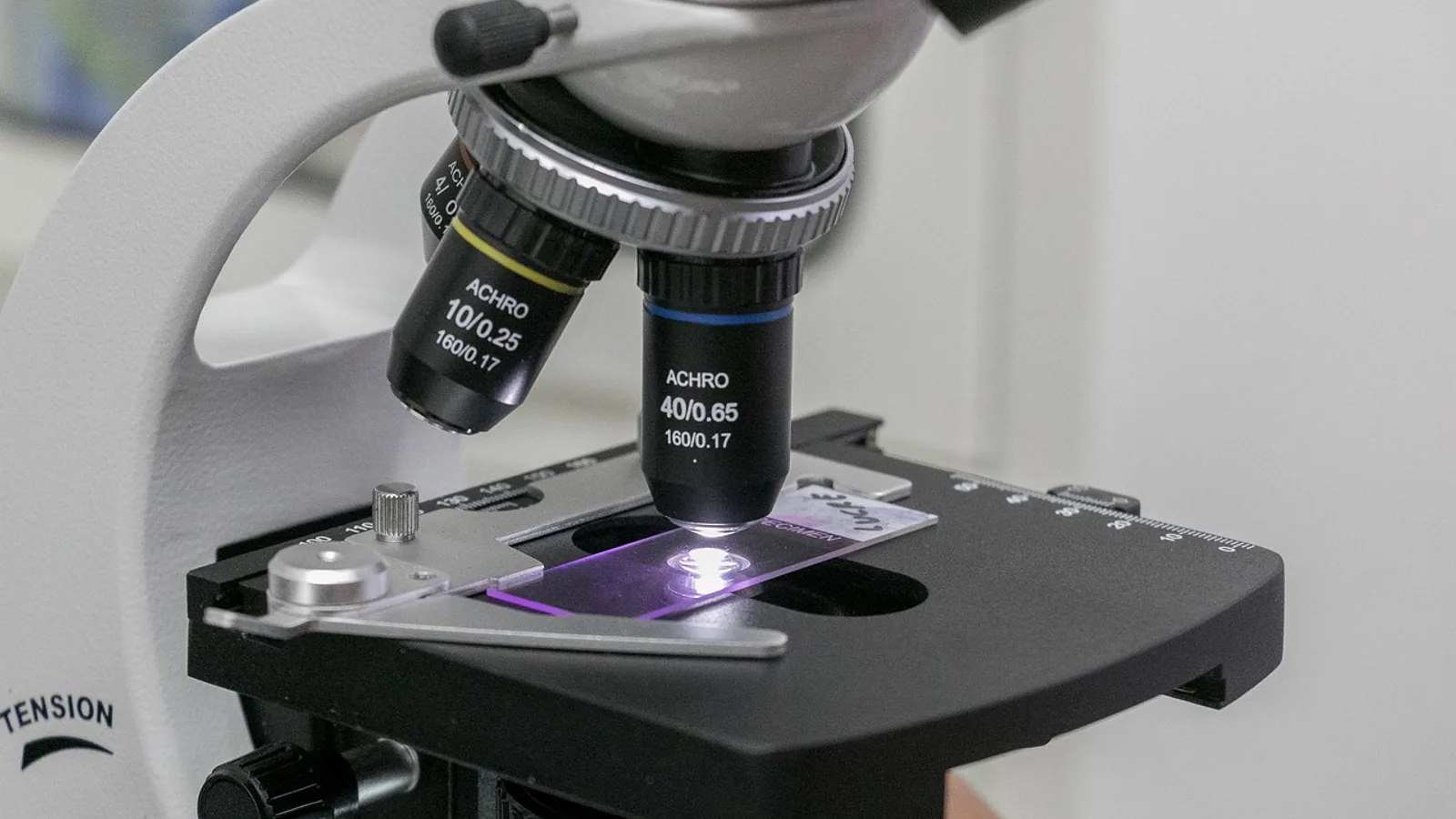
Protein polymerization is the reversible or irreversible assembly of many identical or related protein subunits into a long, repeating filament held together by noncovalent bonds. It is the physical basis for the cytoskeleton, for force generation during cell division and migration, and for the pathological fibrils seen in neurodegeneration.
Polymerization matters because it converts a pool of soluble monomers into a structured, dynamic material with new mechanical properties. A cell can build a 7 nm actin cable, a 25 nm microtubule spindle, or a 10 nm amyloid fibril from the same basic principle: many weak contacts summed into a stable lattice. Understanding the kinetics of that assembly, and the nucleotide hydrolysis that drives it, is central to cell biology, pharmacology, and biophysics.
The Three Phases of Polymerization
Almost every polymerizing protein system follows the same kinetic scheme, first formalized for actin and tubulin.
Nucleation
Nucleation is the slow, rate-limiting formation of a small stable oligomer, or nucleus, from free subunits. Because two or three subunits must collide in the correct orientation, nucleation is thermodynamically unfavorable and dominates the lag phase of a polymerization curve.
Nucleation is not always spontaneous. Cells use nucleating proteins to bypass the slow step. Formins assemble filament nuclei by sequentially binding three actin monomers, and the flexible FH1 domain delivers profilin-actin complexes to the FH2 dimer to accelerate this process [1]. A composite nucleator can bind both the fast-growing barbed end and the slow-growing pointed end of the same filament, coordinating elongation and controlling cable thickness [2]. Myosin 15 acts as a nucleation-promoting factor in cochlear hair cells, and a deafness-causing mutation in its actin-binding interface abolishes that nucleation activity [3]. Septins, a fourth cytoskeletal component, show cooperative polymerization that can be tuned by ionic strength and membrane adsorption [4].
Elongation
Once a nucleus exists, subunits add to its ends. Elongation is usually fast and roughly linear with time until monomers are depleted. Formins accelerate elongation by processively stepping along the barbed end, and their FH1 domains deliver profilin-actin to the growing tip [5]. The mode of subunit addition matters: a bias toward FH1-mediated delivery shortens the lifetime of the formin at the filament end and limits filament length [5]. Formin tail domains can act as a switch, inhibiting or enhancing processive elongation depending on which splice variant is expressed [6].
Steady State and Treadmilling
At steady state, monomer addition at one end balances loss at the other. This is treadmilling, a flux of subunits through the polymer with no net change in filament length. Treadmilling requires that the two ends have different affinities for subunits, which is why filament polarity is fundamental.
Critical Concentration and Nucleotide Hydrolysis
The critical concentration, abbreviated Cc, is the free subunit concentration at which the polymer neither grows nor shrinks. Below Cc, filaments depolymerize. Above Cc, they grow. Each end of a polar filament has its own Cc.
Nucleotide hydrolysis changes Cc. Actin binds ATP, and tubulin binds GTP. After incorporation, the nucleotide is hydrolyzed to ADP or GDP, and the resulting subunit has a higher Cc and a weaker lattice contact. This creates a "cap" of nucleotide-bound subunits at the growing end that stabilizes the polymer.
For microtubules, GTP-bound ends show elevated and more persistent clustering of protofilaments, which favors formation of the straight lattice that accommodates new dimers. Ends trapped with long, stiff GDP-protofilaments are more prone to shortening [7]. The size of the GTP cap is a balance between GTP-tubulin addition and subsequent hydrolysis, and the cap is smaller during mitosis for a given growth rate [8]. All-atom simulations of microtubule tips have been accelerated using equation-free multiscale methods to compare GDP- and GTP-complexed conformations [9].
A Comparison of Filament Systems
| System | Subunit | Nucleotide | Polarity | Dynamics | Primary function |
|---|---|---|---|---|---|
| Actin filament | Actin monomer, 42 kDa | ATP | Yes, barbed and pointed ends | Treadmilling, nucleation by formins and Arp2/3 | Cell shape, motility, muscle contraction |
| Microtubule | Alpha-beta tubulin heterodimer | GTP | Yes, plus and minus ends | Dynamic instability, GTP cap | Spindle, intracellular transport, cilia |
| Amyloid fibril | Intrinsically disordered or misfolded protein | None required | Typically no functional polarity | Nucleation-dependent, lag phase, irreversible | Pathological aggregate in neurodegeneration |
| Septin filament | Septin complex | GTP | Yes | Cooperative, membrane-tunable | Cytokinesis, membrane barriers |
Actin filaments are about 7 nm in diameter. Microtubules are about 25 nm. Amyloid fibrils are about 10 nm and give a characteristic cross-beta X-ray diffraction pattern.
Example 1: Actin
Actin is the most studied polymerizing protein. Monomeric G-actin binds ATP and assembles into F-actin, a double helical filament with a fast-growing barbed end and a slow-growing pointed end.
Nucleation and elongation control
Cells control actin assembly with nucleators. The Arp2/3 complex creates branched networks that push against membranes [10]. Formins create unbranched cables. Human FHOD3L nucleates actin and rapidly but briefly elongates filaments after a temporary pause, and its elongation activity, not its nucleation, capping, or bundling activity, is required for sarcomere formation in cardiomyocytes [11]. Adherens junction proteins alpha-catenin, vinculin, and VASP assemble hierarchically under actomyosin contractility and then act synergistically to promote nucleation, elongation, and bundling while inhibiting Arp2/3 branching [10].
The pointed end
The pointed end was long considered passive. Recent work shows it has active roles in assembly, capping, and disassembly, and pointed-end polymerization by the bacterial effector VopF challenged the barbed-end-centric view [12]. The proline-rich domain of fission yeast Wsp1p binds actin filaments with micromolar affinity and slows elongation by Mg-ATP-actin monomers by half [13].
Actin in specialized structures
Actin polymerization builds curved and ring-shaped structures across evolution, including the axonal membrane periodic skeleton of circumferential actin rings interconnected by spectrin tetramers [14]. Assembly can be parsed into conserved stages: spatial confinement, membrane anchoring, nucleation, elongation, scaffolding, cross-linking, turnover, and force production [14].
Example 2: Tubulin and Microtubules
Microtubules are hollow cylinders of 13 protofilaments, each a linear chain of alpha-beta tubulin heterodimers. They exhibit dynamic instability, switching stochastically between growth and shrinkage.
GTP hydrolysis in the beta-tubulin monomer is coupled to this behavior. The GTP cap at the plus end stabilizes the lattice. EB1 is a standard marker for the GTP cap, and live-cell imaging of EB1 comets shows that cap size for a given growth rate is reduced during mitosis [8]. Microtubule dynamics is a therapeutic vulnerability: the SKP2 inhibitor SKPin C1 binds tubulin and inhibits microtubule polymerization, selectively disrupting spindle assembly in VHL-deficient renal cell carcinoma [15]. VHL loss alters microtubule dynamics by promoting growth speed while reducing stability, and destabilizers reduce GTP-tubulin and acetylated microtubule levels [15].
Microtubule-associated proteins tune assembly. Tau modulates protofilament number and can increase microtubule diameter under high paclitaxel-to-tubulin ratios [16]. G-alpha-i2 interacts with EB1 to control microtubule dynamics during neural crest migration, and nocodazole treatment restores Rac1 activity and cell morphology in knockdown embryos [17]. Crowding also matters: microtubule elongation rates decrease with low-molecular-weight crowdants and increase with high-molecular-weight crowdants, effects linked to viscosity and nucleation [18].
Example 3: Amyloid Fibrils
Amyloid polymerization is mechanistically distinct from cytoskeletal assembly. It is nucleation-dependent, shows a pronounced lag phase, and is essentially irreversible under physiological conditions. The lag phase reflects the slow formation of a nucleus, after which elongation is rapid and can be seeded by preformed fibrils.
Amyloid fibrils are about 10 nm wide and produce a cross-beta X-ray diffraction pattern, meaning beta strands run perpendicular to the fibril axis. Examples include amyloid-beta in Alzheimer disease, alpha-synuclein in Parkinson disease, and the prion protein in transmissible spongiform encephalopathies. Prion replication is a templated conformational change, the clearest example of seeded polymerization in biology.
Because amyloid assembly is nucleation-dependent, the lag phase is the target of many experimental and theoretical studies. Seeding assays, in which a small amount of preformed fibril is added, shorten or eliminate the lag phase and are a standard way to demonstrate that a protein forms amyloid.
How Polymerization Is Measured
Light scattering and turbidity
Light scattering detects the increase in particle size as monomers assemble. It is label-free, works at high protein concentrations, and reports bulk polymerization in real time. Turbidity at 340 nm is a common low-cost variant.
Fluorescence
Fluorescence is the most sensitive approach. Pyrene-actin fluorescence increases several-fold when actin polymerizes, making it the classic actin assembly assay. For tubulin, intrinsic tryptophan fluorescence and fluorescent GTP analogs are used. Total internal reflection fluorescence microscopy allows single filament observation, which is how formin stepping and filament nucleation were dissected [10].
Sedimentation
Sedimentation separates polymer from monomer. Ultracentrifugation pellets filaments and leaves monomers in the supernatant, giving a direct measure of the polymer fraction at equilibrium. This is the standard method for determining critical concentration.
Electron microscopy
Electron microscopy provides direct structural information. Cryo-electron microscopy resolved five structural states of the formin INF2 and two of DIA1 bound to F-actin, showing how profilin-actin is delivered to the barbed end and how the incoming monomer transitions into the filament conformation [19]. Cryo-electron tomography combined with coarse-grained modeling revealed how bent protofilaments cluster at microtubule ends [7].
A Pathway View of Polymerization
The flowchart below summarizes the decision path from monomer to stable polymer, including the branch point where nucleotide hydrolysis determines whether the polymer grows or depolymerizes.
flowchart TD
    A[Free subunit with nucleotide] --> B[Nucleation]
    B --> C{Nucleus stable}
    C -->|No| D[Return to monomer pool]
    C -->|Yes| E[Elongation at both ends]
    E --> F[Nucleotide hydrolysis in lattice]
    F --> G{Cap present}
    G -->|Yes| H[Continued growth]
    G -->|No| I[Depolymerization or treadmilling]
    H --> J[Steady state]
    I --> J
    J --> K[Filament or fibril function]
Common Mistakes and Limitations
Confusing polymerization with aggregation. Cytoskeletal polymerization is ordered, reversible, and nucleotide-driven. Amyloid formation is nucleation-dependent and effectively irreversible. The two require different assays and different interpretations.
Assuming one critical concentration. A polar filament has two, one per end. Reporting a single Cc without specifying the end is a common error.
Ignoring the lag phase. A flat early signal in a polymerization assay often reflects nucleation, not a failed experiment. Adding seeds or nucleators is the correct control.
Treating nucleotide hydrolysis as the driving force. Hydrolysis is not required for assembly. It changes the off-rate and enables treadmilling and dynamic instability, but ATP-actin and GTP-tubulin can polymerize without hydrolysis.
Overinterpreting bulk assays. Light scattering and turbidity report the total polymer mass, not filament number or length. Two conditions with identical turbidity can have very different filament architectures.
Forgetting crowding and viscosity. In vitro buffer conditions do not match the cytoplasm. Crowdants of different molecular weights have opposite effects on microtubule elongation rates [18].
Extrapolating from one cell type. Microtubule dynamics and GTP cap size are cell-cycle regulated and differ between cell types [8].
Individual experimental systems and clinical samples vary, and any diagnostic or therapeutic interpretation needs professional evaluation.
Quick Review
- Polymerization proceeds through nucleation, elongation, and steady state, with treadmilling at steady state.
- Critical concentration is the free subunit concentration at which a filament end neither grows nor shrinks.
- ATP-actin and GTP-tubulin hydrolyze their nucleotide after incorporation, creating a stabilizing cap.
- Actin filaments are about 7 nm, microtubules about 25 nm, and amyloid fibrils about 10 nm with a cross-beta pattern.
- Amyloid polymerization is nucleation-dependent and shows a lag phase that seeding can eliminate.
- Formins, Arp2/3, and myosin 15 are actin nucleators with distinct architectures and functions.
- Light scattering, fluorescence, sedimentation, and electron microscopy are the core methods for measuring assembly.
The Thermodynamic Logic Behind Subunit Assembly
Before any kinetic model makes sense, it helps to understand why polymerization happens at all. A single actin monomer contacting another actin monomer forms a handful of weak interactions, each contributing only a small amount of favorable free energy. That contact is easily broken by thermal motion. But when dozens of subunits line up in register, the same weak contacts are repeated hundreds of times along the lattice. The cumulative binding energy scales with the number of contacts, while the entropic penalty of immobilizing one more subunit stays roughly constant. This is the classic principle of multivalency: many weak bonds summed across a repeating interface produce an assembly that is stable on biological timescales even though no single bond is strong.
This explains a feature that surprises students. Polymerization is not driven by a special "strong" bond between subunits. It is driven by repetition. It also explains why polymerization is so sensitive to concentration. At low subunit concentration, the probability that a free monomer finds a filament end is low, and the entropic cost of adding it is not offset. At high concentration, collisions are frequent and the balance tips toward growth. The critical concentration is simply the concentration at which those two tendencies cancel.
The same logic applies to the nucleation step, which is why nucleation is slow. Forming a dimer from two monomers means paying the entropic cost of immobilizing both while collecting only one set of contacts. Forming a trimer collects two sets of contacts. Only when the oligomer reaches a size where the accumulated contacts reliably outweigh the entropic penalty does it become a stable nucleus that can grow. This is why nucleation is described as the rate-limiting step and why cells invest in nucleating proteins that preorganize subunits into a favorable geometry.
Reading a Polymerization Curve Like a Kineticist
A standard polymerization experiment plots polymer mass or a fluorescence signal against time. The shape of that curve carries more information than most beginners extract from it. Learning to read it is one of the most transferable skills in this field.
The curve typically has three regions. The lag phase is flat or nearly flat. During this window, nuclei are forming but the total polymer mass is too small to detect. The length of the lag phase is inversely related to the nucleation rate: efficient nucleators shorten it, and poor nucleation conditions lengthen it. If you change a condition and the lag phase changes but the elongation slope does not, you have affected nucleation specifically.
The elongation phase is the steep, roughly linear rise. Its slope reports the net rate of subunit addition, which depends on the concentration of free monomer, the number of growing ends, and the on-rate constant. If you change a condition and the slope changes but the lag phase does not, you have affected elongation or the number of ends rather than nucleation.
The plateau is the steady state, where free monomer has dropped to the critical concentration and net growth stops. The height of the plateau reports the total amount of polymerizable protein in the sample. If the plateau is lower than expected, either some protein is inactive or the critical concentration is higher than assumed.
This three-region reading gives you a diagnostic framework. A condition that abolishes the lag phase entirely suggests preformed seeds or a contaminating nucleator. A condition that produces a normal lag phase but a reduced plateau suggests a change in critical concentration or a fraction of dead protein. A condition that produces a normal lag phase and slope but a plateau that slowly declines suggests depolymerization or filament severing. Each pattern points to a different mechanistic explanation, and each can be tested with a follow-up experiment.
Worked Example: Determining a Critical Concentration by Sedimentation
Critical concentration is one of the most frequently cited numbers in a polymerization paper, and it is worth walking through how it is actually obtained, because the procedure reveals assumptions that are easy to overlook.
The experiment begins with purified monomer at a known concentration. You prepare a series of samples spanning a range of total protein concentrations, then allow each to reach equilibrium under polymerizing conditions. Equilibrium matters: if you pellet before the reaction has plateaued, you are measuring a kinetic intermediate, not a thermodynamic endpoint.
Each sample is then subjected to high-speed ultracentrifugation. Filaments are large and sediment to the bottom of the tube as a pellet. Monomers are small and remain in the supernatant. You then quantify protein in both fractions, typically by measuring absorbance or by running a gel with standards.
For each sample you calculate the concentration of protein in the supernatant, which represents the free monomer remaining at equilibrium, and the concentration in the pellet, which represents polymer. You then plot polymer concentration on the y-axis against total protein concentration on the x-axis. The data points fall on a line that intersects the x-axis at a positive value. That intercept is the critical concentration. Below it, no polymer forms. Above it, the amount of polymer increases linearly with total protein, because the free monomer is buffered at the critical concentration.
The subtlety is that this analysis assumes a single critical concentration. For a polar filament with two ends, the true situation is more complex. The polymer that forms at steady state reflects the balance at both ends, and the apparent critical concentration you measure is a weighted average that depends on which end dominates under your conditions. This is why careful papers specify the buffer, the nucleotide, and the presence or absence of capping proteins when they report a critical concentration. A number reported for the barbed end in the presence of a pointed-end capper is not the same as a bulk measurement.
Worked Example: Interpreting a Seeding Experiment
Seeding is the most direct experimental test that a polymerization reaction is nucleation-dependent. The design is simple but the interpretation requires care.
You set up two parallel reactions with identical protein concentration and buffer. To one you add a small amount of preformed polymer, typically a few percent of the total protein by mass. To the other you add an equivalent volume of buffer. You then monitor polymerization over time.
If the reaction is nucleation-dependent, the seeded sample shows a dramatically shortened or absent lag phase, while the unseeded sample retains its lag. The elongation slopes of the two reactions should be similar once both are growing, because the intrinsic elongation rate does not depend on how the nuclei were formed. This is the signature of seeding: it bypasses nucleation without changing elongation.
The control that matters most is the nature of the seed. A proper seeding experiment uses seed that is structurally identical to the polymer being studied. Adding preformed fibrils of one protein to a solution of a different protein should not seed unless the two are cross-seeding, which is itself an interesting but separate result. If your seed accelerates polymerization of an unrelated protein, you should suspect a contaminant, a buffer artifact, or nonspecific aggregation rather than genuine templated seeding.
Seeding is also the conceptual basis for understanding prion replication, where the templated conformational change propagates the misfolded state. In that case the seed is not just a structural nucleus but an information-carrying template that converts the conformation of newly recruited protein. This is why prion diseases are infectious in a way that simple amyloid accumulation is not.
Comparing Nucleators: Formins, Arp2/3, and Beyond
The article above lists several actin nucleators, but the functional differences between them are worth developing because they determine filament architecture and therefore cellular function.
Formins are dimeric proteins that nucleate unbranched filaments and then remain processively attached to the barbed end as it elongates. Their defining feature is the FH2 domain, which forms a ring around the barbed end and steps along as subunits add. The FH1 domain, which is proline-rich, recruits profilin-actin complexes and delivers them to the growing tip. This delivery mechanism makes formins efficient elongators, but it also means the filament length they produce depends on the balance between delivery rate and the lifetime of the formin at the end. A bias toward FH1-mediated delivery shortens the time the formin remains associated, which limits how long the filament can grow under its control [5]. Formin tail domains add another layer of regulation, acting as switches that can inhibit or enhance processive elongation depending on splice variant [6].
The Arp2/3 complex works on a completely different principle. It nucleates a new filament as a branch off the side of an existing filament, producing a dendritic network rather than a bundle. This branched architecture is what pushes the leading edge of a migrating cell forward, because many short filaments growing against the membrane generate force collectively. The Arp2/3 complex requires a nucleation-promoting factor to activate it, and the resulting branch angle is characteristic.
Myosin 15 represents a third category: a motor protein repurposed as a nucleator. In cochlear hair cells it promotes actin nucleation, and a deafness-causing mutation in its actin-binding interface abolishes that activity [3]. This is a useful reminder that nucleation is not confined to dedicated nucleator families. Any protein that can hold two or three actin monomers in the correct orientation can, in principle, lower the nucleation barrier.
Septins form a fourth system with distinct behavior. Their polymerization is cooperative, meaning that assembly accelerates as more subunits join, and it is tunable by ionic strength and by adsorption to membranes [4]. This membrane sensitivity is functionally important because septins form barriers and scaffolds at specific cellular locations rather than assembling uniformly throughout the cytoplasm.
The practical takeaway is that when you observe actin polymerization in a cell or in a reconstituted system, the architecture you see tells you which nucleator is likely responsible. Long unbranched cables point to formins. Dense branched networks point to Arp2/3. Rings and barriers point to septins. Learning to read architecture as a signature of the underlying nucleator is a skill that transfers directly from imaging experiments to mechanistic interpretation.
The Pointed End Deserves More Attention
For decades the pointed end of the actin filament was treated as a passive terminus, and most models of actin dynamics focused entirely on the barbed end. That view has been revised. The pointed end has active roles in assembly, capping, and disassembly, and pointed-end polymerization by the bacterial effector VopF directly challenged the barbed-end-centric model [12]. This matters because it means that a complete description of actin dynamics must account for both ends, and that pathogens have evolved to exploit the pointed end as a way to manipulate host cytoskeletal behavior.
The regulation of pointed-end dynamics is also more subtle than a simple on-off switch. The proline-rich domain of fission yeast Wsp1p binds actin filaments with micromolar affinity and slows elongation by Mg-ATP-actin monomers by roughly half [13]. This is a partial inhibition rather than a block, which means the pointed end can be tuned rather than simply capped. For experimentalists, this has a practical implication: if you are measuring elongation rates and you have not controlled for pointed-end binding proteins, your measured rate may reflect a mixture of barbed-end and pointed-end contributions.
Microtubule Dynamics in Context
Microtubules add a layer of complexity that actin filaments do not have: dynamic instability. Instead of treadmilling smoothly, a microtubule can switch stochastically between growth and rapid shrinkage. The switch from growth to shrinkage is called catastrophe, and the switch back is called rescue. These transitions are governed by the state of the GTP cap at the plus end.
The GTP cap is not a static structure. It is a steady-state balance between the addition of GTP-bound tubulin dimers and the hydrolysis of GTP to GDP within the lattice. When the cap is large, the lattice is stabilized and growth continues. When hydrolysis outpaces addition and the cap shrinks, the GDP-rich lattice becomes unstable and catastrophe follows. Live-cell imaging of EB1, which marks the GTP cap, shows that cap size for a given growth rate is reduced during mitosis [8]. This is a beautiful example of how a cell can tune a biophysical parameter to change its behavior: by shrinking the cap during mitosis, the cell makes microtubules more dynamic, which is exactly what is needed to build and remodel a spindle.
The structural basis of this behavior is becoming clearer. GTP-bound ends show elevated and more persistent clustering of protofilaments, which favors the straight lattice that accommodates new dimers, whereas ends trapped with long, stiff GDP-protofilaments are more prone to shortening [7]. All-atom simulations have been accelerated using equation-free multiscale methods to compare GDP- and GTP-complexed conformations, giving atomistic insight into why the nucleotide state changes mechanical behavior [9].
Microtubule-associated proteins add another layer. Tau modulates protofilament number and can increase microtubule diameter under high paclitaxel-to-tubulin ratios [16]. G-alpha-i2 interacts with EB1 to control microtubule dynamics during neural crest migration, and nocodazole treatment restores Rac1 activity and cell morphology in knockdown embryos [17]. These examples show that microtubule dynamics is not a fixed property of tubulin but a tunable parameter that cells adjust through a network of interacting proteins.
Crowding, Viscosity, and the Problem with Buffer
One of the most common sources of irreproducibility in polymerization experiments is the difference between in vitro buffer and the actual cytoplasm. The cytoplasm is crowded with macromolecules at concentrations that would be considered absurd in a test tube. This crowding has measurable effects on polymerization kinetics.
The effects are not intuitive. Microtubule elongation rates decrease with low-molecular-weight crowdants and increase with high-molecular-weight crowdants, and these effects are linked to viscosity and nucleation rather than to simple excluded volume [18]. This means that adding a crowding agent to your buffer does not automatically make it more cytoplasm-like. The molecular weight of the crowdant matters, and the direction of the effect can reverse depending on which crowdant you choose.
For experimentalists, the practical lesson is to report crowding conditions explicitly and to avoid assuming that a result obtained in dilute buffer will hold in a crowded environment. If your goal is to understand behavior in cells, you need either to replicate crowding conditions carefully or to validate your in vitro findings with cellular measurements.
Troubleshooting Common Polymerization Assays
Even a well-designed polymerization experiment can fail in ways that are hard to diagnose. The following patterns cover the most frequent problems and their likely causes.
No signal at all. The most common cause is inactive protein. Actin and tubulin are sensitive to denaturation during purification and storage, and a preparation that has lost activity will simply not polymerize. Test with a positive control: a known good preparation or a condition that reliably polymerizes. If the positive control works and your sample does not, the problem is the sample. If neither works, the problem is the assay or the buffer.
Signal that never plateaus. If your polymerization curve keeps rising without reaching a plateau, you may have a nucleotide regeneration problem, a contaminating nucleator, or a protein that is slowly aggregating rather than polymerizing. Check whether the signal is reversible by cooling or diluting. True polymerization is reversible under appropriate conditions; aggregation is not.
Lag phase that is too long. This usually means nucleation is inefficient. Check the nucleotide state of your protein, the ionic conditions, and whether you have inadvertently removed a required nucleator. Adding seeds is a diagnostic: if seeding eliminates the lag, nucleation was the problem.
Plateau that is too low. Either some protein is inactive or the critical concentration is higher than expected. Measure the critical concentration directly by sedimentation rather than assuming a literature value. Buffer conditions, especially ionic strength and nucleotide, can shift it substantially.
High variability between replicates. Polymerization is sensitive to temperature, mixing, and the surface properties of the cuvette or plate. Standardize all of these. For fluorescence assays, check for photobleaching and inner filter effects at high protein concentrations.
Signal that appears immediately. If polymerization starts without a lag phase, you likely have preformed seeds or aggregates in your starting material. Clarify by centrifugation before starting the experiment.
Common Misconceptions Worth Correcting
Several misconceptions recur so often that they are worth stating explicitly.
The first is that nucleotide hydrolysis provides the energy for polymerization. It does not. ATP-actin and GTP-tubulin can polymerize with nonhydrolyzable analogs. Hydrolysis changes the off-rate and enables treadmilling and dynamic instability, but the assembly itself is driven by the free energy of subunit-subunit contacts. Confusing the two leads to incorrect predictions about what happens when hydrolysis is blocked.
The second is that there is a single critical concentration for a filament. A polar filament has two, one for each end. Reporting a single number without specifying the end and the conditions is a common error that makes results hard to compare across labs.
The third is that amyloid formation and cytoskeletal polymerization are the same kind of process. They share the nucleation-elongation framework, but they differ in reversibility, in nucleotide dependence, and in biological consequence. Amyloid assembly is effectively irreversible under physiological conditions and is not regulated by the cell in the way that actin and tubulin assembly are.
The fourth is that bulk assays report filament number. They do not. Light scattering and turbidity report total polymer mass. Two samples with identical turbidity can have very different numbers and lengths of filaments, which matters enormously for understanding function.
The fifth is that in vitro results translate directly to cells. Crowding, viscosity, and the presence of regulatory proteins all change the kinetics. A result obtained in dilute buffer is a starting point, not a conclusion.
Practical Applications Across Fields
Understanding protein polymerization has direct practical consequences in several areas.
In pharmacology, microtubule dynamics is a validated drug target. The SKP2 inhibitor SKPin C1 binds tubulin and inhibits microtubule polymerization, selectively disrupting spindle assembly in VHL-deficient renal cell carcinoma [15]. This is an example of how a specific cellular context, in this case VHL loss, can create a therapeutic window for a polymerization inhibitor. VHL loss alters microtubule dynamics by promoting growth speed while reducing stability, and destabilizers reduce GTP-tubulin and acetylated microtubule levels [15]. The lesson is that the same polymerization machinery can be differentially sensitive to drugs depending on the cellular background.
In neuroscience, actin polymerization underlies the structural plasticity of synapses and the maintenance of axonal architecture. The axonal membrane periodic skeleton, composed of circumferential actin rings interconnected by spectrin tetramers, is a striking example of how polymerization builds a stable, repeating structure with a defined geometry [14]. Assembly can be parsed into conserved stages: spatial confinement, membrane anchoring, nucleation, elongation, scaffolding, cross-linking, turnover, and force production [14]. This staged view is useful because it identifies points where mutations or drugs can disrupt the structure.
In developmental biology, microtubule dynamics controls cell migration. The interaction between G-alpha-i2 and EB1 during neural crest migration is a specific example, and the fact that nocodazole treatment restores Rac1 activity and cell morphology in knockdown embryos shows how tightly migration is coupled to microtubule behavior [17].
In biophysics and structural biology, cryo-electron microscopy has transformed our understanding of how nucleators work at the atomic level. The resolution of five structural states of the formin INF2 and two of DIA1 bound to F-actin showed how profilin-actin is delivered to the barbed end and how the incoming monomer transitions into the filament conformation [19]. Cryo-electron tomography combined with coarse-grained modeling revealed how bent protofilaments cluster at microtubule ends [7]. These structural approaches complement kinetic measurements and together provide a complete picture of polymerization.
In synthetic biology and materials science, the principles of polymerization are being used to design self-assembling protein materials. The same rules that govern actin and tubulin, namely nucleation, elongation, and the balance between weak contacts and entropic cost, apply to engineered systems. Understanding the natural systems is a prerequisite for designing new ones.
Putting It Together
Protein polymerization is a case study in how biology extracts complex behavior from simple physical principles. A pool of soluble subunits becomes a dynamic material through nucleation, elongation, and steady-state turnover. Nucleotide hydrolysis adds a timing mechanism that enables treadmilling and dynamic instability. Regulatory proteins tune every step, from nucleation to capping to severing. The result is a system that can build a spindle, push a membrane, maintain an axon, or, when regulation fails, form a pathological fibril.
For students, the most valuable takeaway is the framework: read the curve, identify the step, test with the right control, and interpret with the assumptions in mind. For working researchers, the most valuable takeaway is that the details matter. The end you measure, the crowdant you use, the nucleotide state of your protein, and the cell type you study all change the answer. Polymerization is not a single phenomenon but a family of related processes, and understanding which one you are looking at is the first step toward understanding what it does.
Frequently Asked Questions
What is the difference between polymerization and aggregation?
Polymerization is ordered, reversible assembly of subunits into a repeating filament, usually with nucleotide hydrolysis. Aggregation is nonspecific clumping. Amyloid formation sits between the two: it is ordered but effectively irreversible.
Why do polymerization curves have a lag phase?
The lag phase reflects nucleation, the slow formation of a stable oligomer from free subunits. Once nuclei exist, elongation is fast. Adding preformed seeds shortens or removes the lag.
What is critical concentration?
Critical concentration is the free subunit concentration at which the rate of subunit addition equals the rate of loss at a filament end. Above it the end grows, below it the end shrinks.
Do actin and tubulin need ATP or GTP to polymerize?
No. Both can polymerize with nonhydrolyzable nucleotide analogs. Hydrolysis changes the off-rate and enables treadmilling and dynamic instability, but it is not required for assembly itself.
Why are amyloid fibrils considered polymers?
They are linear, repeating assemblies of protein subunits held by noncovalent bonds, with a defined cross-beta structure about 10 nm wide. The assembly is nucleation-dependent, which distinguishes it kinetically from cytoskeletal polymerization.
Can polymerization be measured without labeling?
Yes. Light scattering and turbidity are label-free and report bulk polymer mass in real time. Sedimentation gives an equilibrium polymer fraction, and electron microscopy provides direct structural information.
Related Articles
- Protein Denaturation: Mechanisms, Examples, and Biological Relevance
- Post Translational Protein Modification: Mechanisms and Examples
- Inducible Operon: Definition, Mechanism, and Examples
- Repressible Operon: Definition, Mechanism, and Examples
- Genomic Imprinting: Definition, Mechanism, and Examples
- Channel Proteins: Definition, Function, and Examples
- Chloramphenicol Acetyltransferase: Mechanism & Uses
Sources
- Mechanism of formin-mediated filament nucleation from profilin-actin.
- Multifaceted "composite" actin nucleator orchestrates polymerization via dynamic assembly.
- Myosin-based nucleation of actin filaments contributes to stereocilia development critical for hearing.
- Cooperativity in septin polymerization is tunable by ionic strength and membrane adsorption.
- The mode of subunit addition regulates the processive elongation of actin filaments by formin.
- Formin tails act as a switch, inhibiting or enhancing processive actin elongation.
- Microtubule dynamics are defined by conformations and stability of clustered protofilaments.
- The microtubule GTP-tubulin cap size is modulated during cell division.
- Data-driven equation-free dynamics applied to many-protein complexes: The microtubule tip relaxation.
- Actomyosin-dependent assembly of the mechanosensitive machinery from adherens junctions triggers actin polymerization and organization.
- Human formin FHOD3-mediated actin elongation is required for sarcomere integrity in cardiomyocytes.
- Renaissance at the actin filament pointed end: Mechanisms of assembly, capping and depolymerization.
- The proline-rich domain of fission yeast WASp (Wsp1p) interacts with actin filaments and inhibits actin polymerization.
- Curving Actin Across Systems: Do Axonal Actin Rings Share Common Underlying Mechanisms?
- Microtubule dynamics is a therapeutic vulnerability in VHL-deficient renal cell carcinoma.
- A model of the microtubule cytoskeleton of the axon initial segment in mixtures of tubulin, tau, and GTP.
- Interaction of Gαi2 with EB1 controls microtubule dynamics and Rac1 activity in Xenopus neural crest cell migration.
- Physical effects of crowdant size and concentration on collective microtubule polymerization.
- Mechanisms of actin filament severing and elongation by formins.