Facilitated Diffusion: Channels vs Carriers
By Dr. Zubair Khalid, DVM, MS, PhD ·
Facilitated diffusion is the passive movement of a solute across a biological membrane through a transmembrane protein, driven only by the solute's electrochemical gradient and requiring no metabolic energy. It is the middle ground between simple diffusion and active transport, offering the selectivity of a binding site without the energy cost of a pump.
This mechanism matters because it is everywhere in biology. Glucose enters your erythrocytes and neurons through facilitated diffusion. Water crosses the plasma membrane of your kidney cells through aquaporin channels. Transcription factors find their target genes by a facilitated diffusion search along DNA. In every case, the cell gets speed and specificity without spending a single ATP molecule.
What Is Facilitated Diffusion?
Facilitated diffusion is a form of passive transport. Unlike simple diffusion, which lets small nonpolar molecules slip directly through the lipid bilayer, facilitated diffusion requires a dedicated membrane protein. The solute binds to or enters the protein, and the protein provides a protected path across the hydrophobic core of the membrane. The driving force is always the electrochemical gradient. If the gradient reverses, net movement reverses too.
The concept emerged from studies of sugar entry into red blood cells in the middle of the twentieth century. Glucose entered erythrocytes far faster than simple diffusion through the lipid bilayer could explain, yet the process did not consume energy and could not concentrate glucose inside the cell. The resolution was a membrane protein that binds glucose and shuttles it across. That protein, now called GLUT1, became the founding member of a large family of carriers.
The key features of facilitated diffusion are:
- It is passive. No ATP, no proton motive force, no light energy.
- It is protein-mediated. The protein confers selectivity and speed.
- It obeys the gradient. Transport stops when the solute reaches equilibrium.
- It can be saturated. At high substrate concentrations, all carrier binding sites are occupied and the rate plateaus.
- It is specific. Each transporter or channel recognizes a narrow set of substrates.
For a student, the simplest way to picture facilitated diffusion is a turnstile. A turnstile does not push you through the gate. It only opens for someone with the right token, and you still walk through under your own power. The token is the solute, and the turnstile is the transporter.
A useful diagram of facilitated diffusion contains three elements: a lipid bilayer drawn as two rows of phospholipids, a transmembrane protein spanning the bilayer, and an arrow showing the direction of the concentration gradient. For a carrier protein, the diagram usually shows the protein in two conformations, open to one side and then open to the other. For a channel, the diagram shows a continuous pore with a selectivity filter near the extracellular end. These diagrams are models. The real membrane is crowded with many proteins, and the real transporter moves constantly under thermal energy.
Summary Table: Channels vs Carriers vs Active Transport
| Feature | Channel protein | Carrier protein | Active transporter |
|---|---|---|---|
| Energy source | None, gradient only | None, gradient only | ATP hydrolysis or ion gradient |
| Direction of net movement | Down the gradient | Down the gradient | Against the gradient |
| Conformational change | Gating only, no large reorientation | Alternating access, large conformational change | Alternating access, often with phosphorylation |
| Typical transport rate | Very fast, about 10^7 to 10^8 ions per second | Slower, about 10^2 to 10^4 molecules per second | Variable, generally slow |
| Saturation | Not saturated under normal conditions | Shows Michaelis-Menten saturation | Shows saturation |
| Examples | Potassium channels, aquaporins, nuclear pore complexes | GLUT glucose transporters, PnuT thiamine transporter, urea transporters | ABC transporters, Na+/K+ ATPase, SGLT |
The rates in this table are textbook approximations. The key contrast is between the continuous pore of a channel and the alternating access of a carrier.
Facilitated Diffusion vs Active Transport
The single most important distinction is thermodynamic. Facilitated diffusion moves a solute down its electrochemical gradient and never accumulates the solute against that gradient. Active transport moves a solute against its gradient and therefore requires an external energy source.
For charged solutes, the driving force has two components. The concentration gradient pushes solute from high to low concentration. The membrane potential pushes charged solute toward the side with opposite charge. Together they form the electrochemical gradient. A passive channel or carrier always moves net solute in the direction set by this combined gradient.
There are two flavors of active transport. Primary active transport uses ATP directly. The Na+/K+ ATPase hydrolyzes ATP to pump three sodium ions out and two potassium ions in. ABC transporters use ATP binding and hydrolysis to move substrates such as lipids, drugs, and monolignols across membranes. In plant lignin biosynthesis, monolignols cross the plasma membrane by passive diffusion, by ABC transporter-mediated active efflux, or by vesicle-mediated transport [1]. Secondary active transport uses the energy of one solute moving down its gradient to drive another solute against its gradient. The sodium-glucose cotransporter SGLT1 uses the inward sodium gradient to pull glucose into the cell against its concentration gradient.
The experimental test for facilitated diffusion is simple in principle. If transport still occurs when ATP is depleted and when ion gradients are collapsed, the process is probably passive. Esquivel and White used exactly this logic to show that azole antifungal drugs enter the rice blast fungus Magnaporthe oryzae by facilitated diffusion. Accumulation of radiolabeled fluconazole was not ATP-dependent and not pH-dependent, which ruled out primary and secondary active transport [2]. In the same way, Jaehme and coworkers showed that the bacterial thiamine transporter PnuT is a facilitator because the direction of transport was determined solely by the thiamine gradient [3].
Facilitated diffusion and active transport are not mutually exclusive. Both mechanisms often handle the same substrate in the same organism. Glucose enters the intestinal epithelial cell by secondary active transport through SGLT1 at the apical surface and leaves the cell by facilitated diffusion through GLUT2 at the basolateral surface. The two systems work in series because each one is thermodynamically suited to its job. The same logic appears in plants, where monolignols use passive, active, and vesicular routes depending on the direction and magnitude of transport required [1].
Channel Proteins
A channel protein is a transmembrane protein that forms a continuous aqueous pore. When the pore is open, solutes that fit the selectivity filter diffuse through at rates limited only by diffusion. The channel does not bind the solute and carry it across. It simply provides a tunnel.
How Channels Work
Most channels are not permanently open. They have gates that respond to voltage, ligand binding, mechanical force, or light. The gating transition is a conformational change, but it is a switch between open and closed states. It is not the same as the alternating access cycle of a carrier, where the binding site itself moves from one side of the membrane to the other.
The selectivity filter is the narrowest part of the pore. In potassium channels, the filter is lined with carbonyl oxygen atoms that exactly replace the water molecules around a dehydrated potassium ion. A sodium ion is too small to contact all four carbonyls at once, so it is not stabilized and does not pass. This is standard structural biology and explains why channels can be highly selective even though they do not undergo large conformational changes.
Aquaporins use a different trick. The narrowest part of the aquaporin pore is about 2.8 angstroms wide, just wide enough for a single water molecule. A pair of conserved asparagine residues forms hydrogen bonds with the water molecule in the center of the pore, forcing it to flip orientation. This prevents protons from hopping through the water chain, so the channel conducts water but not protons.
Channel-Facilitated Diffusion in the Laboratory
Pagliara, Dettmer, and Keyser studied a synthetic model of channel-facilitated diffusion and found that particle binding at the channel entrance can actually boost the flux through the pore [4]. At first glance this seems counterintuitive. A particle bound at the mouth of a channel should block it. The authors showed that under the right conditions, the bound particle acts as a gateway that increases the local concentration of the permeating species and raises the translocation rate. This work is a reminder that the simple picture of a passive pore is incomplete. The entrance region of a channel is a functional domain, not just a hole.
Examples of Channels
- Potassium channels allow rapid, selective potassium efflux during the repolarization phase of the action potential.
- Sodium channels mediate the depolarization phase of the action potential in neurons and muscle.
- Aquaporins allow water to cross membranes while excluding protons and ions.
- Nuclear pore complexes allow nucleocytoplasmic exchange of proteins and RNA, and they participate in the facilitated diffusion of small molecules and even some proteins.
The nuclear pore complex is a special case. It is a large protein assembly, not a single polypeptide pore, and it allows passive diffusion of small molecules while using energy-dependent mechanisms for large cargo. The nuclear pore is also the gateway for transcription factors, which connects the membrane-transport sense of facilitated diffusion to the DNA-search sense covered later in this article.
Carrier Proteins
A carrier protein, also called a transporter or permease, binds a specific solute and undergoes a conformational change that moves the solute across the membrane. This is the alternating access model. The binding site faces one side of the membrane in the outward-open state, binds the solute, then the protein closes and opens to the other side, releasing the solute.
The Alternating Access Cycle
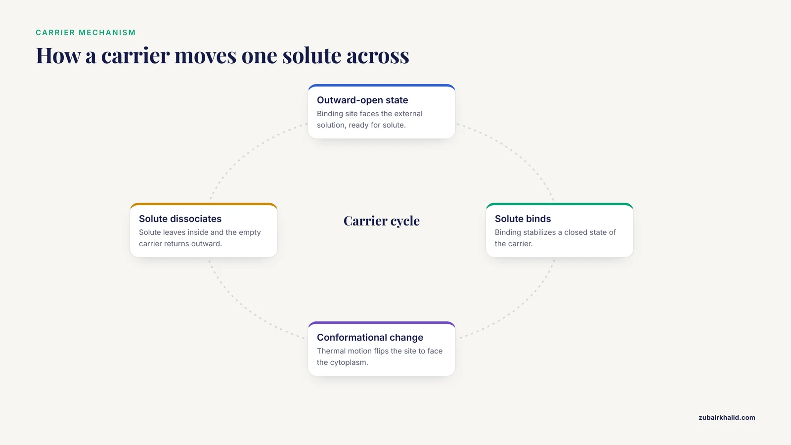
The cycle has four steps. First, the carrier is in the outward-open conformation, with the binding site exposed to the external solution. Second, the solute binds to the site, which stabilizes a closed state. Third, the protein undergoes a large conformational change so that the binding site now faces the cytoplasm. Fourth, the solute dissociates, and the empty carrier returns to the outward-open state. The energy for these conformational changes comes from thermal motion, not from ATP. This is why the process is passive.
Kinetics of Carriers
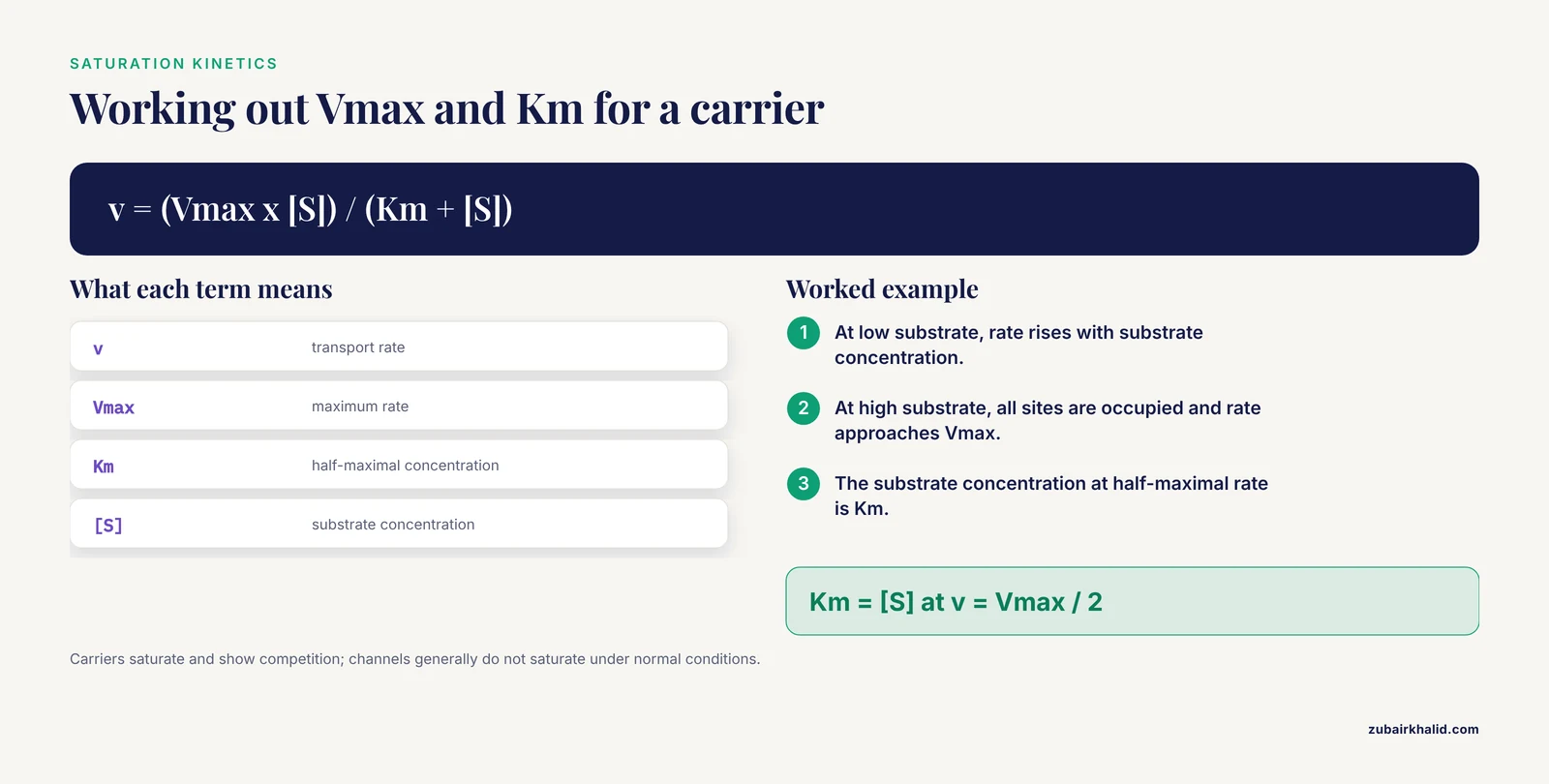
Because carriers have a finite number of binding sites, they show saturation kinetics. At low substrate concentrations, the transport rate is proportional to the substrate concentration. At high concentrations, all sites are occupied and the rate approaches a maximum, called Vmax. The substrate concentration at half-maximal rate is called the Km. This behavior is described by the same Michaelis-Menten equation used for enzymes, and it is the clearest experimental signature of carrier-mediated transport.
Carriers are also susceptible to competition. A structural analog that binds the site but is not transported will inhibit transport of the real substrate. This is how the thiamine analogs pyrithiamine and oxythiamine were used to probe the PnuT transporter [3].
Glucose Transporters: The Classic Carriers
The GLUT family of glucose transporters is the best studied group of facilitated diffusion carriers. Joost and coworkers reviewed the structure-function relationships of these proteins and emphasized that the family members share a predicted topology of 12 transmembrane helices [5]. Different GLUT isoforms have different tissue distributions, substrate specificities, and kinetic properties.
- GLUT1 transports glucose in erythrocytes and across the blood-brain barrier.
- GLUT2 is a low-affinity transporter in the liver, kidney, and pancreatic beta cells.
- GLUT4 is the insulin-responsive transporter in muscle and adipose tissue. Insulin stimulation causes GLUT4-containing vesicles to fuse with the plasma membrane, a central event in glucose homeostasis and cell signaling.
Sato and coworkers localized facilitated-diffusion glucose transporters in rat pancreatic islets by immunohistochemistry [6]. Their work showed that the islet cells express specific GLUT isoforms, which is relevant to glucose sensing and insulin secretion. Briczinski and coworkers demonstrated that glucose transport in Bifidobacterium animalis subsp. lactis occurs by facilitated diffusion [7]. This bacterial example shows that the mechanism is not restricted to eukaryotes, and it matters for the food industry because bifidobacteria are used as probiotics. In lactobacilli, the uptake of D-xylose has been linked to the phosphoenolpyruvate:mannose phosphotransferase system, which the authors described as a mechanism of facilitated diffusion [8]. That system is unusual because it couples sugar transport to phosphoenolpyruvate, but it illustrates the diversity of bacterial uptake strategies.
Plant Monosaccharide Transporters
Yamada and coworkers identified ESL1, an abiotic stress-inducible monosaccharide transporter in Arabidopsis thaliana [9]. ESL1 is localized to the tonoplast, the membrane surrounding the vacuole. Its transport activity is independent of a proton gradient, which distinguishes it from the proton-coupled sugar transporters in the same plant. The Km for glucose is extraordinarily high, meaning the transporter has low affinity and operates best when sugar concentrations are high. Under drought and high salinity, plants accumulate sugars in the vacuole as a stress response, and ESL1 appears to move those sugars across the tonoplast by facilitated diffusion [9].
Thiamine Uptake by PnuT
Jaehme and coworkers purified the PnuT transporter from Shewanella woodyi and reconstituted it into liposomes [3]. They showed that the electrochemical gradient of thiamine alone determines the direction of transport. The transporter binds thiamine and the analogs pyrithiamine and oxythiamine, but it does not recognize the phosphorylated forms thiamine monophosphate or thiamine pyrophosphate. This is consistent with a metabolic trapping mechanism. The cell phosphorylates thiamine once it enters the cytoplasm, so the free thiamine concentration inside stays low and the gradient keeps driving uptake [3].
Urea Transporters
Wood and coworkers studied pulsatile urea excretion in the gulf toadfish Opsanus beta [10]. Urea moves across the gill by a specific facilitated diffusion transport system that is activated when the fish switches from ammonia excretion to urea excretion. The transporter is saturable and selective, and it allows the fish to dump a large pulse of urea without losing water or ions. This is a clean physiological example of a carrier that is regulated on demand.
Synthetic Carriers
Martínez-Crespo and coworkers built a calixpyrrole cavitand, a synthetic cup-shaped molecule, that acts as a carrier for the amino acid L-proline. The molecule forms a 1:1 complex with proline and shuttles it across liposomal membranes and into living human cells. The transport follows a mobile carrier mechanism, which is the same principle as a biological carrier protein. This work is a proof of principle that synthetic molecules can supplement or replace natural transporters, with potential therapeutic applications.
Facilitated Diffusion in the Nucleus: Protein-DNA Search
The term "facilitated diffusion" has a second, equally important meaning in molecular biology. It describes the process by which DNA-binding proteins find their specific target sites on DNA. Transcription factors, DNA repair enzymes, and other regulatory proteins cannot rely on pure three-dimensional diffusion to locate a single target among millions of base pairs. The search would take too long. Instead, they combine 3D diffusion through the nucleoplasm with 1D sliding along the DNA double helix [12][13].
The Search Mechanism
In the classical model, a DNA-binding protein diffuses in 3D until it encounters DNA at a random position. It then binds weakly and slides along the helix in 1D, sampling bases as it moves. After some distance, it dissociates and resumes 3D diffusion. This cycle of sliding and jumping is called facilitated diffusion [12][13]. Hachmo and Amir showed that the process can be analyzed naturally with conditional probability, which provides an intuitive way to understand why the combination of 1D and 3D search is so much faster than either alone [13].
Loverdo and coworkers quantified the contributions of hopping and jumping in facilitated diffusion [14]. Hopping refers to short excursions where the protein leaves the DNA but rebinds nearby. Jumping refers to longer excursions that land the protein at a distant site on the same or a different DNA molecule. Both modes expand the search radius of a single binding event. A third mode, intersegmental transfer, allows a protein with two DNA-binding sites to move directly from one DNA segment to another without fully dissociating.
Search and Recognition States
A DNA-binding protein has to do two contradictory things. During the search, it must bind DNA weakly and slide quickly. At the target, it must bind tightly and specifically. Bauer and Metzler modeled this with a generalized facilitated diffusion model that includes separate search and recognition states [15]. Leven and Levy extended the model and showed that the two states are negatively coupled. A protein that slides very fast is unlikely to recognize its target very fast, because the same interactions that speed sliding destabilize the specific recognition complex [16]. The cell solves this problem by tuning the frustration of the protein-DNA interface so that the total time for search plus recognition is minimized [16].
Crowding and Chromatin Structure
The nucleus is crowded. It contains up to 400 g/L of macromolecules, mostly proteins [12]. Dey and Bhattacherjee performed large-scale simulations to ask how this crowding affects the search. They found that nonspecifically interacting bulk crowders change the search mode. The protein floats along the DNA under the influence of protein-crowder interactions, rather than being guided purely by DNA electrostatics [12]. They also identified a surprising mechanism involving genomic crowders, which are proteins already bound to DNA. A genomic crowder can form a transient bridge with the searching protein and transport it over a DNA segment, speeding up the search [12].
The same group studied human uracil DNA glycosylase (hUNG), a DNA repair enzyme that searches for uracil lesions [17]. hUNG has a 93-residue disordered tail at its N-terminus. The simulations showed that crowding, the shape of the enzyme, and the disordered tail work together to enhance facilitated diffusion. The disordered tail is not a rigid domain. It samples many conformations and helps the enzyme navigate around crowders [17].
Liu, Cherstvy, and Metzler added another layer of realism. In cells, the cytoplasm and nucleoplasm are viscoelastic, so protein diffusion is often anomalous rather than Brownian [18]. Their simulations showed that an optimal search time exists even when bulk diffusion is anomalous, and they quantified how the balance between 3D and 1D search shifts with the binding affinity [18].
Chromatin is not a naked linear DNA molecule. It is folded into a fractal structure. Bénichou and coworkers built a model of facilitated diffusion on fractal chromatin and showed that the mechanism still works, and that the search time can be minimized by tuning the protein-DNA affinity [11]. Smrek and Grosberg extended this idea to crumpled fractal DNA globules, which mimic the compact conformation of chromatin in the nucleus [19]. The conclusion is that facilitated diffusion is robust to the geometry of the DNA substrate.
Why This Matters for Cell Signaling
Transcription factors are the endpoint of many signaling cascades. A signaling pathway is only as fast as the transcription factor that carries the signal from the cytoplasm to its target gene. Facilitated diffusion is what makes gene regulation fast enough to be biologically useful. It is also what makes it specific. The two-state model of search and recognition explains why a transcription factor does not get stuck on the first piece of DNA it touches [15][16].
Facilitated Diffusion in Cell Signaling
Facilitated diffusion is woven into several major signaling pathways. The transporter is often the regulated step that controls how much signal enters the cell.
Glucose Sensing in Pancreatic Beta Cells
The pancreatic beta cell uses GLUT2, a low-affinity facilitated diffusion carrier, to monitor blood glucose [6]. Because GLUT2 has a high Km, its transport rate tracks the glucose concentration over the physiological range. When glucose rises, more glucose enters the cell, glycolysis accelerates, and the ATP-to-ADP ratio rises. The rise in ATP closes ATP-sensitive potassium channels, depolarizes the membrane, and triggers insulin secretion. The transporter itself is passive, but it sits at the start of a signaling cascade that controls whole-body glucose homeostasis.
Insulin Action and GLUT4
Insulin lowers blood glucose by stimulating glucose uptake into muscle and adipose tissue. The target is GLUT4, a facilitated diffusion carrier stored in intracellular vesicles. Insulin signaling triggers a cascade that ends with the fusion of GLUT4 vesicles with the plasma membrane. The number of active carriers at the surface increases, so the rate of facilitated diffusion rises. This is a beautiful example of regulation by protein trafficking rather than by gating. The transporter is always passive. The cell controls how many transporters are present.
Abscisic Acid Signaling and ESL1
In plants, the stress hormone abscisic acid induces the expression of ESL1, the tonoplast monosaccharide transporter [9]. Under drought and high salinity, the plant accumulates sugars in the vacuole to raise osmotic pressure and protect cellular structures. ESL1 provides the facilitated diffusion route for those sugars. The signaling pathway controls the abundance of the transporter, and the transporter controls the passive flux.
Transcription Factor Search as a Signaling Endpoint
Many signaling pathways end in the nucleus, where a transcription factor must find its target gene. The facilitated diffusion search described in the previous section is the terminal step of those pathways. The speed of that search determines how quickly the cell can respond to a signal. The two-state model shows that the search and recognition steps are balanced so that the pathway ends in a specific, stable binding event rather than a nonspecific encounter [15][16].
How Facilitated Diffusion Is Studied
Researchers use a combination of biochemistry, biophysics, and computation to study facilitated diffusion.
Liposome Reconstitution
The cleanest way to prove that a transporter works by facilitated diffusion is to purify it and put it into a synthetic membrane. Jaehme and coworkers reconstituted PnuT into liposomes and showed that the direction of thiamine transport was set entirely by the thiamine gradient [3]. Because the liposome contains no ATP and no other proteins, any transport observed must be passive and protein-mediated.
Radiolabeled Uptake Assays
Esquivel and White measured the accumulation of radiolabeled fluconazole in Magnaporthe oryzae [2]. By comparing uptake in the presence and absence of ATP synthesis inhibitors and proton gradient disruptors, they could distinguish active transport from facilitated diffusion. The same approach is used for glucose, amino acids, and drugs in many organisms.
Electrophysiology
For ion channels, the patch clamp technique is the gold standard. A glass micropipette is sealed to a small patch of membrane, and the current flowing through single channels is recorded. The records show discrete opening and closing events, and the amplitude of each event reports the conductance of a single channel. This technique is standard textbook knowledge and is the reason we know so much about channel gating.
Fluorescence and Single-Molecule Tracking
Single-molecule tracking experiments follow individual transcription factors as they slide along DNA. These experiments are mentioned by Liu and coworkers as the experimental counterpart to their simulations [18]. The trajectories reveal the dwell times, diffusion coefficients, and hopping events that define facilitated diffusion.
Computer Simulations
Molecular dynamics and coarse-grained simulations are essential because the search process is too fast and too crowded to observe directly. Dey and Bhattacherjee used large-scale simulations with tuned models of protein and DNA to identify the floating and bridged-complex mechanisms [12][17]. Bauer and Metzler and Leven and Levy used analytical models to make predictions about search and recognition kinetics [15][16].
Physiological and Clinical Relevance
Facilitated diffusion is not a laboratory curiosity. It is the mechanism behind glucose homeostasis, drug uptake, vitamin absorption, and stress responses.
- Thiamine uptake in bacteria depends on PnuT, and the metabolic trapping mechanism keeps the gradient pointed inward [3].
- Azole antifungals enter the rice blast fungus by facilitated diffusion [2]. Understanding this influx is relevant to drug resistance, because reduced intracellular accumulation is a common resistance mechanism.
- Urea excretion in gulf toadfish uses a regulated facilitated diffusion transporter [10].
- Plant stress responses depend on monosaccharide transporters such as ESL1 to move sugars into the vacuole [9].
- Synthetic carriers for amino acids may one day supplement defective natural transporters in human disease.
The lignin example is a useful contrast. Monolignols, the building blocks of lignin, cross plant membranes by passive diffusion, ABC transporter-mediated active efflux, and vesicle-mediated secondary active transport [1]. The plant uses all three mechanisms because the direction and magnitude of transport must be controlled precisely during cell wall polymerization. This shows that passive and active mechanisms are not mutually exclusive. They are deployed side by side to meet different physiological demands.
Common Mistakes and Limitations
Students and working researchers make the same mistakes over and over. Here are the ones worth correcting.
- Confusing facilitated diffusion with active transport. The test is simple. Does the process require ATP or an ion gradient? If not, it is passive. The azole uptake work by Esquivel and White is a model of how to answer this question experimentally [2].
- Thinking channels and carriers work the same way. Channels are pores. Carriers are alternating access machines. The kinetic signatures are different. Carriers saturate and show competition. Channels generally do not saturate under physiological conditions.
- Assuming all glucose transporters are identical. GLUT1, GLUT2, and GLUT4 have different affinities, different tissue distributions, and different regulatory mechanisms [6]. The low affinity of GLUT2 is what allows the pancreatic beta cell to sense glucose over a wide range of concentrations.
- Treating the two meanings of facilitated diffusion as unrelated. The membrane-transport sense and the DNA-search sense are both examples of proteins using thermal energy to move molecules along a gradient or along a polymer. The same mathematical ideas, such as binding, dissociation, and diffusion, apply to both.
- Believing that facilitated diffusion cannot be regulated. It can. GLUT4 is stored in vesicles and inserted into the membrane in response to insulin. The urea transporter in toadfish gills is activated on demand [10]. ESL1 is induced by drought and high salinity [9]. Passive transport does not mean unregulated transport.
- Overlooking the role of crowding. The nucleus is packed with macromolecules, and crowding changes the search mechanism in ways that are not obvious from dilute-solution experiments [12][18][17].
- Forgetting that a diagram is a model. A textbook diagram of facilitated diffusion shows one protein and one solute. The real membrane contains hundreds of proteins, and the real nucleus contains thousands of DNA-bound crowders. The diagram is a starting point, not the whole story.
A final limitation applies to all transport studies. A transporter that works by facilitated diffusion in a liposome may behave differently in a living cell, where the membrane potential, the lipid composition, and the cytoplasmic environment are different. Reconstitution experiments are powerful because they isolate the transporter, but they are also artificial by design [3].
Quick Review
- Facilitated diffusion is passive, protein-mediated transport down an electrochemical gradient. It requires no ATP.
- Channels are aqueous pores that allow very fast transport and do not undergo alternating access.
- Carriers bind solutes and undergo conformational changes, showing saturation and competition kinetics.
- GLUT glucose transporters, PnuT, ESL1, and urea transporters are canonical carriers. Potassium channels and aquaporins are canonical channels.
- Active transport moves solutes against their gradient using ATP or an ion gradient. ABC transporters and SGLT are examples.
- In the nucleus, facilitated diffusion describes the combination of 3D diffusion and 1D sliding that lets transcription factors and DNA repair proteins find their targets [12][13].
- Crowding, chromatin structure, and the tradeoff between search and recognition states shape the efficiency of the DNA search [12][18][17][11][16].
Frequently Asked Questions
What is the difference between simple diffusion and facilitated diffusion?
Simple diffusion moves small nonpolar molecules directly through the lipid bilayer. Facilitated diffusion moves polar or charged molecules through a transmembrane protein. Both are passive, but facilitated diffusion is selective and can be saturated.
What is the difference between a channel and a carrier protein?
A channel forms a continuous aqueous pore and transports solutes very quickly. A carrier binds a solute and changes shape to move it across the membrane, which is slower and shows saturation kinetics.
Does facilitated diffusion require ATP?
No. Facilitated diffusion is driven entirely by the electrochemical gradient of the transported solute. If ATP is required, the process is active transport.
What are some examples of facilitated diffusion in the human body?
Glucose uptake by GLUT transporters, water movement through aquaporins, and ion movement through gated ion channels are the main examples. Insulin-regulated GLUT4 insertion into muscle and fat cell membranes is a signaling-controlled example.
Why do carrier proteins show saturation kinetics?
Carriers have a finite number of binding sites. At high substrate concentrations, all sites are occupied and the transport rate reaches a maximum. Channels, which do not bind and release each solute, do not show the same saturation behavior.
What does facilitated diffusion mean for DNA-binding proteins?
For transcription factors and DNA repair enzymes, facilitated diffusion is the combination of 3D diffusion through the nucleoplasm and 1D sliding along DNA. It lets proteins find specific target sites much faster than pure 3D diffusion would allow [12][13].
Sources
- Molecular mechanisms and regulation of lignin synthesis, transmembrane transport, and cell wall polymerization in plants.
- Accumulation of Azole Drugs in the Fungal Plant Pathogen Magnaporthe oryzae Is the Result of Facilitated Diffusion Influx
- PnuT uses a facilitated diffusion mechanism for thiamine uptake
- Channel-facilitated diffusion boosted by particle binding at the channel entrance.
- Structure-function relationship of glucose transporters catalyzing facilitated diffusion
- Immunohistochemical localization of facilitated-diffusion glucose transporters in rat pancreatic islets.
- Transport of Glucose by Bifidobacterium animalis subsp. lactis Occurs via Facilitated Diffusion
- Transport of d-Xylose in Lactobacillus pentosus, Lactobacillus casei, andLactobacillus plantarum: Evidence for a Mechanism of Facilitated Diffusion via the Phosphoenolpyruvate:Mannose Phosphotransferase System
- Functional Analysis of an Arabidopsis thaliana Abiotic Stress-inducible Facilitated Diffusion Transporter for Monosaccharides*
- Pulsatile urea excretion in gulf toadfish (Opsanus beta): evidence for activation of a specific facilitated diffusion transport system.
- Facilitated diffusion of proteins on chromatin.
- Structural Basis of Enhanced Facilitated Diffusion of DNA Binding Proteins in Crowded Cellular Milieu
- Conditional probability as found in nature: Facilitated diffusion
- Quantifying hopping and jumping in facilitated diffusion of DNA-binding proteins.
- Generalized facilitated diffusion model for DNA-binding proteins with search and recognition states.
- Quantifying the two-state facilitated diffusion model of protein-DNA interactions
- Mechanism of Facilitated Diffusion of DNA Repair Proteins in Crowded Environment - A Case Study with Human Uracil DNA Glycosylase.
- Facilitated Diffusion of Transcription Factor Proteins with Anomalous Bulk Diffusion.
- Facilitated diffusion of proteins through crumpled fractal DNA globules.