EGFP vs GFP: What Changed and When to Use Each
By Dr. Zubair Khalid, DVM, MS, PhD ·

Enhanced green fluorescent protein (EGFP) is a modified version of the wild-type green fluorescent protein (GFP) from the jellyfish Aequorea victoria, carrying the amino acid substitutions F64L and S65T together with a humanized coding sequence. EGFP and GFP share the same beta-barrel architecture and the same chromophore chemistry, but EGFP matures faster, folds more efficiently at 37 °C, and is excited strongly at 488 nm, the wavelength most fluorescence microscopes and flow cytometers already provide.
That single wavelength change reshaped how labs use fluorescent reporters. A protein that once required ultraviolet excitation and produced a weak, slowly maturing signal in mammalian cells became a routine tag that could be imaged on any standard FITC filter cube, quantified by flow cytometry, and detected on a western blot with a commercial antibody. The confusion that follows is predictable: catalogs, papers, and plasmid maps use "GFP" as shorthand for EGFP, while older literature uses "GFP" for the genuine jellyfish protein. Designing an experiment with the wrong assumption about which one is in the tube leads to wasted clones, failed fusions, and misinterpreted images. The comparison below separates the two molecules at the level of sequence, protein behavior, and bench practice.
Summary Comparison
| Property | Wild-type GFP (avGFP) | EGFP |
|---|---|---|
| Source | Aequorea victoria, isolated 1962 | Engineered from avGFP in the mid-1990s |
| Length | 238 amino acids | 239 amino acids |
| Molecular mass | About 27 kDa | About 27 kDa |
| Key substitutions | None | F64L, S65T, plus extensive silent codon changes |
| Excitation maxima | About 395 nm (major) and 475 nm | About 488 nm (single dominant peak) |
| Emission maximum | About 509 nm | About 507 to 509 nm |
| Molar extinction coefficient | Lower at 488 nm | About 55,000 M⁻¹ cm⁻¹ |
| Quantum yield | About 0.8 for the 395 nm species | About 0.60 |
| Folding at 37 °C | Poor | Efficient |
| Chromophore maturation | Slow, efficient only below 30 °C | Fast, including at 37 °C |
| Mammalian expression | Weak (codon bias, poor folding at 37 °C) | Strong (humanized codons) |
| Photostability | Moderate | Better under 488 nm illumination |
| Typical uses | Historical work, blue-light excitation schemes | Routine tagging, reporters, transgenic models |
What GFP Is and Where It Came From
Osamu Shimomura isolated a green-fluorescent protein from the jellyfish Aequorea victoria in 1962 while studying the bioluminescent protein aequorin. Aequorin emits blue light, and GFP absorbs that blue light and re-emits it as green, a natural resonance energy transfer pair. The protein itself is 238 amino acids, and its structure, solved in 1996, is a compact barrel of 11 beta strands wrapped into a cylinder roughly 3 nm in diameter and 4 nm tall, with a short alpha helix threaded through the center.
The chromophore sits inside that helix and is built from three consecutive residues, Ser65, Tyr66, and Gly67. After the protein folds, these three residues undergo a series of reactions that require molecular oxygen: cyclization of the backbone amide, dehydration, and oxidation of the tyrosyl ring. The product is a rigid p-hydroxybenzylideneimidazolinone ring system, and its conjugation is what produces the visible green color. The barrel is not decorative. It excludes water and reactive oxygen species from the chromophore, which is why GFP fluorescence survives exposure to detergents and why the protein is unusually resistant to proteases and heat.
That stability comes at a cost in the wild-type protein. Wild-type GFP absorbs light at two maxima, roughly 395 nm from the protonated chromophore and 475 nm from the deprotonated phenolate form, and it emits at about 509 nm. The 395 nm peak is the larger of the two, and it sits in the ultraviolet range. Ultraviolet excitation is phototoxic to live cells, poorly transmitted by standard glass optics, and unavailable on most flow cytometers. Worse, illuminating at 395 nm drives the chromophore through excited-state proton transfer, so the two species interconvert during imaging. The result is a signal that changes with illumination history, which makes ratiometric or quantitative work difficult.
Wild-type GFP also performs badly in mammalian cells for a second, separate reason: its codons are skewed toward the jellyfish's usage rather than the human one, which reduces protein yield. (A cryptic splice site in the wild-type gene is a problem in plants, where it is spliced out of many transcripts.) A mammalian cell transfected with a wild-type GFP plasmid fluoresces weakly not because the protein is a poor fluorophore but because there is too little of it. This distinction matters because the two problems required different solutions, and EGFP addresses both.
One further quirk of the natural protein is worth knowing. Nucleotide substitutions that appeared in early GFP plasmids changed amino acid 68, and most of the "wild-type" GFP in modern repositories carries the substitution Q80R relative to the original jellyfish clone. When a paper compares "GFP" to EGFP, the reference molecule is usually this lab-adapted variant, not the literal jellyfish sequence.
What Changed in EGFP
EGFP combines two protein-level changes with one nucleic acid-level change. Each solves a different problem, and the combination is what makes the variant useful.
F64L: better folding at 37 °C
Position 64 lies adjacent to the chromophore in the folded barrel. The phenylalanine-to-leucine substitution at this position improves the efficiency with which the protein reaches its native conformation, particularly at the elevated temperatures of a mammalian incubator. Wild-type GFP folds well at room temperature but a large fraction of molecules misfold at 37 °C. F64L raises the proportion of correctly folded protein, which increases the number of functional chromophores per cell without changing the total amount of transcription.
S65T: shifting excitation to 488 nm
Position 65 is the first residue of the chromophore itself. The serine-to-threonine substitution locks the chromophore into the deprotonated phenolate form, which is the species that absorbs at about 488 nm. The consequences stack neatly. First, the 395 nm peak almost disappears, so nearly all absorbed light produces the bright green emission species and the signal no longer fluctuates with excited-state proton transfer. Second, the chromophore becomes more resistant to acid quenching, which is why EGFP is often described as acid-tolerant relative to wild-type. Third, the oxidation step of maturation proceeds faster, so new protein becomes fluorescent sooner after synthesis.
The S65T mutation is the single most consequential difference between GFP and EGFP for practical purposes. It is what allows a standard 488 nm argon laser, LED light source, or FITC filter cube to excite the protein efficiently, and it is why EGFP became a default rather than a specialty reagent.
Human codon optimization and splice removal
The third change is silent at the protein level. The coding region was recoded to use codons preferred in human cells, which raises translation efficiency. Reports of this work describe roughly 190 nucleotide changes without any corresponding amino acid change beyond F64L and S65T. The 5′ end was also placed behind a strong Kozak consensus sequence (GCCACCATGG is a common form) to improve ribosome initiation.
The combined effect is a protein that behaves as though it is the same fluorophore in a better package: same emission color, same barrel, same oxygen-dependent maturation, but roughly an order of magnitude more useful signal per transfected cell.
A short lineage note
The EGFP variant most labs order today descends from the "GFPmut1" series described by Cormack, Valdivia, and Falkow, combined with the humanized sequence work from the Tsien laboratory and others. Commercial plasmids such as the pEGFP series popularized the name. This matters because the label "EGFP" in a catalog is a claim about a specific mutation set, not a general synonym for "bright green." Variants such as GFPmut2, GFPmut3, Emerald, and mNeonGreen all exist in the same color family and behave differently.
The EGFP Sequence and Plasmid Architecture
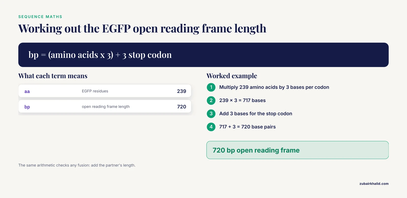
An EGFP open reading frame is 720 base pairs, encoding 239 amino acids and a stop codon. The exact sequence of any particular plasmid depends on the flanking elements, so the first practical step in any cloning project is to open the map, confirm the reading frame, and check the junctions.
Reading frame and fusion junctions
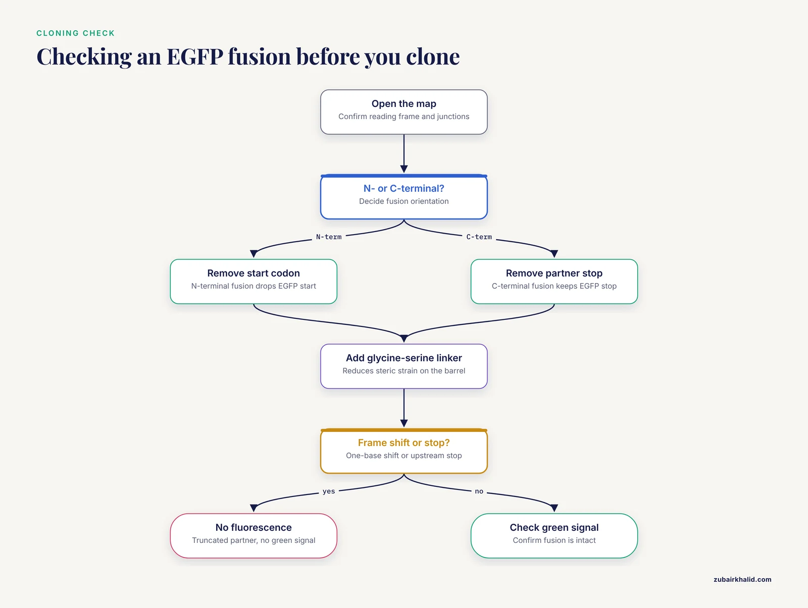
For an N-terminal fusion, the EGFP coding sequence must lack its own start codon and must sit downstream of, and in frame with, the partner protein. For a C-terminal fusion, the partner's stop codon must be removed and EGFP's stop codon retained. The most common errors in EGFP cloning are a one-base frame shift at the junction and the accidental retention of an upstream stop codon, both of which produce a truncated partner and no fluorescence. A short flexible linker, typically glycine-serine repeats, between the partner and EGFP reduces the chance that the fusion partner sterically distorts the barrel and blocks chromophore maturation.
What a typical EGFP plasmid contains
Most mammalian EGFP plasmids share a common layout: a strong constitutive promoter such as CMV, EF-1 alpha, or CAG, a multiple cloning site positioned either upstream or downstream of the EGFP open reading frame, a polyadenylation signal from SV40 or bovine growth hormone, a bacterial origin of replication, and an antibiotic resistance gene for selection in E. coli. Many also carry a separate eukaryotic selection cassette for neomycin (G418) or puromycin, either as a second expression unit or as a fusion to a drug-resistance gene. The pEGFP-N1 style map is the familiar example.
Plasmids intended for bacterial expression look different. A pET-series vector places EGFP under a T7 promoter and adds an N-terminal hexahistidine tag for purification. Work with E. coli BL21(DE3) carrying pET28(a)-EGFP showed that lowering the IPTG inducer concentration to 0.005, 0.010, or 0.025 mM maintained or even increased EGFP yields compared with higher induction, which is a practical route to cheaper protein production [1]. The lesson generalizes: EGFP is easy to overexpress to toxic, inclusion-body-forming levels, and gentler induction often gives more soluble protein.
Coordinated expression with 2A peptides
Because EGFP is small and easy to assay, it is often used as the visible marker for a second gene of interest. A viral 2A sequence placed between the two open reading frames allows a single transcript to produce two separate proteins through ribosomal skipping during translation. This was demonstrated for simultaneous overexpression of the vgf gene and EGFP both in SH-SY5Y neuroblastoma cells and in the hypothalami of Siberian hamsters and mice, with stable dual expression in vivo [2]. The technique is now standard for building reporter lines where a separate internal ribosome entry site would be too large or too weakly expressed.
Delivery formats
EGFP has been introduced into cells and animals by nearly every method available: plasmid transfection, retroviral and lentiviral transduction, adeno-associated virus, electroporation, sperm-vector methods, and lipofection combined with restriction enzyme-mediated insertion. Porcine fetal fibroblasts transfected by retrovirus vector carried both EGFP and a neomycin resistance gene into nuclear transfer embryos [3], and a separate sperm-vector approach produced a transgenic pig expressing a human albumin and EGFP fusion in the liver, skin, and hooves [4]. Sperm plasmid DNA lipofection combined with REMI has also been used to generate transgenic chickens expressing EGFP [5]. The breadth of these delivery routes is a reminder that the fluorescent protein is rarely the limiting factor. The promoter, the integration site, and the silencing behavior of the construct usually determine whether the animal actually glows.
EGFP Fluorescence: What the Photons Do
EGFP absorbs maximally near 488 nm and emits maximally near 507 nm, a Stokes shift of roughly 19 nm. Reported molar extinction coefficients cluster around 55,000 M⁻¹ cm⁻¹ with a quantum yield near 0.60, giving a brightness roughly comparable to fluorescein but considerably more photostable in a live-cell context.
The chromophore and pH
The EGFP chromophore populates the anionic phenolate form at physiological pH because its pKa is close to 6.0. Below that pH, the chromophore protonates, conjugation is interrupted, and fluorescence drops sharply. A systematic study of EGFP across a pH range found the protein highly stable at alkaline pH and quenched as pH moved from neutral to acidic. Fluorescence loss above pH 6.0 was almost completely reversible on return to neutral, while the return from pH 5.0 to 2.0 was only partially reversible, indicating that acid exposure causes some persistent structural damage [6]. This profile makes EGFP a usable natural pH sensor for the neutral-to-acidic range and also explains a common imaging artifact: secretory pathway compartments and lysosomes are acidic, so EGFP fusions that traffic there look dimmer than nuclear or cytosolic fusions made at the same level.
A second spectral peak
The first descriptions of EGFP reported a single fluorescence form, in contrast to the two absorbing species of wild-type GFP. Crystallography later revealed two conformations in the EGFP structure, raising the question of how structure and spectra relate. Three-dimensional fluorescence spectroscopy resolved part of the puzzle by identifying a second fluorescence peak for EGFP, which was not intrinsic protein autofluorescence or a spectroscopy artifact. The second peak was pH dependent, sensitive to high temperature, and linearly related to EGFP concentration, tying it to protein structure rather than to a contaminant [7]. For most imaging applications the second peak is negligible, but it matters for spectral unmixing and for any quantitative work that relies on a clean single-component spectrum.
Two-photon excitation
EGFP is not the brightest fluorophore under two-photon illumination. A physical model of green fluorescent protein spectra predicted that homologs blue-shifted in one-photon absorption would have larger two-photon cross sections, and a panel of 12 blue-shifted GFP homologs proved up to 2.5 times brighter than EGFP under two-photon excitation [8]. Multiphoton spectroscopy of EGFP has also characterized its acid-base behavior in detail, which helps when interpreting signals from tissue where pH is not controlled. For deep-tissue imaging, a blue-shifted homolog or a red fluorescent protein is usually the better choice than EGFP, despite EGFP's superior brightness under one-photon 488 nm excitation.
Switching and preservation
EGFP has been made reversibly switchable by electrochemical means. Applying negative and positive potentials to EGFP immobilized on indium tin oxide coated glass drove the protein between dim and bright states at the single-molecule level. The switching was fast, reversible, and tolerated hundreds of cycles before photobleaching, pointing toward uses in advanced fluorescence microscopy [9]. Separately, formulation work has extended the shelf life of the protein in solution. Imidazolium chloride ionic liquids at 0.100 mol/L preserved EGFP fluorescence for one to three months longer than phosphate-buffered saline or sodium chloride, and protected activity against the detergent SDS, guanidine hydrochloride, and hydrogen peroxide [10]. These are specialty applications, but they illustrate that EGFP fluorescence is a property of a folded protein, and anything that unfolds the protein turns the light off.
The EGFP Protein: Folding, Stability, and Aggregation
EGFP folds into the same 11-strand barrel as wild-type GFP, with the chromophore buried at the center. Its folding pathway has been studied by spectroscopic methods that separate the appearance of secondary structure from the appearance of fluorescence. The two do not happen at the same time: the barrel can form before the chromophore has finished oxidizing, which is why newly synthesized EGFP is briefly non-fluorescent. Spectroscopic comparison of EGFP folding and aggregation showed that the protein can populate partially folded states that associate with one another, particularly at high concentration or elevated temperature. In practice this means overexpression can generate fluorescent aggregates rather than diffuse signal, and a punctate EGFP pattern is not automatically evidence of a localization signal.
Two structural details are often misremembered. First, wild-type GFP and EGFP both have a weak tendency to dimerize at the interface between barrels. For most reporter uses the tendency is irrelevant, but for quantitative work where tagging stoichiometry matters, a monomeric variant carrying the A206K substitution is the correct choice. Second, the chromophore requires molecular oxygen to mature. In hypoxic tissue, EGFP fluorescence underreports the amount of protein present, which is a real confounder in tumor xenograft imaging and in any experiment where oxygen tension varies across the sample.
At about 27 kDa, the protein is too large to pass through gap junctions, whose size limit is about 1 kDa. In cortex, EGFP expressed in astrocytes under a GFAP promoter was transferred to neurons in deeper layers, and the transfer was not blocked by inhibitors of endocytosis or exosome release. It required F-actin, and the authors concluded that EGFP moved between cells through tunneling nanotubes, an actin-dependent cell-cell contact [11]. This is a striking example of a reporter protein behaving as a cargo. It is also a caution: a neighboring cell that looks green has not necessarily been transfected.
EGFP Applications
EGFP is used wherever a bright, stable, oxygen-dependent green signal is useful. The list below covers the applications that appear most often in the primary literature.
Cell and tissue labeling
The most common use is straightforward: tag a protein, a cell type, or an entire organism and follow the green signal. Transgenic pigs generated by sperm vector methods expressed an EGFP-human albumin fusion in multiple tissues [4]. Embryos produced by nuclear transfer from transfected porcine fetal fibroblasts carried both EGFP and a neomycin resistance gene [3]. Transgenic chickens expressing EGFP were generated by lipofection of sperm plasmid DNA combined with restriction enzyme-mediated insertion [5]. In each case the fluorescent protein served as a heritable, visible marker of successful genetic modification.
Reporter viruses and neutralization tests
EGFP can be inserted into a viral genome to make a reporter virus. A recombinant Newcastle disease virus carrying the EGFP gene in the first genomic position was used to build an EGFP-based neutralization test in which neutralizing antibody titers were scored as the reciprocal of the highest serum dilution that still produced green fluorescence. The assay gave conclusive results within 24 hours and required no additional staining step, making it much faster than conventional neutralization [12]. Reporter viruses of this kind are a general strategy, not a Newcastle disease virus specialty.
Immune cell function
EGFP-labeled target cells allow killing to be measured by loss of fluorescence. In an EGFP-K562 flow cytometric method, natural killer cell cytotoxic activity was quantified by tracking the disappearance of green target cells [13]. The readout is fast, avoids radioisotopes, and works on any instrument with a 488 nm laser and a 530 nm bandpass filter.
Calcium and second messenger studies
EGFP can be used as a bystander reporter in cells loaded with a small-molecule indicator. Rotavirus enterotoxin NSP4 fused to EGFP was expressed in cells loaded with Fura-2 to measure cytoplasmic calcium, with the EGFP signal identifying the expressing population and the Fura-2 ratio reporting calcium concentration [14]. The green channel and the blue Fura-2 channel must be separated carefully because the emission spectra overlap.
Biosensors
The barrel of EGFP can be engineered to report on analytes. One strategy splits EGFP into two fragments and inserts a metal-binding loop between beta strands 9 and 10, so that binding of the target changes the conformation and restores fluorescence. This produced an E. coli biosensor that emitted light only in the presence of cadmium ions [15]. The same logic underlies a wide range of EGFP-based reporters.
Silencing readouts
Because EGFP fluorescence is easy to quantify in live animals, it is a convenient endpoint for RNA interference. Contactless high-intensity pulsed electromagnetic field delivery of siRNA against EGFP in B16F10-EGFP tumors reduced the fluorescent tumor area by up to 39 percent six days after transfer, with silencing confirmed at both mRNA and protein levels [16]. The design is a general template: express EGFP in the target tissue, deliver the silencing reagent, and use fluorescence as the primary readout.
Immunogenicity considerations
Therapeutic uses of EGFP require attention to the immune response. EGFP contains an H2-Kd restricted cytotoxic T lymphocyte epitope that is recognized in BALB/c mice, which is relevant for any study using EGFP-expressing cells in that strain [17]. The picture is strain dependent: in C57BL/6 mice, EGFP was found to be minimally immunogenic [18]. For short-term imaging in immunocompetent animals, immune rejection is usually a minor issue. For long-term engraftment or repeated administration, strain background and expression level matter.
EGFP Fluorescence in Western Blotting
EGFP is one of the easiest proteins to detect on a western blot, provided the membrane, antibody, and imaging channel are chosen with its properties in mind.
Predicted size and migration
EGFP has a predicted mass of about 27 kDa. In practice it often migrates slightly above the predicted position on standard SDS-PAGE because the barrel structure is compact and binds somewhat less SDS than a random-coil protein of the same length. A fusion protein adds the partner's mass and, if the partner is large or highly charged, the fusion can run well away from a simple sum of the components. A pre-stained ladder covering 10 to 250 kDa is adequate for most EGFP work.
Antibodies
Anti-GFP antibodies raised against the full-length protein recognize EGFP because the epitopes are largely shared. Polyclonal anti-GFP sera are the more tolerant choice for denatured blots, and monoclonal anti-GFP antibodies that recognize both native and denatured protein are widely available. For immunoprecipitation, single-domain antibody reagents that bind the folded barrel are gentler than conventional antibodies and preserve EGFP fluorescence. Because EGFP is compact and stable, it also survives many lysis buffers that would destroy less robust proteins.
Sample preparation
Standard Laemmli sample buffer with a reducing agent and boiling is compatible with EGFP, but excessive heating of some EGFP fusions promotes aggregation, visible as a high-molecular-weight smeared band or material retained in the well. If the fusion is prone to aggregation, reducing the boiling time to a few minutes at 95 °C or heating at 70 °C helps. EGFP fluorescence also survives SDS-PAGE, which allows in-gel fluorescence to be read directly before transfer and used as a quick check that the fusion is intact and not degraded.
Channel selection and imaging
The most common mistake in fluorescent western blotting of EGFP is choosing a secondary antibody dye that emits in the same channel. EGFP emits near 507 nm, and many near-infrared and green-emitting secondary dyes overlap that region. Use secondary antibodies labeled with far-red fluorophores at 680 and 800 nm, or use chemiluminescence, and keep the EGFP channel separate. If EGFP is being used as a loading control, note that its signal comes from the fusion protein, not from a host protein, so it only reports on expression of that construct.
Controls
Include lysate from untransfected cells to confirm that the band at about 27 kDa is specific, and probe for a housekeeping protein such as GAPDH or tubulin on the same membrane. Because anti-GFP antibodies can detect endogenous GFP-family proteins weakly in some tissues, the untransfected lane is not optional. If the blot will be stripped and reprobed, do not let the membrane dry out, because EGFP's compact structure makes it resistant to some stripping protocols.
When to Use Which
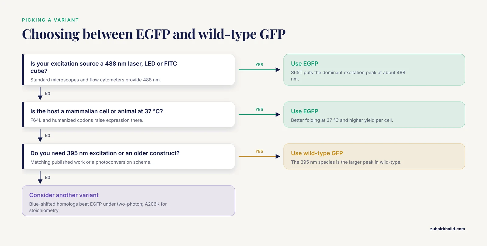
Choosing between the two is straightforward once the question is framed around the instrument and the host cell.
Use EGFP when the excitation source is a 488 nm laser, LED, or FITC filter cube, when the host is a mammalian cell or an animal maintained at 37 °C, or when the construct must be expressed at high enough levels to be visible by eye under a benchtop microscope. Those conditions cover most modern work.
Use a wild-type or near wild-type GFP only for specific reasons: matching an older published construct, working with an instrument set up for 395 nm excitation, or needing the 395 nm species for a photoconversion or ratiometric scheme. Even then, most of the "GFP" in circulation is a lab-adapted variant rather than the genuine jellyfish sequence.
Consider an alternative variant when the experiment stresses EGFP. For two-photon deep-tissue imaging, a blue-shifted homolog or a red fluorescent protein is often brighter [8]. For anaerobic or hypoxic samples, a fluorescent protein that matures without oxygen, such as a flavin-based or a red variant with different oxygen requirements, avoids the maturation problem entirely. For pH-sensing, the native pH sensitivity is an asset. For quantitative stoichiometry, a monomeric A206K variant is the correct backbone. For super-resolution or tracking, the electrochemically switchable behavior of EGFP offers an option [9].
Common Mistakes and Limitations
Treating "GFP" as interchangeable with "EGFP" in a sequence file or a cloning design is the single most frequent error. The two differ in codons and in two amino acids, and a primer designed against wild-type GFP may not amplify an EGFP plasmid or may introduce unwanted changes at positions 64 and 65.
Assuming EGFP is monomeric. It is not. A weak dimerization interface exists at the barrel surface, and it matters for experiments where tagged protein stoichiometry is being measured.
Forgetting the oxygen requirement. EGFP needs molecular oxygen to mature, so dim fluorescence in hypoxic or anaerobic conditions reflects maturation failure rather than low expression. The same applies to cells grown in thick, poorly oxygenated layers.
Ignoring pH. EGFP fluorescence drops steeply below pH 6.0 and only partially recovers after exposure to pH 5.0 or lower [6]. Any fusion that enters a secretory or endolysosomal compartment will look dimmer than a cytosolic fusion made at the same level.
Interpreting punctate EGFP as localization. Fluorescent aggregates form at high expression, and partially folded intermediates can associate. Confirm a localization pattern with an independent method before building a model on it.
Reading neighbor cells as transfection. EGFP moves between cells through F-actin-dependent tunneling nanotubes in neural tissue [11]. A green neighbor is not proof of successful transfection.
Using the brightest protein for two-photon work. EGFP is not the brightest under two-photon excitation, and blue-shifted homologs can outperform it substantially [8].
Overlooking immunogenicity in immune-competent models. EGFP carries a defined CTL epitope in BALB/c mice [17], although it is minimally immunogenic in C57BL/6 [18]. The strain matters.
Masking the western blot channel. EGFP emission at about 507 nm overlaps with many green-emitting secondary dyes. Use far-red detection channels and confirm that the EGFP signal itself is not being read as antibody signal.
Freezing and thawing protein lysates. EGFP is stable, but repeated cycles degrade fusion proteins and produce a characteristic 27 kDa degradation band that is easily mistaken for a second protein.
Quick Review
- EGFP is wild-type GFP with F64L, S65T, and a humanized coding sequence. It is not a different fluorophore, it is the same chromophore in a better-behaved protein.
- S65T shifts the dominant excitation to 488 nm, which is what made EGFP compatible with standard lasers and filter cubes.
- F64L improves folding at 37 °C, and codon optimization raises expression in mammalian cells.
- EGFP excitation is about 488 nm and emission about 507 nm, with an extinction coefficient near 55,000 M⁻¹ cm⁻¹ and a quantum yield near 0.60.
- EGFP fluorescence is pH sensitive and oxygen dependent. Both properties cause real artifacts more often than they cause theoretical problems.
- The open reading frame is about 720 bp and the protein is about 27 kDa. Anti-GFP antibodies detect EGFP on a western blot if the imaging channel is chosen carefully.
- A206K gives monomeric EGFP. Use it whenever tag-induced dimerization would confound the result.
Related Guides
- Troubleshooting Western Blotting: Useful when EGFP western blot signal is weak or inconsistent
- Western Blot Troubleshooting No Bands: Causes and Fixes: Helps diagnose absent EGFP bands after western blot transfer
- How to Validate Antibodies for Quantitative Western Blot: Guides anti-GFP antibody validation for quantitative EGFP western blots
- Optimizing Exposure Time for Western Blot: Optimize exposure time when imaging EGFP western blots
- Densitometry and Band Analysis for Quantitative Western Blot: Quantify EGFP band intensity with densitometry best practices
- Quantitative Western Blotting: Essential for quantitative comparisons of EGFP expression by western blot
- Fluorescence-Based Methods for Studying Protein-Protein Interactions: Fluorescence methods for studying EGFP-tagged protein interactions
- Sample Storage for Western Blot: Sample storage guidance preserves EGFP fusion protein for later analysis
- Dot Blot vs. Western Blot: When to Use Each Technique for Protein Detection: For choosing between dot blot and western blot for protein detection
- Negative Controls in Western Blotting: Why They Matter and How to Use Them: Negative controls are essential for validating antibody specificity
- Total Protein Staining for Western Blot Normalization: Total protein staining provides a robust loading control alternative
- Housekeeping Proteins as Loading Controls: Housekeeping proteins are common loading controls but need validation
- How Much Protein to Load for Western Blot?: Protein loading amount affects signal linearity and detection
- Optimizing Protein Loading for Quantitative Western Blot: Optimizing loading improves quantitative accuracy of western blots
Frequently Asked Questions
Is EGFP the same as GFP?
No. EGFP is an engineered derivative of wild-type GFP. It carries the F64L and S65T amino acid substitutions plus a humanized coding sequence, and these changes alter its excitation spectrum, folding, and expression level. The name "GFP" is often used loosely for EGFP in modern catalogs and papers.
Why is EGFP brighter than wild-type GFP at 488 nm?
The S65T mutation locks the chromophore into the deprotonated form that absorbs near 488 nm, so a standard laser excites the protein efficiently instead of the ultraviolet-preferring wild-type species. Human codon optimization and better folding at 37 °C also increase the amount of fluorescent protein per cell.
What are the EGFP excitation and emission wavelengths?
EGFP absorbs maximally at about 488 nm and emits maximally at about 507 nm, with the emission often reported between 507 and 509 nm. This pair fits FITC filter sets and the 488 nm laser line on standard flow cytometers.
Can an anti-GFP antibody detect EGFP on a western blot?
Yes. Antibodies raised against full-length GFP recognize EGFP because the two proteins share most of their sequence. Pick secondary reagents that emit outside the green channel, and include an untransfected lysate lane as a negative control.
Is EGFP immunogenic?
It depends on the host strain and the context. EGFP contains an H2-Kd restricted CTL epitope recognized in BALB/c mice, while it was found to be minimally immunogenic in C57BL/6 mice. Short-term imaging studies rarely encounter a problem, but long-term engraftment in immune-competent animals can.
When should I use GFP instead of EGFP?
Use a wild-type or near wild-type GFP when you need to match an older published construct or when your instrument is configured for 395 nm excitation. For almost all new mammalian work with 488 nm optics, EGFP or a more modern variant is the better default.
Sources
- Improving the cost effectiveness of enhanced green fluorescent protein production using recombinant Escherichia coli BL21 (DE3): Decreasing the expression inducer concentration
- The use of a viral 2A sequence for the simultaneous over-expression of both the vgf gene and enhanced green fluorescent protein (eGFP) in vitro and in vivo
- Expression of enhanced green fluorescent protein (EGFP) and neomycin resistant (NeoR) genes in porcine embryos following nuclear transfer with porcine fetal fibroblasts transfected by retrovirus vector
- Production of a transgenic pig expressing human albumin and enhanced green fluorescent protein.
- Use of Sperm Plasmid DNA Lipofection Combined with REMI (Restriction Enzyme-Mediated Insertion) for Production of Transgenic Chickens Expressing eGFP (Enhanced Green Fluorescent Protein) or Human Follicle-Stimulating Hormone1
- Reversible and irreversible fluorescence activity of the Enhanced Green Fluorescent Protein in pH: Insights for the development of pH-biosensors.
- Revealing a new fluorescence peak of the enhanced green fluorescent protein using three-dimensional fluorescence spectroscopy
- Blue-Shifted Green Fluorescent Protein Homologues Are Brighter than Enhanced Green Fluorescent Protein under Two-Photon Excitation
- Electrochemical fluorescence switching of enhanced green fluorescent protein.
- Imidazolium-based ionic liquids as additives to preserve the Enhanced Green Fluorescent Protein fluorescent activity
- Astrocyte-to-neuron transportation of enhanced green fluorescent protein in cerebral cortex requires F-actin dependent tunneling nanotubes
- Development of a novel Newcastle disease virus (NDV) neutralization test based on recombinant NDV expressing enhanced green fluorescent protein
- A novel enhanced green fluorescent protein (EGFP)-K562 flow cytometric method for measuring natural killer (NK) cell cytotoxic activity.
- Cytoplasmic calcium measurement in rotavirus enterotoxin-enhanced green fluorescent protein (NSP4-EGFP) expressing cells loaded with Fura-2.
- A Biosensor Platform for Metal Detection Based on Enhanced Green Fluorescent Protein
- Electrotransfer of siRNA to Silence Enhanced Green Fluorescent Protein in Tumor Mediated by a High Intensity Pulsed Electromagnetic Field
- Immunogenicity of enhanced green fluorescent protein (EGFP) in BALB/c mice: identification of an H2-Kd-restricted CTL epitope
- The enhanced green fluorescent protein (eGFP) is minimally immunogenic in C57BL/6 mice