c-Myc Gene: Function, Regulation, and Cancer Role
By Dr. Zubair Khalid, DVM, MS, PhD ·
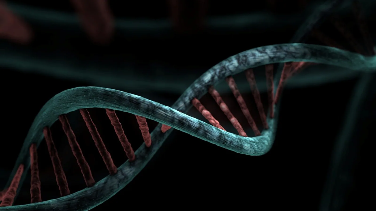
The c-myc gene (official symbol MYC) encodes a basic helix-loop-helix leucine zipper (bHLH-LZ) transcription factor that heterodimerizes with its partner Max to bind E-box DNA elements and activate gene programs controlling cell cycle entry, biomass accumulation, and metabolism. Deregulation of the myc proto-oncogene protein, through translocation, amplification, or failed protein turnover, is one of the most common events in human cancer and converts a tightly controlled growth factor into a constitutive driver of proliferation.
Why c-Myc Matters
No other oncogene is activated across as many tumor types as MYC. The MYC family oncoproteins, which include MYC, MYCN, and MYCL, act as master transcriptional regulators that orchestrate gene expression programs governing nearly all aspects of tumor development, and they are frequently linked to aggressive tumor behavior, poor prognosis, and therapy resistance [1]. Because the protein is intrinsically disordered and lacks enzymatic activity, it was long considered undruggable, but the first direct MYC inhibitor evaluated in humans, OMO-103, recently showed promising antitumor activity in phase I clinical trials [1]. For a student, c-Myc is the cleanest example of how a single transcription factor can couple growth signals to the entire biosynthetic machinery of a cell, and how breaking one regulatory node can produce cancer.
The MYC Gene Family: What c-Myc Is and What It Is Not
The name "MYC" comes from myelocytomatosis, the avian retroviral disease in which the gene was first identified. Three canonical family members exist in humans, and they share the same domain architecture and the same obligate partner:
| Feature | MYC (c-Myc) | MYCN (N-Myc) | MYCL (L-Myc) |
|---|---|---|---|
| Locus | 8q24.21 | 2p24.3 | 1p34.2 |
| Protein class | bHLH-LZ transcription factor | bHLH-LZ transcription factor | bHLH-LZ transcription factor |
| Obligate partner | Max | Max | Max |
| DNA element | E-box (CACGTG) | E-box | E-box |
| Classic tumor association | Burkitt lymphoma, many carcinomas | Neuroblastoma, medulloblastoma | Small cell lung cancer |
| Normal expression window | Broad, most proliferating tissues | Neural and embryonic | Neural and lung |
Two clarifications prevent the most common confusion. First, c-Myc, N-Myc, and L-Myc are true paralogs, meaning they arose by gene duplication and retain related sequence and function. They are not interchangeable in every tissue, but they can substitute for one another in experimental systems because they share Max and E-box specificity. Second, several proteins carry "MYC" in their name without being MYC paralogs at all. MYCBP, MYCT1, and MYCL1-adjacent loci are distinct genes, and MYC-associated factor X (MAX) is the partner, not a family member. When a paper reports "MYC amplification," it means the 8q24 locus unless the authors state otherwise.
Protein Architecture: How c-Myc Binds DNA
c-Myc is a modular protein. Reading from the N-terminus:
- Transactivation domain (TAD). Residues roughly 1 to 143 contain the transcriptional activation function and the conserved MYC boxes (MBI, MBII). MBII is the docking site for most coactivators and is the region most often mutated in loss-of-function alleles.
- Central acidic and proline-rich region. This segment contributes to cofactor recruitment and is heavily phosphorylated.
- bHLH-LZ domain. The basic region contacts DNA. The helix-loop-helix and leucine zipper mediate dimerization. The leucine zipper is a coiled coil that holds two helices together.
The bHLH-LZ region is intrinsically disordered on its own. It becomes ordered only when it pairs with Max. A striking demonstration of this came from intracellular covalent cyclization experiments in which a single helix-stabilizing constraint placed within helix two induced helicity and increased thermal stability, enabling sequence-specific E-box binding in the absence of Max [2]. That result shows the DNA-binding competence of c-Myc is a conformational problem, not simply an affinity problem, and it establishes conformational reprogramming as a route to interrogate otherwise intractable disordered proteins [2].
The Myc-Max Heterodimer
Max is a small bHLH-LZ protein that is constitutively expressed and unusually stable. It can homodimerize, and Max homodimers repress E-box targets. Myc cannot homodimerize efficiently, so the functional switch is stoichiometric: when Myc outnumbers Max-binding competitors, the heterodimer dominates and E-box targets are activated. When Myc is low, Max homodimers and Mad-family proteins occupy the same sites and repress them. This is why Myc dosage, not merely Myc presence, determines transcriptional output.
The heterodimer is the physically relevant unit in cells. In Burkitt lymphoma models, knockdown of thrombospondin 2 reduced c-MYC expression, weakened MYC/MAX dimerization, and reduced MYC binding to target gene promoters, with parallel declines in CDK2, CDK4, CDK6, and cyclin D1 protein [3]. That experiment is a useful template: dimerization, promoter occupancy, and target protein levels can each be measured separately.
What c-Myc Actually Does: Target Gene Programs
c-Myc binds thousands of genomic sites, but its biological output clusters into a few coherent programs. The table below lists representative targets with the function each one serves.
| Target gene | Protein function | Program |
|---|---|---|
| CCND1 (cyclin D1) | G1 cyclin, activates CDK4/6 | Cell cycle entry |
| CDK4 | Catalytic partner of cyclin D | Cell cycle entry |
| CDK2, CDK6 | Cell cycle kinases | Cell cycle progression |
| ODC1 (ornithine decarboxylase) | Rate-limiting polyamine synthesis | Biomass and growth |
| LDHA (lactate dehydrogenase A) | Converts pyruvate to lactate | Aerobic glycolysis |
| Ribosomal RNA and ribosomal protein genes | Ribosome assembly | Protein synthesis capacity |
| EIF4E, translation factors | Cap-dependent translation initiation | Protein synthesis capacity |
| TRIB1 | Pseudokinase adaptor, c-Myc co-regulator | Feed-forward loop [4] |
| WSB1 | E3 ligase substrate adaptor, Wnt activator | Feed-forward loop [5] |
| ALOX5 | Arachidonate 5-lipoxygenase | Lipid signaling, macrophage polarization [6] |
| FTH1, GPX4 | Iron storage and glutathione peroxidase | Ferroptosis suppression [7] |
| CASC19 (lncRNA) | Scaffold recruiting HDAC1 | Epigenetic repression of NPM1 [8] |
Three points about this table deserve emphasis. First, the cell cycle and ribosome arms explain why Myc is sufficient to drive proliferation in serum-starved cells. Second, the metabolic arm explains the Warburg phenotype: LDHA induction shifts pyruvate away from oxidative phosphorylation even when oxygen is present. Third, the list contains several genes whose products feed back on Myc itself, which is why Myc biology is described as network-embedded rather than linear.
Feed-Forward and Reciprocal Loops
The TRIB1 gene sits on chromosome 8q24 in close proximity to MYC, so the two are frequently co-amplified in human cancers. Bioinformatic analyses show a positive correlation between MYC and TRIB1 expression across multiple cancer types, and in prostate cancer cells c-Myc binds the proximal TRIB1 promoter and activates its transcription, while TRIB1 depletion lowers c-Myc levels and alters c-Myc target gene expression [4]. The same reciprocal logic appears in cervical cancer, where c-Myc directly activates WSB1 transcription and WSB1 is required for c-Myc-driven activation of Wnt/β-catenin signaling, forming a positive feedback loop [5]. In gastric cancer, c-Myc transcriptionally activates the long non-coding RNA CASC19, which then recruits histone deacetylase 1 to the NPM1 promoter and sustains H3K27 deacetylation [8]. Each of these loops widens the gap between a transient growth signal and a durable oncogenic state.
Regulation of c-Myc: Three Layers
Layer 1: Transcription Initiation
MYC transcription responds to growth factors through the MAPK and PI3K pathways, to Wnt/β-catenin signaling, and to STAT3. In non-small cell lung cancer, replication factor C subunit 5 interacts with and activates STAT3, acting as a co-transcriptional activator that upregulates c-Myc, and GATA3 acts upstream as a negative regulator of RFC5 [9]. In cervical cancer, NTMT1 promotes cell cycle progression via MYC upregulation, and NTMT1 expression correlates with activation of MYC, E2F1, CDK1, and CCNB1 in epithelial-enriched regions [10]. These examples illustrate a general principle: many oncogenic pathways converge on the MYC promoter rather than mutating MYC itself.
Layer 2: Protein Stability
The c-Myc protein has a half-life measured in minutes under normal conditions. Two phosphosites govern its destruction. Phosphorylation at threonine 58 by GSK-3 primes phosphorylation at serine 62 by ERK, and the doubly phosphorylated protein is recognized by the Fbw7 ubiquitin ligase and sent to the proteasome. Mutations at threonine 58 are among the most common MYC coding changes in lymphoma because they block this recognition step and stabilize the protein. Anything that raises ERK or PI3K output therefore raises Myc protein even without changing MYC mRNA.
Layer 3: Post-Translational Rewiring
Stability is not the only post-translational control. A targeted O-GlcNAcylation chimera that recruits O-GlcNAc transferase to c-Myc suppresses HeLa cell proliferation, rewires c-Myc genomic occupancy, and reprograms expression of the downstream oncogene MALAT1 in an O-GlcNAcylation-dependent manner [11]. This shows that modifying c-Myc's regulatory state, rather than its abundance, is a distinct and pharmacologically accessible layer of control [11].
A broader framework places c-Myc among transcription factors that occupy discrete regulatory regimes (homeostatic, adaptive, and survival) connected by threshold-like transitions, converting continuous redox inputs into distinct changes in promoter occupancy and transcriptional programs [12]. This context-sensitive view explains why the same factor can behave differently in different tissues.
Oncogene Activation Mechanisms
Three mechanisms convert the c-myc gene into a cancer driver, and they are not mutually exclusive.
Chromosomal Translocation: t(8;14) in Burkitt Lymphoma
Burkitt lymphoma carries a reciprocal translocation between chromosome 8 and chromosome 14, written t(8;14)(q24;q32). The MYC coding sequence on chromosome 8 is placed under the control of the immunoglobulin heavy chain enhancer on chromosome 14. Variant translocations t(2;8) and t(8;22) place MYC near light chain loci. The result is constitutive, high-level MYC transcription in the B cell lineage. Critically, the MYC protein itself is normal. The disease is a regulatory accident, not a structural mutation, which is why Burkitt lymphoma is the textbook example of enhancer hijacking. In Burkitt lymphoma cell lines, knockdown of THBS2 inhibited PI3K/AKT phosphorylation, lowered c-MYC expression, weakened MYC/MAX dimerization, and reduced MYC binding to target promoters, confirming that the translocated allele still depends on upstream signaling [3].
Gene Amplification
Amplification produces extra copies of the 8q24 region. Because TRIB1 lies nearby, MYC and TRIB1 are often co-amplified, and this co-amplification occurs in a large percentage of human cancers [4]. Amplification is common in melanoma, where increased MYC activity occurs by amplification of its gene locus or by upstream signaling leading to stabilization and overexpression [13]. It also appears in salivary duct carcinoma, where comprehensive genomic profiling revealed amplification of ERBB2 and MYC in a patient whose disease had progressed on multiple prior therapies [14].
Amplified MYC DNA is not always chromosomal. Extrachromosomal DNAs carrying MYC form nuclear clusters in COLO320-DM colorectal cancer cells, and BRD4 binding sites on these circles drive phase separation into in-trans contact domains enriched for PVT1-MYC fusion contacts, which explains preferential upregulation of the fusions while the canonical MYC copy remains unaffected [15]. The BET inhibitor JQ1 reverses this phase separation and reduces MYC transcription in a switch-like manner [15]. This is a mechanistically distinct route to overexpression that depends on copy number plus three-dimensional organization.
Protein Stabilization
Stabilization raises Myc protein without changing gene copy number or mRNA. It occurs through T58 mutations, through loss of Fbw7, or through upstream kinase activation. In melanoma, increased MYC activity arises either from locus amplification or from upstream signaling leading to stabilization and overexpression [13]. In clear cell renal cell carcinoma, c-Myc directly binds the ALOX5 promoter and activates transcription, and restoring ALOX5 in c-Myc-silenced cells partially rescues the malignant phenotype and M2 macrophage polarization [6]. That rescue experiment is the standard way to prove a target sits downstream of Myc rather than beside it.
c-Myc in Apoptosis and Stress
The most counterintuitive fact about c-Myc is that strong activation can kill cells. Deregulated Myc drives proliferation and simultaneously sensitizes cells to apoptosis, particularly when survival signals are limiting. This dual behavior is why MYC activation alone often produces hyperplasia rather than frank malignancy, and why secondary lesions that disable apoptosis are common in Myc-driven tumors.
Recent work reframes this. MYC proteins engage in multiple protein complexes that resolve transcription-associated stress and function as both DNA- and RNA-binding proteins, and through these activities they enhance the stress resilience of proliferating cells and enable tumor cells to sustain nonphysiological, oncogenic gene expression programs [16]. In other words, Myc creates replication and transcription stress by forcing high output, then supplies part of the machinery that manages that stress. The apoptotic threshold is the balance between the two.
Downstream targets modulate this balance directly. MYC binds the promoters of FTH1 and GPX4 and enhances their transcriptional activity, and MYC knockdown increases intracellular Fe2+ and reactive oxygen species and sensitizes colorectal cancer cells to oxaliplatin, while re-expression of either FTH1 or GPX4 partially restores resistance [7]. Here Myc is suppressing ferroptosis, a distinct cell death program, which adds a third outcome alongside proliferation and apoptosis.
How c-Myc Is Studied in Practice
Several standard assays appear repeatedly in the primary literature and are worth knowing by name.
- Chromatin immunoprecipitation followed by quantitative PCR (ChIP-qPCR). Proves direct promoter binding. Used to show MYC occupancy at the TRIB1 promoter [4], at WSB1 [5], at ALOX5 [6], and at FTH1 and GPX4 [7].
- Dual-luciferase reporter assays. Confirm that a promoter fragment confers Myc-dependent transcription, used alongside ChIP for WSB1 [5] and for FTH1 and GPX4 [7].
- Co-immunoprecipitation (Co-IP). Detects the MYC/MAX dimer and other protein partnerships [3].
- Cleavage under targets and release using nuclease (CUT&RUN). Maps genome-wide MYC occupancy, used with RNA sequencing to identify genes induced only when both MYC and TAF2 are overexpressed [17].
- CRISPR activation screens. Gain-of-function screening identifies modifiers of Myc-driven disease. A next-generation mouse whole-genome CRISPRa platform using 10 sgRNAs per transcriptional start site was used in an in vivo screen to identify accelerators of MYC-driven lymphomagenesis, with high validation rates [18].
The combination matters. Binding without a functional readout is weak evidence, and a phenotype without a binding assay leaves the mechanism open.
c-Myc in Human and Animal Cancers
MYC is deregulated in most human cancers and drives tumor progression by affecting all the hallmarks of cancer [13]. The breadth is the point. In hepatocellular carcinoma, MYC and TAF2 are frequently overexpressed and co-amplified, and combined overexpression of TAF2 and MYC significantly augmented proliferation, migration, and invasion compared with either gene alone, with RASGEF1A induced and the MEK/ERK pathway robustly activated [17]. In pancreatic ductal adenocarcinoma, IGF2BP3 supports gene programs related to DNA replication, cell cycle, apoptosis, TNF signaling, and epithelial-mesenchymal transition, and m6A, m5C, and pseudouridine modifications are enriched within MYC gene targets [19]. In prostate cancer, TMEM59L expression is negatively regulated by androgen receptor signaling and elevated in enzalutamide-resistant tissue, where it drives a neuroendocrine-like phenotype and stemness through a MUC1/β-catenin/MYC axis [20].
Animal cancers matter here too. The gene was discovered in avian myelocytomatosis, and MYC-driven lymphomagenesis remains a workhorse model in mice, where CRISPRa screening has been used specifically to find accelerators of the disease [18]. Canine lymphomas show comparable MYC involvement, which is why comparative oncology uses them as spontaneous models. In every species, the same three activation mechanisms appear.
Quick Review
- c-Myc is a bHLH-LZ transcription factor that must dimerize with Max to bind E-box DNA.
- Myc-Max activates proliferation, ribosome biogenesis, and metabolic targets such as cyclin D1, CDK4, ODC1, and LDHA.
- MYCN and MYCL are true paralogs with the same architecture and partner. MAX is the partner, not a family member.
- Activation occurs by translocation t(8;14), gene amplification at 8q24, or protein stabilization via blocked T58 phosphorylation.
- Myc both drives proliferation and lowers the apoptotic threshold, and it suppresses ferroptosis through FTH1 and GPX4.
- Direct binding is proven by ChIP-qPCR and reporter assays, and function is proven by rescue experiments.
- Feed-forward loops with TRIB1, WSB1, and CASC19 lock the oncogenic state in place.
Pathway Overview
The flowchart below traces the main decision path from growth signal to Myc-driven proliferation, including the points where regulation fails in cancer.
flowchart TD
    A[Growth factor signal] --> B[MAPK and PI3K activation]
    B --> C[MYC transcription at 8q24]
    C --> D[Myc protein synthesis]
    D --> E{Thr58 phosphorylated}
    E -->|Yes| F[Fbw7 ubiquitination and degradation]
    E -->|No| G[Myc accumulates]
    G --> H[Myc Max heterodimer]
    H --> I[E box target activation]
    I --> J[Cell cycle entry]
    I --> K[Ribosome and biomass]
    I --> L[Glycolysis and metabolism]
    J --> M[Proliferation]
    K --> M
    L --> M
    N[Translocation or amplification] --> C
Common Mistakes and Limitations
Treating Myc as a simple on-off switch. Myc output is graded and stoichiometric. What matters is the ratio of Myc to Max and to Mad-family repressors, not the presence or absence of Myc protein.
Confusing MYC with MYCN or MYCL. They are paralogs with overlapping function but distinct loci and tumor associations. A neuroblastoma paper reporting MYCN amplification is not reporting c-Myc amplification.
Assuming translocation changes the protein. In t(8;14) the MYC coding sequence is intact. The defect is regulatory, which is why the translocated allele still responds to upstream signaling [3].
Reading promoter binding as proof of regulation. ChIP-qPCR shows occupancy. A rescue experiment, such as restoring ALOX5 in c-Myc-silenced cells [6] or re-expressing FTH1 or GPX4 after MYC knockdown [7], is what establishes that the target mediates the phenotype.
Ignoring the apoptotic arm. Myc activation without survival signals can kill cells. Models that only measure proliferation miss half the biology.
Overlooking extrachromosomal DNA. Copy number from standard sequencing can miss MYC carried on extrachromosomal circles, where three-dimensional contacts rather than raw copy number drive expression [15].
Limitations. Most mechanistic data come from immortalized cell lines and xenograft models, which do not fully reproduce human tumor architecture or immune context. Findings in one tissue do not automatically transfer to another, as the redox regime framework makes clear [12]. Individual patient tumors require clinical evaluation by a qualified professional, and no laboratory marker substitutes for that assessment.
Frequently Asked Questions
What does the c-myc gene do?
It encodes a transcription factor that partners with Max to bind E-box DNA and switch on gene programs for cell cycle entry, ribosome production, and metabolism. Cells use it to convert growth signals into coordinated proliferation.
Is c-Myc the same as N-Myc?
No. They are paralogs from separate loci. c-Myc is on chromosome 8q24, and N-Myc is on 2p24. Both use Max and E-boxes, but their normal expression windows and tumor associations differ.
How does c-Myc cause cancer?
It is overexpressed or stabilized so that growth and metabolic programs stay on without external signals. This happens by translocation, amplification, or blocked protein degradation, and it is usually reinforced by feed-forward loops.
What is the t(8;14) translocation?
It is a swap between chromosome 8 and chromosome 14 that places the intact MYC gene next to immunoglobulin heavy chain enhancers. The result is constitutive MYC transcription in B cells and is the hallmark of Burkitt lymphoma.
Why is c-Myc considered undruggable?
The protein is intrinsically disordered and has no enzymatic active site, so conventional small molecules have little to grab. Recent strategies instead target its conformational state, its post-translational modifications, or its partner interactions.
Does c-Myc always promote cell survival?
No. Strong Myc activation also sensitizes cells to apoptosis, and it suppresses ferroptosis through targets such as FTH1 and GPX4. The net outcome depends on which survival and death pathways are active in that cell.
Related Articles
- Proto Oncogenes: From Normal Genes to Cancer Drivers
- Oncogene Definition: How Mutated Genes Drive Cancer
- Histone Deacetylase: Function, Mechanism, and Role in Gene Regulation
- RNA Therapeutics for Gene Regulation: A Comparison of ASOs, siRNAs, and mRNA-Based Approaches for Targeting Oncogenes and Tumor Suppressors
- Spliceosome Proteins: Composition, Function, and Regulation
- Molecular Basis of Cancer: From Oncogenes to Tumor Suppressors
- Acetylation Group: Role in Proteins and Drugs
- Genetic Transduction: How Phages Move Bacterial Genes
Sources
- Targeting MYC-Driven Cancers: From Oncogenic Addiction to Therapeutic Vulnerability.
- Conformational reprogramming of c-Myc via intracellular cyclisation.
- Down-regulation of THBS2 inhibits the malignant progression of Burkitt lymphoma by blocking the PI3K/AKT/c-MYC pathway.
- Bidirectional Regulation of c-Myc and TRB1 Establishes Oncogenic Reciprocal Regulation.
- Hyperthermia sensitizes cervical cancer to chemoradiotherapy by downregulating c-Myc/WSB1/β-catenin expression.
- Transcriptional activation of ALOX5 by c-Myc drives malignant progression and M2 macrophage polarization in clear cell renal cell carcinoma.
- Ferroptosis suppression by MYC confers oxaliplatin resistance in colorectal cancer via FTH1 and GPX4.
- Unraveling the c-Myc-CASC19/HDAC1-NPM1 epigenetic axis: A novel regulatory circuitry and therapeutic target in gastric carcinogenesis.
- RFC5 and STAT3 form a transcriptional complex to drive NSCLC progression via c-Myc.
- NTMT1 (METTL11A) promotes MYC-dependent proliferation and downstream HLA-A suppression in cervical cancer.
- Targeted O-GlcNAcylation enables functional rewiring of c-Myc.
- Systemic redox switching of transcription factors: A context-sensitive framework for cell fate decisions.
- MYC as a key target for melanoma therapy.
- Efficacy of S-1 Monotherapy for Salivary Duct Carcinoma With MYC Amplification.
- Phase separation of ecDNA condensates establishes in-trans contact domains that boost selective MYC regulatory interactions.
- Tales of destruction and repair: the changing view of MYC proteins in tumorigenesis.
- Molecular mechanism of augmentation of oncogenic activity of MYC by TAF2 in HCC.
- A high-density CRISPR activation platform for mapping cancer dependencies and resistance pathways ex vivo and in vivo.
- IGF2BP3-mediated regulation of the RNA isoform and modification landscape in pancreatic cancer.
- TMEM59L promotes neuroendocrine differentiation and enzalutamide resistance in prostate cancer by regulating MUC1/β-catenin/MYC axis.