Rapid Pathogen Screening: Methods, Uses, and Limits
By Dr. Zubair Khalid, DVM, MS, PhD ·
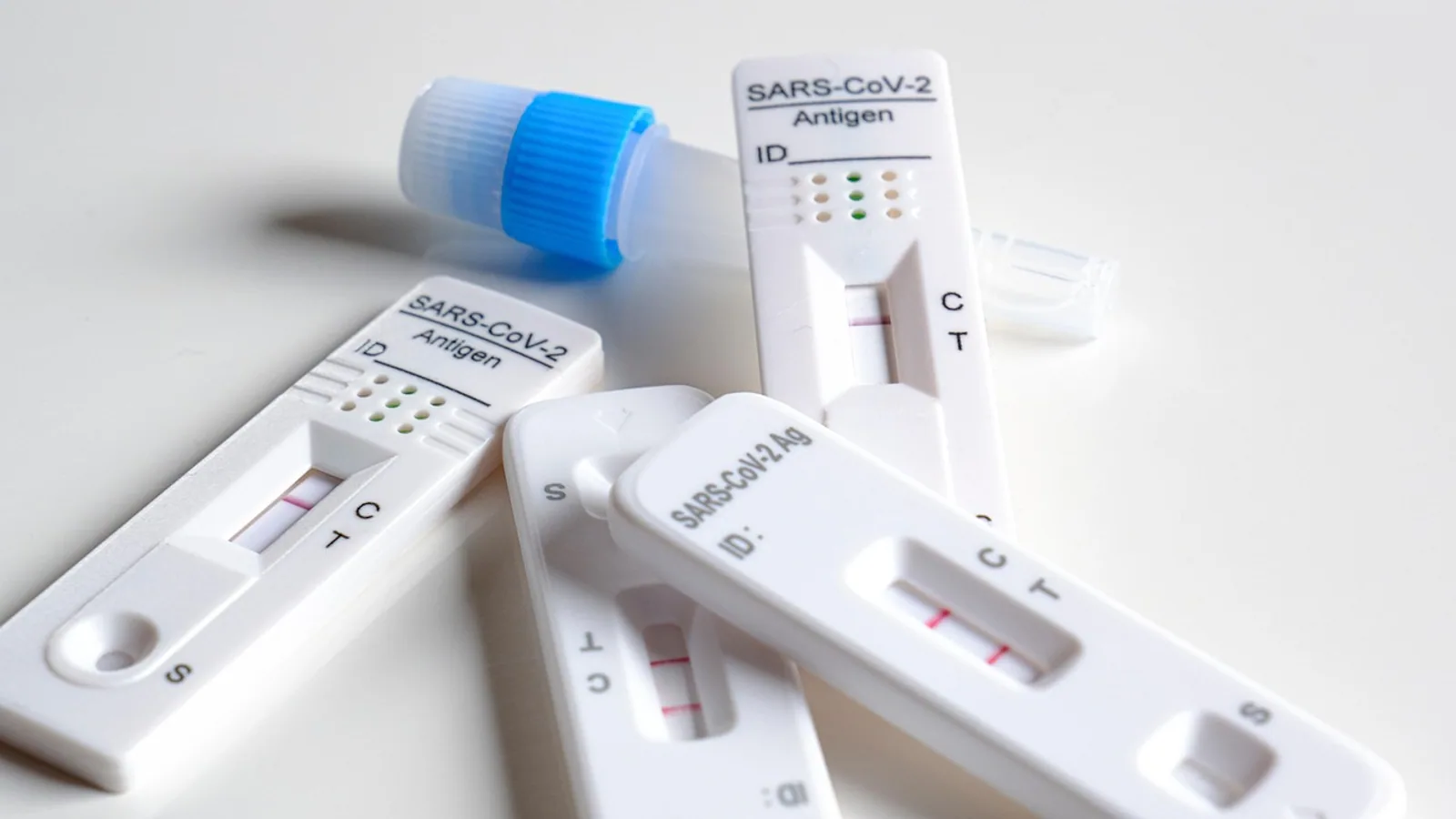
Rapid pathogen screening covers any laboratory or point-of-care workflow that identifies an infectious agent faster than conventional culture. It spans three method families that differ in speed, sensitivity, and the information they return: antigen immunoassays that read a protein on the pathogen surface in 15 to 30 minutes, nucleic acid amplification tests such as polymerase chain reaction that read a gene target in roughly 1 to 4 hours, and culture that grows the organism over 24 to 72 hours.
Each family answers a different question. An antigen test answers "is the pathogen protein present, right now, at this sampling site." A PCR test answers "is pathogen nucleic acid present, and at what approximate quantity." Culture answers "is there a living organism, what is it exactly, and which drugs will kill it." Rapid pathogen screening earns its place when the first two questions decide the next clinical action, and culture takes over when the third is the deciding factor.
This article explains the principle behind each method, the reagents and controls that make an assay valid, how to read a result, where each method fails, and how to combine them into a decision workflow. It is written for laboratory staff, veterinary and medical professionals, and technical readers who need bench-level detail.
What Rapid Pathogen Screening Achieves
The core value of rapid screening is time to a result that changes management. Conventional culture is the reference method for most bacterial infections, but it is limited by a long turnaround time and reduced sensitivity, especially in patients who already received antibiotics [1]. A positive culture can take a day, and a full identification plus susceptibility panel can take two to three days. Rapid screening compresses that window for the decisions that cannot wait.
Three broad capabilities define the field.
- Detection speed. Antigen lateral flow assays produce a visible line within 15 minutes. A cellulose nanobead lateral flow assay for Nipah virus nucleocapsid protein returned results within 15 minutes with an analytical limit of detection of 1 ng/mL [2]. Nucleic acid tests run from sample to answer in 1 to 4 hours depending on extraction and amplification format. A fully automated whole-blood sample-to-answer system completed extraction plus 40 PCR cycles in under 30 minutes [3].
- Analytical sensitivity. PCR platforms detect very low copy numbers. A multiplex digital PCR assay for lower respiratory pathogens reached a limit of detection of 5 copies per test across 33 targets, with reliable quantification from 10 to 100,000 copies per reaction [4]. A multiplex RT-PCR and capillary electrophoresis platform for feline respiratory pathogens achieved a limit of detection of 1 copy per microliter and a 1.6 hour turnaround [5].
- Breadth within one run. Multiplexing allows several pathogens to be screened from a single sample. A six-target multiplex real-time PCR assay detected influenza A and B, respiratory syncytial virus, human rhinovirus, human adenovirus, and Mycoplasma pneumoniae from one tube with 100 percent specificity against a 57-pathogen panel and detection limits between 248 and 394 copies/mL [6].
What rapid screening does not achieve is the same thing culture achieves. It cannot tell you which antimicrobial will work. It cannot distinguish living organisms from dead ones in all cases, since nucleic acid and antigen can persist after the organism is no longer viable. It detects what the assay was designed to detect, and nothing more.
The Three Method Families Compared
The comparison table below is the reference for method selection. Sensitivity ranges are typical performance envelopes for each technology class, not guarantees for any specific product. Real sensitivity depends on specimen type, organism burden, transport time, and the patient or animal population tested.
| Method | Typical turnaround time | Typical sensitivity | Typical specificity | Best use |
|---|---|---|---|---|
| Antigen immunoassay | 15 to 30 minutes | 50 to 90 percent | Moderate to high, depends on cross-reactivity | Field triage, point-of-care decisions, outbreak screening where speed outweighs missed cases |
| Nucleic acid amplification (PCR, dPCR, isothermal) | 1 to 4 hours | 90 to 100 percent | High, dependent on primer and probe design | Confirmation of screening results, low-burden infections, multiplex panels, resistance gene detection |
| Culture | 24 to 72 hours | Variable, reduced by prior antimicrobial exposure | High for viable organisms | Antimicrobial susceptibility testing, isolate recovery, outbreak typing |
Two rules follow from this table and should govern method selection.
- Antigen tests are less sensitive than PCR. A negative antigen result does not rule out infection when pretest probability is moderate to high. Antigen assays detect a protein that must be present in sufficient quantity at the sampling site, so low-burden infections and poor sampling produce false negatives.
- Culture is required for susceptibility testing. Molecular methods can detect resistance genes, and a multiplex PCR panel showed excellent consistency with antimicrobial susceptibility test results for AmpC and KPC carbapenemase genes [7], but gene presence is not the same as phenotypic resistance. Only culture-based susceptibility testing, performed to Clinical and Laboratory Standards Institute (CLSI) methods, provides minimum inhibitory concentrations for drug selection.
Antigen Immunoassays: Principle and Practice
How Antigen Tests Work
An antigen immunoassay uses antibodies to capture a pathogen protein. The dominant format is the lateral flow immunoassay, in which a sample migrates along a nitrocellulose strip and binds to a labeled antibody conjugate. If the target antigen is present, a capture line forms. A second control line confirms that the strip ran correctly.
Antibody quality determines everything. Development of a Nipah virus nucleocapsid antigen test illustrates the process: anti-Nipah antibodies were isolated from human synthetic phage-display libraries, reformatted as full-length immunoglobulins, and screened by ELISA and immunofluorescence. Four lead antibodies advanced to a sandwich-pairing matrix, which identified an optimized capture-detection configuration. Bio-layer interferometry confirmed binding affinities in the nanomolar range, with dissociation constants of 39.50 nM and 26.36 nM for the two selected antibodies [2].
Target selection matters as much as antibody selection. A lateral flow immunoassay for N9 subtype avian influenza was built against the neuraminidase protein rather than hemagglutinin, because hemagglutinin-targeting tests lose reliability as the virus drifts antigenically. The neuraminidase-targeting assay showed no cross-reactivity with N1, N2, or N6 subtypes, with limits of detection of 10 to the 3.3 TCID50 per 0.1 mL for whole virus and 98 ng/mL for recombinant neuraminidase protein [8].
Clinical Performance of Antigen Screening
Antigen tests perform best when organism burden is high. A point prevalence study of community-dwelling older adults in Germany used multi-pathogen rapid antigen self-tests detecting adenovirus, SARS-CoV-2, influenza A and B, and respiratory syncytial virus on two dates. Of 5,118 tests, 93.9 percent were negative and the overall detection rate was 5.7 percent. Adenovirus was the most frequently detected pathogen on both dates, followed by SARS-CoV-2 and influenza A. Among participants with a positive result, at least one acute respiratory infection symptom was reported by 47.7 percent in June and 58.7 percent in December [9].
That symptom proportion is instructive. Roughly 40 to 50 percent of antigen-positive participants reported no symptoms, which reflects both the sensitivity limits of antigen testing in low-burden states and the reality that antigen positivity does not always mean active clinical disease.
Antigen tests also extend beyond lateral flow. A terbium-keton complex fluorescent immunoassay for methicillin-resistant Staphylococcus aureus antigen showed a concentration-dependent fluorescence signal with a 15-minute response time in milk, hand swabs, and cattle drinking water [10].
Reading an Antigen Result
| Result | Interpretation | Action |
|---|---|---|
| Control line only | No antigen detected | Negative. If clinical suspicion is high, confirm by PCR |
| Control line plus test line, any intensity | Antigen detected | Positive. Treat as presumptive, confirm by PCR when the result drives therapy |
| No control line | Test invalid | Repeat with a new device and a fresh sample |
| Faint test line at the reading time limit | Low antigen burden | Report as positive with a low-signal note, or confirm by PCR |
Read the strip within the manufacturer's stated window. A line that appears after the window is not a valid result. A line that fades is not a negative result.
Nucleic Acid Amplification: Principle and Practice
How PCR and Isothermal Methods Work
Nucleic acid amplification tests copy a pathogen-specific DNA or RNA sequence until it reaches a detectable level. Polymerase chain reaction cycles between two or three temperatures to denature, anneal primers, and extend a new strand. Isothermal methods such as loop-mediated isothermal amplification and recombinase polymerase amplification hold a single temperature, which simplifies the instrument but can complicate primer design.
Digital PCR partitions the reaction into thousands of individual droplets or wells and counts positive partitions, which converts a threshold-based signal into an absolute copy number without a standard curve. A 33-plex digital PCR assay quantified bacteria, viruses, fungi, and atypical pathogens in bronchoalveolar lavage fluid from 10 to 100,000 copies per test with linearity above R-squared 0.99, and the full workflow from pretreatment to result took 2.5 hours [4].
Sample preparation often takes longer than amplification. A fully automated whole-blood platform integrated robotic tube decapping and aliquoting, magnetic bead nucleic acid extraction, ultrafast real-time PCR, and a compact fluorescence detector. The complete process from DNA extraction through 40 PCR cycles finished within 30 minutes, with a limit of detection of 100 CFU/mL [3].
Controls for Nucleic Acid Testing
Every amplification run needs three control types.
- Positive control. A known template at a defined concentration. It confirms that primers, probes, polymerase, and thermal cycling all work. A failed positive control invalidates the run.
- Negative control. Nuclease-free water or buffer processed through the entire extraction and amplification workflow. It detects contamination in reagents or the environment.
- Internal amplification control. A non-target template spiked into each sample. It detects inhibitors that carry over from the specimen matrix and would otherwise cause a false negative. Extraction-independent formats reduce this risk. A multiplex PCR integrated with a paper lateral flow strip was designed directly from isolates to eliminate DNA extraction and cut assay time, and the whole multiplex assay ran on a single strip within an hour [11].
An extraction control is also worth including when the workflow uses column or bead purification. It confirms that nucleic acid was recovered from the specimen.
Reading a PCR Result
Real-time PCR reports a cycle threshold, the cycle at which fluorescence crosses a threshold. A low cycle threshold means a high target concentration. A high cycle threshold near the limit of detection means a low target concentration, which can be clinically relevant or can represent contamination. Endpoint PCR reports presence or absence after gel electrophoresis or lateral flow detection. Digital PCR reports copies per unit volume and avoids the cycle threshold interpretation problem entirely.
When a real-time PCR result sits near the limit of detection, repeat the extraction from a fresh aliquot of the original specimen. Do not simply rerun the same nucleic acid, because a repeat on the same material cannot distinguish a real low-burden infection from a one-time contamination event.
Culture: Principle and Practice
How Culture Works
Culture places a specimen onto or into a growth medium that supplies nutrients and selects for or against certain organisms. A living organism multiplies until visible colonies or turbidity appear. Culture is the reference method for bloodstream infections, urinary tract infections, and most bacterial infections [1][11].
Culture has a plateau in speed that no optimization fully removes. Blood culture requires 24 to 72 hours [3]. Urinary tract infection culture requires 24 to 48 hours for pathogen identification [11]. Molecular methods sidestep this delay but they do not replace the susceptibility information culture provides.
Where Culture Still Wins
The advantages of culture are phenotype and isolate. A cultured organism can be tested against a panel of antimicrobials to generate minimum inhibitory concentrations, which is the only direct evidence of what will work clinically. A cultured organism is a physical isolate that can be archived, typed by whole-genome sequencing, and compared across an outbreak.
Culture also captures organisms that molecular panels do not include. In a comparison of multiplex PCR against conventional culture for postoperative intra-abdominal infections, multiplex PCR detected pathogens at higher rates than culture at both patient and specimen level and covered a broader pathogen spectrum [7]. That advantage cuts both ways: a broad molecular panel may report a pathogen that culture would have dismissed as a contaminant.
Where Culture Falters
Prior antimicrobial exposure suppresses growth without eliminating the organism, which lowers culture sensitivity and can delay positivity. Fastidious organisms and fungi are harder to recover than common bacteria. A pilot study comparing culture-independent digital PCR with paired blood culture in suspected bloodstream infection found that digital PCR detected a broader spectrum of pathogens, 34 species versus 28, with improved identification of fastidious organisms and fungi, particularly Candida species. Digital PCR sensitivity was 75.0 percent and specificity 53.8 percent against blood culture, with clinically plausible explanations for many of the discordant PCR-positive results [1]. The discordant positives are the relevant detail: some represent true infections that culture missed, and some represent contamination or transient nucleic acid.
Method Selection: A Decision Workflow
The flowchart below traces the decision path from a suspected infection to a method choice, then to confirmation and susceptibility testing.
flowchart TD
    A[Suspected infection] --> B{Need result within 30 minutes}
    B -->|Yes| C[Antigen test at point of care]
    B -->|No| D{Need susceptibility data}
    C --> E{Antigen result positive}
    E -->|Yes| F[Treat as presumptive and confirm]
    E -->|No| G{Clinical suspicion high}
    G -->|Yes| H[Confirm by PCR]
    G -->|No| I[Reassess clinical picture]
    D -->|Yes| J[Culture plus susceptibility testing]
    D -->|No| K[PCR or multiplex panel]
    F --> K
    H --> K
    K --> L{Resistance gene or persistence question}
    L -->|Yes| J
    L -->|No| M[Report molecular result]
    J --> M
    M --> N[Integrate results and decide]
The logic is straightforward. Antigen first when minutes matter. PCR first when sensitivity matters or when the panel is broad. Culture in parallel whenever susceptibility testing or isolate recovery is needed, even when a faster method has already returned a result.
Materials, Reagents, and Working Conditions
The table below lists common reagents and conditions across the three method families. Working concentrations are typical and should be confirmed against the specific assay's instructions.
| Item | Method family | Typical working condition | Purpose |
|---|---|---|---|
| Anti-target capture antibody | Antigen | Coated on nitrocellulose or plate well | Binds the pathogen antigen |
| Labeled detection antibody | Antigen | Colloidal gold, latex, or fluorescent conjugate | Generates the visible or measured signal |
| Running buffer | Antigen | Manufacturer-supplied, phosphate or Tris based | Carries sample along the strip and controls pH |
| Lysis buffer | Nucleic acid | Manufacturer-supplied, often chaotropic salt based | Releases nucleic acid and inactivates nucleases |
| Proteinase K | Nucleic acid | 10 to 20 mg/mL stock, used at micrograms per reaction | Digests proteins and improves nucleic acid recovery |
| Dithiothreitol | Nucleic acid, respiratory specimens | Used as a liquefaction pretreatment | Provides balanced RNA and DNA recovery from viscous specimens [4] |
| Primers and probes | Nucleic acid | 0.1 to 1 micromolar each in the reaction | Define the target amplicon and generate signal |
| Thermostable polymerase | Nucleic acid | 1 to 2.5 units per reaction | Extends new DNA strands |
| Master mix with dNTPs | Nucleic acid | 200 micromolar each dNTP | Supplies building blocks for amplification |
| Enrichment broth | Culture | Buffered peptone water or lactose broth | Revives stressed organisms before plating [12] |
| Selective agar | Culture | Organism-specific formulation | Isolates the target from mixed flora |
| Quality control organism panel | All | Known positive and negative strains | Validates each run and each new lot |
Safety Note
Lysis buffers that contain guanidine salts are hazardous and can release toxic gas if mixed with bleach. Never combine nucleic acid extraction waste with bleach. Treat all clinical specimens as potentially infectious, work in a biosafety cabinet when the procedure generates aerosols, and dispose of sharp items in puncture-resistant containers. Antigen running buffers and culture media are generally low hazard, but antibody conjugates may contain preservatives such as sodium azide, which is toxic if ingested and can form explosive metal azides in copper plumbing. Follow the safety data sheet for every reagent.
Step-by-Step Procedure for a General Rapid Screening Workflow
The numbered steps below apply to a generic rapid screening workflow. Adjust volumes and times to the specific assay.
- Verify specimen suitability. Confirm the specimen type matches the assay's validated matrix. An assay validated on nasopharyngeal swabs is not automatically valid on saliva. An assay validated on whole blood is not automatically valid on serum. Record specimen collection time.
- Verify chain of custody and integrity. Reject specimens with leaked transport medium, missing labels, or delays beyond the assay's validated stability window.
- Bring reagents to working temperature. Remove assay components from refrigerated storage and let them equilibrate. Cold reagents slow antibody binding and can shift the limit of detection.
- Label and organize. Label the device or plate with specimen identifiers before adding any material. Mislabeled sample order is the most common source of wrong results.
- Prepare the sample. For antigen tests, add the specimen to the extraction buffer in the volume the manufacturer states. For nucleic acid tests, perform lysis and extraction, or use a direct-from-specimen format if the assay supports it. For culture, inoculate enrichment broth and incubate for the required time before plating.
- Add sample to the device. Apply the sample at the designated port or well. Touching the nitrocellulose strip or the well wall can distort liquid flow and produce an invalid result.
- Run the reaction. Start timing at the moment the sample is added. Antigen assays read at a fixed time, typically within 15 minutes [2][10]. Real-time PCR runs 30 to 45 cycles at programmed temperatures. Isothermal assays hold a single temperature, often 37 degrees C, for the full reaction time [13].
- Read the result within the valid window. For lateral flow, read at the manufacturer's stated time. For PCR, read the amplification curve and cycle threshold. For culture, examine plates at 24 hours and again at 48 to 72 hours before calling a negative.
- Interpret with controls. Confirm the positive control is positive, the negative control is negative, and the internal control amplified for every sample. If any control fails, the run is invalid and must be repeated.
- Record and report. Note the result, the assay lot, and the cycle threshold or line intensity where relevant. A low-signal positive carries less weight than a strong positive.
- Archive or refer. Retain the extracted nucleic acid or the culture isolate for confirmation or susceptibility testing.
Why Each Critical Step Matters
- Temperature equilibration affects reaction kinetics and can shift a borderline result in either direction.
- Sample volume accuracy changes the effective concentration of the target and can push a positive into the negative range.
- Timed reading windows exist because signal can develop non-specifically after the window closes, and because a positive line can fade past the window.
- Control validation is the only way to distinguish a true negative from an inhibited or failed reaction.
- Culture hold time matters because slow-growing organisms can appear after the first 24-hour read.
Expected Results and How to Act on Them
| Scenario | Antigen | PCR | Culture | Recommended action |
|---|---|---|---|---|
| Symptomatic, high pretest probability | Positive | Not done | Not done | Treat and confirm by PCR if the result drives therapy |
| Symptomatic, high pretest probability | Negative | Positive | Pending | Treat, because antigen tests are less sensitive than PCR |
| Asymptomatic screening | Positive | Negative | Not done | Repeat the antigen test and consider PCR contamination or low-burden carriage |
| Suspected bacterial infection, susceptibility needed | Not done | Positive with resistance gene detected | Pending | Start empiric therapy, adjust when susceptibility returns |
| Chronic or recurrent infection | Negative | Negative | Positive | Culture wins because it detects viable organisms that molecular methods may not amplify on a given panel |
Troubleshooting
| Symptom | Likely cause | Fix |
|---|---|---|
| No control line on a lateral flow strip | Device failure, insufficient sample volume, strip not fully wetted, or expired reagent | Repeat with a new device, verify sample volume and buffer ratio, check the expiry date |
| Faint test line that fades before the read window | Low antigen burden or slow capillary flow | Read exactly at the window, consider PCR confirmation, do not over-interpret a fading line |
| Faint test line that appears after the read window | Non-specific binding or crossing conjugate | Report negative and repeat if clinically indicated |
| Positive control fails in PCR | Degraded primers, degraded polymerase, or a programming error | Repeat with a new reagent lot and verify the thermal program |
| Negative control amplifies | Contamination in reagents, tips, or the workspace | Decontaminate the workspace, use fresh aliquots, use filter tips and uracil-N-glycosylase where supported |
| Internal control fails in some samples only | Reaction inhibition from the specimen matrix | Dilute the nucleic acid, re-extract, or use a direct-from-specimen format |
| A sample with low cycle threshold on a negative control well | Carryover contamination from a previous high-titer specimen | Sequence the amplicon or re-extract from a fresh specimen aliquot |
| Culture plates positive for an unexpected organism | Mixed flora or contamination during plating | Repeat from a fresh specimen and use selective media |
| Culture negative but molecular positive | Prior antimicrobial exposure, fastidious organism, or nucleic acid persistence | Interpret with clinical data, and consider repeating culture after antimicrobial washout when feasible |
| Antigen negative but clinical picture strongly suggests infection | Below the antigen test's limit of detection | Confirm by PCR before ruling out |
Variations and Alternative Formats
The three method families have variations that shift their performance envelope.
- Isothermal amplification. Recombinase polymerase amplification runs at a single low temperature and can be paired with lateral flow detection. A duplex recombinase polymerase amplification and lateral flow assay for fish pathogens detected 10 DNA copies per reaction with a total runtime of 35 minutes in a disposable chip read by smartphone [13]. The advantage is field deployability. The limitation is primer design complexity and less predictable multiplexing than PCR.
- Loop-mediated isothermal amplification. An alternative to PCR for food pathogen screening, evaluated alongside real-time PCR for Salmonella detection in meat analog products. Pre-enrichment with buffered peptone water, lactose broth, or modified lactose broth produced similar positive rates following culture confirmation, ranging from 76 to 86 percent, and all screening methods produced similar proportions of positive detection outcomes [12].
- Nucleic acid lateral flow. Combining PCR with a paper lateral flow readout reduces instrument requirements. A multiplex PCR and paper lateral flow assay for urinary tract pathogens used threshold-guided amplification to report clinically significant bacterial loads of 10 to the fifth CFU per mL or greater and returned results in under an hour [11].
- Digital PCR. Partitioning the reaction converts a relative signal into an absolute count. Digital PCR detected pathogens at higher rates than urine or respiratory culture in a lower respiratory tract infection study, 96.19 percent versus 83.81 percent positive, with a particular advantage in polymicrobial infections, 85.71 percent versus 40.00 percent [4].
- Sequencing-based identification. Nanopore sequencing of blood culture samples before instrument positivity identified fungal species to the species level within 1.5 hours from sequencing initiation, with accurate identification from as few as 4,000 reads and a total turnaround of approximately 7 hours [14].
- Direct-from-specimen lateral flow immunoassay. A multiplex lateral flow immunoassay detected five major carbapenemase enzymes directly from blood, urine, and respiratory specimens with 100 percent specificity for NDM, KPC, and IMP enzymes and 100 percent sensitivity for *bla*KPC in Klebsiella pneumoniae and for *bla*OXA-48 and *bla*IMP in Acinetobacter baumannii. Turnaround dropped from 30 to 70 hours to about 50 minutes, and sensitivity for *bla*VIM was significantly diminished [15].
Storage and Stability
Antigen lateral flow devices typically tolerate room temperature storage, and an N9 subtype lateral flow immunoassay maintained a six-month shelf life without loss of sensitivity [8]. Keep devices sealed until use and away from direct heat.
Nucleic acid reagents require cold storage and repeated freeze-thaw cycles degrade enzymes. Aliquot polymerase and master mix to avoid repeated freezing. Store extracted nucleic acid at 4 degrees C for short-term use and at minus 20 or minus 80 degrees C for longer storage. Avoid repeated freeze-thaw of nucleic acid because it shears long fragments and lowers yield.
Culture media have variable shelf life and should be protected from drying and from light if they contain light-sensitive components. Prepared plates stored at 4 degrees C should be brought to room temperature before inoculation and used within the labeled period.
For all methods, record the lot number and expiry date, and verify each new lot with the quality control panel before reporting patient results.
Common Mistakes and Limitations
Treating a negative antigen test as a rule-out. Antigen tests are less sensitive than PCR. A negative result in a symptomatic patient with high pretest probability does not rule out infection and should prompt confirmation.
Using PCR to guide antimicrobial selection. PCR detects nucleic acid, not susceptibility. Resistance gene panels inform empiric choices, but gene presence does not equal phenotypic resistance. Culture-based susceptibility testing remains the standard.
Reading a lateral flow strip outside the window. A line that appears after the stated read time is not a valid positive. A line that fades before the window closed is not automatically negative.
Skipping controls to save time. A run without valid controls produces uninterpretable results. The time saved is lost to repeat testing and to wrong clinical decisions.
Ignoring the specimen matrix. Assays are validated for specific specimen types. Substituting a different matrix without validation changes the sensitivity and can introduce inhibitors.
Over-reading a high cycle threshold. A cycle threshold near the assay's limit of detection can be a true low-burden infection, a contamination event, or a carryover from a previous sample. Repeat from a fresh specimen aliquot before acting.
Assuming a broad molecular panel is always better. Broader panels detect more organisms, including contaminants and colonizing flora. A multiplex PCR detected more pathogens than culture in intra-abdominal drainage fluid, but the result required clinical interpretation to separate relevant pathogens from background [7].
Forgetting that culture and molecular methods measure different things. Molecular methods detect presence. Culture detects viability. A patient exposed to antibiotics may have a positive PCR and a negative culture, and neither result is wrong.
Discounting discordant results. In one blood culture versus digital PCR comparison, many PCR-positive and culture-negative results were clinically plausible, and the likelihood ratio for a positive result was 1.62, indicating only moderate rule-in capability [1]. Discordance is information, not error, until it is resolved.
Assuming a single time point is representative. Pathogen circulation varies by season and by population. A point prevalence estimate from two dates is a snapshot, and detection rates can vary substantially across the year [9].
Individual cases require clinical correlation and a veterinarian or physician, because the meaning of any single result depends on the patient's history, the specimen, and the timing of collection.
Frequently Asked Questions
How sensitive is an antigen test compared with PCR?
Antigen tests are less sensitive than PCR, typically detecting the target only when organism burden is high enough to produce sufficient antigen at the sampling site. PCR detects nucleic acid at much lower copy numbers. A negative antigen test does not rule out infection when clinical suspicion is high.
How long does a PCR result take?
Most PCR workflows return a result in 1 to 4 hours. Isothermal formats can be faster, and automated sample-to-answer platforms have completed extraction and 40 amplification cycles in under 30 minutes. Multistep extraction and batching add time.
Why is culture still needed if PCR is faster?
Culture is required for antimicrobial susceptibility testing. It also provides a live isolate for typing and archiving, and it detects organisms that a targeted molecular panel may not include. PCR detects presence, while culture detects viability and drug response.
Can a PCR test tell me which antibiotic to use?
No. PCR can detect specific resistance genes, and those results inform empiric choices, but gene presence does not always match phenotypic resistance. Only culture-based susceptibility testing provides minimum inhibitory concentrations for drug selection.
What does a faint line on an antigen test mean?
A faint line at the correct read time is a positive result and indicates a low antigen burden. A line that appears after the read window is not valid. Confirm faint positives by PCR when the result changes management.
Why did my negative control amplify in PCR?
Contamination is the most likely cause. Replace reagent aliquots, decontaminate the workspace, use filter tips, and repeat the run. If contamination persists, the problem is usually in a shared master mix or a contaminated pipette.
How should I interpret a positive PCR with a negative culture?
The two methods measure different things. A positive PCR with a negative culture can reflect prior antimicrobial exposure, a fastidious organism that did not grow, or nucleic acid persisting after the organism is no longer viable. Interpret with clinical findings and repeat testing when it changes management.
Can rapid screening replace culture entirely?
No. Rapid screening answers detection questions quickly, but culture answers susceptibility, viability, and isolate questions that molecular and antigen methods cannot. In most settings the methods run in parallel, and the results are integrated before a final decision.
Related Articles
- Colony PCR: A Rapid Method for Screening Recombinant Clones
- Colony PCR: Rapid Screening of Bacterial Transformants
- Pro BNP Test for Feline and Canine Heart Disease: Screening and Limits
- Rapid Microbial Identification Methods for Biopharmaceutical Quality Control
- CRISPR Screening: Methods, Applications, and Pitfalls
- Rapid Identification of Clinically Significant Bacteria
- Thin Layer Chromatography: Definition and Method
- Chicken Treats: Safe Options and Portion Limits
- Can Dogs Eat Coconut? Benefits and Safety Limits
Sources
- Fast Microbiology: A Pilot Study Comparing Culture-Independent Digital PCR and Blood Culture for Pathogen Detection in Sepsis.
- A rapid detection of Nipah virus nucleocapsid antigen in inactivated viral cultures.
- Development of a fully automated sample-to-answer system for rapid molecular detection of pathogens in whole blood.
- Multiplex digital PCR for rapid quantitative detection and dynamic monitoring of respiratory pathogens in bronchoalveolar lavage fluid.
- Quantitative multiplex detection of 9 feline respiratory pathogens with enhanced sensitivity: a cost-effective integrated platform combining RT-PCR/PCR and capillary electrophoresis.
- Development of a multiplex real-time PCR assay with fluorescence probe-melting-curve analysis for one-tube detection of respiratory pathogens.
- Clinical evaluation of multiplex pathogen real-time PCR for early detection of pathogens and antimicrobial resistance genes in intra-abdominal infections.
- Rapid Detection of N9 Subtype Avian Influenza Viruses Using an NA-Targeting Lateral Flow Immunoassay.
- Point prevalence assessments of five respiratory viruses in adults ≥ 50 years of age in Germany by multi-pathogen rapid antigen testing on June 1, 2024, and December 15, 2024.
- A highly sensitive terbium-keton complex-based fluorescent immunoassay for detection of methicillin-resistant Staphylococcus aureus (MRSA) antigen in milk, farmer hand swabs, and cattle drinking water.
- Threshold-guided multiplex PCR-LFA: a step toward UTI pathogen detection.
- Comparison of Pre-Enrichment and Screening Methods for the Detection of Salmonella enterica serovar Agona in Meat Analog Products.
- A field-deployable platform for rapid DNA isothermal amplification and lateral flow detection of fish pathogens.
- Random PCR-based nanopore whole-genome sequencing enables pre-positivity detection of fungal bloodstream infections.
- Direct-from-Specimen Detection of Major Carbapenemases by Carbapenem-Resistant K.N.I.V.O. Detection K-Set: Comparative Analysis of Accuracy and Turnaround Time.