Streptococcus mutans and Tooth Decay
By Dr. Zubair Khalid, DVM, MS, PhD ·
Streptococcus mutans is a Gram-positive, facultatively anaerobic coccus that is the principal bacterial pathogen in the initiation and progression of dental caries (tooth decay) [1][2]. It colonizes tooth surfaces, forms structured biofilms called dental plaque, and metabolizes dietary carbohydrates into organic acids that demineralize enamel and dentin [1][3].
Dental caries remains one of the most prevalent chronic polymicrobial diseases worldwide, affecting people of all ages [3]. Among the more than 600 bacterial species that inhabit the oral cavity, S. mutans is considered the most prolific colonizer contributing to caries [2]. Its central role in the disease stems from its ability to produce glucosyltransferases, synthesize extracellular polysaccharides, and facilitate bacterial adhesion and aggregation [1]. Understanding the biology, virulence mechanisms, laboratory identification, and treatment of S. mutans is essential for clinicians, microbiologists, and researchers working in oral health [1][3].
What Is Streptococcus mutans?
Taxonomy and classification
S. mutans belongs to the genus Streptococcus, a large group of Gram-positive, catalase-negative, facultatively anaerobic cocci that grow in pairs or chains. It is classified within the mutans group of streptococci, a cluster of closely related oral species that share the ability to colonize tooth surfaces and produce dental caries [2]. The species name reflects its discovery in association with dental decay, and it remains the most intensively studied cariogenic organism.
The genome of S. mutans encodes a sophisticated arsenal of virulence factors. These include three glucosyltransferases (GtfB, GtfC, and GtfD), glucan-binding proteins such as GbpB, the two-component signal transduction systems ComDE and VicRK, and the F1F0-ATPase proton pump [4][5][6][7][8]. Together, these systems enable the bacterium to adhere to teeth, form biofilms, produce acid, and tolerate the acidic environment it creates [1][7].
Microscopic appearance and Gram stain
On Gram stain, S. mutans appears as Gram-positive cocci arranged in pairs or chains. The cells are spherical to ovoid, approximately 0.5 to 0.75 µm in diameter. In broth culture, chains may be long and are easily visualized at 1000x magnification under oil immersion. The thick peptidoglycan cell wall retains the crystal violet-iodine complex during the decolorization step, which is the basis for the positive Gram reaction.
The Gram stain is the first step in laboratory identification, but it cannot distinguish S. mutans from other viridans group streptococci. Definitive identification requires culture on selective media, biochemical testing, or molecular methods.
Key characteristics of Streptococcus mutans
| Characteristic | Finding |
|---|---|
| Gram reaction | Positive |
| Cell morphology | Cocci in pairs or chains |
| Oxygen requirement | Facultatively anaerobic |
| Catalase | Negative |
| Hemolysis on blood agar | Alpha-hemolytic |
| Acid from mannitol | Positive |
| Acid from sorbitol | Positive |
| Acid from raffinose | Positive |
| Glucosyltransferase production | Positive (GtfB, GtfC, GtfD) [1][5][9] |
| Extracellular polysaccharide synthesis | Positive [1][10][11] |
| Acid production from sugars | Positive (primarily lactic acid) [1][4] |
| Acid tolerance | Positive (via F1F0-ATPase) [7] |
| Biofilm formation | Positive [1][12][11] |
Streptococcus mutans vs Streptococcus sobrinus
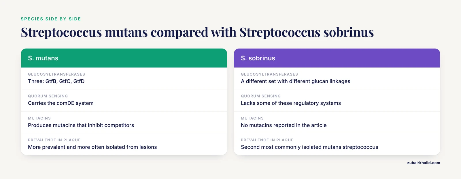
S. sobrinus is the second most commonly isolated mutans streptococcus from human dental plaque and is also strongly associated with dental caries. Both species produce glucosyltransferases and extracellular polysaccharides, but they differ in several clinically relevant ways.
S. mutans produces three glucosyltransferases, GtfB, GtfC, and GtfD, each with distinct product profiles [5][9]. GtfC is linked to both soluble and insoluble glucan synthesis and is a significant factor in the adhesion of S. mutans to tooth surfaces and bacterial cell aggregation within biofilms [9]. S. sobrinus produces a different set of glucosyltransferases, and its glucan products differ in linkage composition. These biochemical differences affect the architecture of the biofilms each species forms.
S. mutans carries the comDE quorum-sensing system, which regulates genetic competence and biofilm formation [6][7]. It also produces mutacins, bacteriocins that inhibit competing oral bacteria. The vicRK two-component system regulates Gtf-mediated adhesion and extracellular polysaccharide synthesis [6][7]. S. sobrinus lacks some of these regulatory systems, which may explain why S. mutans is more prevalent and more frequently isolated from carious lesions [2].
From a clinical perspective, both species contribute to caries, and their relative abundance varies among individuals. Laboratory identification should distinguish between them when species-level information is needed for epidemiological or research purposes. On selective media such as Mitis Salivarius agar with bacitracin, S. mutans and S. sobrinus can be differentiated by colony morphology and sugar fermentation patterns.
How Streptococcus mutans Causes Tooth Decay
Adhesion and colonization of the tooth surface
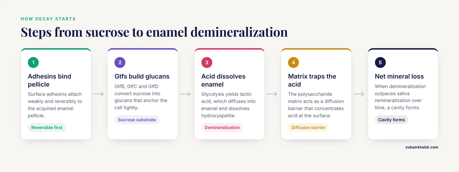
Caries development begins with the colonization of the tooth surface by S. mutans. The tooth is covered by the acquired enamel pellicle, a thin layer of salivary proteins and glycoproteins. S. mutans expresses surface adhesins that bind to components of this pellicle, initiating weak and reversible attachment [1]. This initial attachment is strengthened by the action of glucosyltransferases, which synthesize glucans from sucrose [1][11]. These glucans provide binding sites for S. mutans to adhere tightly to the tooth enamel [11].
Atomic force microscopy studies have provided nanoscale measurements of these adhesion forces [5]. Deletion of the gtfB and gtfC genes reduces the adhesion forces of S. mutans toward substrate and bacterial cell surfaces [5]. Deletion of gtfB also remarkably decreases bacterial adhesion to biofilm surfaces, while gtfC deletion shows no obvious effect at this stage [5]. These findings demonstrate that GtfB and GtfC have distinct and complementary roles in the multi-stage process of biofilm formation [5].
Glucosyltransferases and extracellular polysaccharide synthesis
Glucosyltransferases (Gtfs) are the central virulence enzymes of S. mutans [1][5][9][11]. These enzymes use sucrose as a substrate to synthesize extracellular polysaccharides, primarily glucans [1][9][10]. S. mutans produces three Gtfs with distinct functions:
- GtfB synthesizes insoluble glucans that form the structural scaffold of the biofilm.
- GtfC synthesizes both soluble and insoluble glucans and contributes to bacterial adhesion to tooth surfaces [9].
- GtfD synthesizes soluble glucans that contribute to bacterial aggregation.
The glucans produced by these enzymes serve multiple functions. They provide binding sites for S. mutans and other oral bacteria, creating a cohesive matrix that anchors the biofilm to the tooth surface [1][11]. They also create a diffusion barrier that concentrates acids at the enamel surface [1]. The extracellular polysaccharide matrix is a defining feature of cariogenic biofilms and a primary target for anti-biofilm strategies [1][12][9][11].
Acid production and acid tolerance
S. mutans metabolizes dietary carbohydrates through glycolysis to produce organic acids, primarily lactic acid [1][4]. This acidogenicity is a key virulence trait [7]. The acids diffuse into the enamel and dissolve the hydroxyapatite crystals, a process called demineralization.
What makes S. mutans particularly cariogenic is its acid tolerance. The bacterium can survive and continue metabolizing at pH values below 5.0, conditions that inhibit many competing oral species [7]. The F1F0-ATPase proton pump is essential for this acid tolerance [7]. It extrudes protons from the cytoplasm, maintaining internal pH homeostasis. Compounds that inhibit F1F0-ATPase, such as 3,5-di-tert-butylphenol, reduce acid tolerance and therefore virulence [7].
The two-component signal transduction system also contributes to acid tolerance. The liaS gene is essential for caries pathogenicity, and its inactivation impairs acid tolerance through downregulation of comDE expression [6]. The vicRK system regulates Gtf-mediated adhesion, extracellular polysaccharide synthesis, and acidic metabolite accumulation [6].
Biofilm maturation and the caries process
Within the mature biofilm, S. mutans exists in a structured community embedded in an extracellular polysaccharide matrix [1][12]. The biofilm is not a random collection of bacteria but an organized ecosystem with channels, gradients, and distinct microenvironments [1][12]. The matrix provides mechanical stability, protects bacteria from antimicrobial agents, and retains acids at the tooth surface [1][12].
Membrane vesicles released by S. mutans contribute to biofilm formation [13]. These vesicles carry glucosyltransferases and other proteins that promote adhesion and glucan-dependent biofilm formation [13]. The environmental pH influences the characteristics of these vesicles. At an initial pH of 6.0, the amount of Gtfs on membrane vesicles decreases, guiding low levels of vesicle-dependent biofilm formation [13]. This finding suggests that environmental pH conditions determine the local pathogenic activities of biofilm development in the oral cavity [13].
Cross-kingdom interactions can enhance caries pathogenesis. S. mutans strains expressing the collagen-binding protein Cnm are strongly associated with severe and recurrent dental caries [14]. Cnm enhances early biofilm formation with Candida albicans, a fungus commonly found in the oral cavity [14]. C. albicans binds to Cnm at levels comparable to those of GtfB and higher than those of other S. mutans adhesins [14]. Cnm cannot simultaneously engage collagen and C. albicans, suggesting competition for the same or adjacent binding regions [14]. This dual-binding adhesin promotes coaggregation and dual-species biofilm development, which may explain the clinical enrichment of Cnm-positive S. mutans strains alongside C. albicans [14].
The caries process is cyclical. Each episode of sugar consumption triggers acid production and demineralization. Between episodes, saliva promotes remineralization. When demineralization outpaces remineralization over time, net mineral loss occurs and a cavity forms. Fluoride shifts this balance toward remineralization by promoting fluorapatite formation [3].
Laboratory Identification of Streptococcus mutans
Specimen collection and transport
For laboratory diagnosis, samples are collected from dental plaque using a sterile scaler or curette, or from saliva using a sterile container. Plaque samples should be taken from carious lesions or from the supragingival margin, where S. mutans colonizes most heavily. Saliva samples can be collected by having the patient chew on a sterile paraffin pellet to stimulate saliva flow, then expectorating into a sterile tube. Samples should be transported to the laboratory promptly in a suitable transport medium to maintain bacterial viability.
Culture and isolation
S. mutans grows on blood agar as alpha-hemolytic colonies, producing a green zone of partial hemolysis. For selective isolation from mixed oral samples, Mitis Salivarius agar with bacitracin and sucrose (MSB agar) is the standard medium. On MSB agar, S. mutans forms characteristic raised, granular, dark blue colonies with a frosted-glass or mucoid appearance. The high sucrose content promotes extracellular polysaccharide production, giving colonies a gummy texture that is typical of the species.
Biochemical identification
S. mutans is catalase-negative, which distinguishes it from staphylococci and micrococci. It ferments mannitol, sorbitol, and raffinose with acid production, a feature that separates it from many other viridans group streptococci. It hydrolyzes esculin in the presence of bile. It produces acetoin from glucose (Voges-Proskauer positive). These biochemical reactions, combined with colony morphology and Gram stain, provide reliable species identification in the clinical laboratory.
Molecular methods
PCR-based methods targeting the gtfB, gtfC, and gtfD genes can confirm species identification [4][7][8]. Quantitative PCR (qPCR) can quantify S. mutans in plaque or saliva samples, which is useful for caries risk assessment and monitoring treatment response. Gene expression analysis by reverse transcription quantitative PCR (RT-qPCR) is used in research settings to assess the expression of virulence genes such as gtfB, gtfC, gtfD, gbpB, and genes encoding the F-ATPase [4][8]. These molecular tools have become essential in studies evaluating the efficacy of anti-biofilm agents [7][8].
Treatment and Prevention of Streptococcus mutans Caries
Conventional preventive agents
Fluoride remains the cornerstone of caries prevention [4][3]. It has dual effects: promoting enamel remineralization through the formation of fluorapatite, which is more resistant to acid dissolution than hydroxyapatite, and inhibiting bacterial glycolysis [3]. Fluoride is delivered through toothpaste, mouth rinses, professional varnishes, and community water fluoridation.
Chlorhexidine is a broad-spectrum antimicrobial agent used in mouth rinses and gels [1][2][3]. It is effective against S. mutans and reduces plaque formation, but its long-term use is limited by side effects such as tooth staining and taste disturbance [1]. It can also disrupt the balance of the oral flora, which is a concern with any broad-spectrum antimicrobial [1].
Hydrogen peroxide provides broad-spectrum antimicrobial activity and is incorporated into some mouth rinses [3]. Zinc exhibits multifaceted roles in metabolic inhibition and plaque reduction [3]. Prebiotics such as arginine and xylitol aim to restore ecological balance by favoring health-associated commensal species [3].
Mechanical plaque removal through toothbrushing and flossing remains the primary preventive measure [1][3]. However, it is time-consuming and requires consistent patient compliance. Antibacterial drugs have additional limitations, including short-term effectiveness and the potential for antibiotic resistance [1].
Natural products and phytochemicals
A wide range of natural products has been investigated for anti-biofilm activity against S. mutans [2][9][10][11][7][8][15][16].
Flavonoids, including 2-phenyl chroman flavonoids, can decrease bacterial virulence in the oral cavity by several mechanisms [2]. These include decreasing biofilm adhesivity and formation, inhibiting sortase A (SrtA), and inhibiting glucosyltransferase [2]. Fluoride and chlorhexidine, which show anticaries effects through different mechanisms, can effectively combine with flavonoids in combating dental caries [2].
Chalcone derivatives, which are primary precursors for flavonoid and isoflavonoid biosynthesis, inhibit GtfC and prevent biofilm formation by S. mutans [9]. Molecular docking studies predict interactions between chalcone derivatives and GtfC, and colorimetric assays confirm inhibition of biofilm formation [9].
Raffinose, an oligosaccharide from natural products, strongly inhibits biofilm formation, GTF-related gene expression, and glucan production [11]. Biofilm inhibition on saliva-coated hydroxyapatite discs, which mimic the tooth surface, indicates the applicability of raffinose in oral health [11]. Raffinose may be considered an antibiofilm agent for use in oral supplies and dental materials [11].
Ethyl gallate suppresses S. mutans biofilm formation on polystyrene and glass surfaces and inhibits acidogenicity in a dose-dependent manner [8]. The highest concentration tested (3.53 mM) reduced biofilm formation by 68% on polystyrene and more than 91% on glass [8]. It also prevented a decrease in pH levels by 95% [8]. Gene expression analysis revealed significant changes in gtfC, gtfB, and gbpB expression in treated biofilms [8].
Gallic acid and ethyl gallate from Libidibia ferrea, a plant used in Brazilian folk medicine, show antimicrobial and anti-adherence activities against S. mutans [16]. The ethanolic extract and fractions inhibit biofilm formation, pH drop, and proton permeability [16].
Areca nut essential oil shows antibacterial activity against S. mutans, with the fraction separated at 70°C showing the most prominent bacteriostatic activity [10]. The oil attenuates the adhesion ability of S. mutans by decreasing surface properties and inhibits glucosyltransferase activity [10].
Bergenia crassifolia leaf extracts decrease the viability of bacteria within biofilms and reduce extracellular polysaccharide synthesis by inhibiting Gtf activity [15]. The extracts show no cytotoxicity against normal oral cells [15].
3,5-Di-tert-butylphenol (3,5-DTBP) inhibits biofilm formation at 100 µg/ml without lethal effects on growth [7]. It significantly reduces water-soluble and water-insoluble glucan production, in concurrence with downregulation of gtfBC genes [7]. It arrests lactate dehydrogenase and enolase activity, reducing acidogenicity [7]. It also reduces acid tolerance by impeding F1F0-ATPase [7]. Gene expression analysis revealed downregulation of gtfB, gtfC, gtfD, vicRK, comDE, gbpB, smu0630, and relA [7].
Sulfated vizantin inhibits biofilm maturation without affecting bacterial growth [17]. It dose-dependently inhibits the extracellular release of cell-free glucosyltransferase and enhances the accumulation of cell-associated glucosyltransferase [17]. This disrupts the localization balance between the two forms, resulting in inhibited biofilm maturation [17]. Sulfated vizantin shows no cytotoxicity against human gingival cells [17].
Probiotics and postbiotics
Probiotics are live microorganisms that confer health benefits when administered in adequate amounts [18][3]. They represent an emerging complementary approach targeting the ecological basis of caries [4].
In a controlled animal study using a rat caries model, the probiotic Bifidobacterium animalis subsp. lactis BL-11 reduced total Keyes caries scores by 45%, with dentin lesion scores decreased by up to 73% [4]. S. mutans colonization was reduced by 70.3% and lactic acid production by 58.6% [4]. Enamel microhardness and calcium content were significantly preserved [4]. Histological scoring confirmed markedly reduced dentin demineralization and collagen degradation [4]. Virulence genes governing biofilm formation (gtfB, gtfC, gbpB) and acid production were affected [4].
Lactic acid bacteria strains, including Lactobacillus pentosus and Lactobacillus crispatus, show antimicrobial activity against S. mutans and modulate biofilm formation in vitro [18]. The antimicrobial substances produced by L. pentosus 13-4 may be lipophilic proteins [18].
Postbiotics are non-viable microbial products or metabolic derivatives with biological activity [12]. They include organic acids, exopolysaccharides, bacteriocins, and structural components derived from inactivated probiotic cells [12]. Postbiotics can reduce acidogenic potential, weaken extracellular matrix integrity within biofilms, disrupt bacterial communication systems, and modulate mucosal immune pathways [12]. These effects may limit colonization efficiency and pathogenic behavior of S. mutans while preserving commensal balance [12]. Emerging strategies propose combining postbiotics with bacteriophage-based approaches, immunomodulatory platforms, and innovative delivery systems such as nanoformulations and bioadhesive matrices [12].
Emerging technologies and surface modifications
Antimicrobial peptides, nanoparticles, and non-thermal plasma therapies are being investigated for targeted inhibition of S. mutans biofilm formation [1]. These approaches aim to overcome the limitations of conventional agents, including time consumption, short-term effectiveness, antibiotic resistance, and disruption of oral flora balance [1].
Surface modifications of dental materials can reduce bacterial adhesion and biofilm formation. Photoreactive zwitterionic PMBPAz-MPC coatings on orthodontic PMMA reduce early S. mutans adhesion by 81% before aging and 76% after composite aging [19]. Crystal violet biomass decreased by 44.6% before aging and 37.9% after aging [19]. These coatings also affect protein adsorption and human gingival fibroblast responses [19].
Bacillus velezensis K68, isolated from traditional fermented foods, inhibits biofilm formation mediated by S. mutans [20]. The supernatant from its culture medium inhibited biofilm formation by 84% when S. mutans was cultured for 48 hours [20]. The supernatant contains 1-deoxynojirimycin (1-DNJ), a known GTF expression inhibitor [20]. At 3 ppb, 1-DNJ decreased S. mutans cell surface hydrophobicity by 79.0 ± 0.8% [20]. The supernatant decreased S. mutans adherence by 99.97% under sugar-dependent conditions and 98.83% under sugar-independent conditions [20]. Treated S. mutans exhibited significantly reduced expression of gtfB, gtfC, and gtfD [20].
Common Mistakes and Limitations
- Assuming S. mutans is the only cariogenic organism. Dental caries is a polymicrobial disease involving many acidogenic and aciduric species [1][3]. S. mutans is the primary pathogen, but other bacteria and fungi contribute to disease progression [14][6].
- Overlooking the role of diet. S. mutans requires dietary sugars to produce acid and extracellular polysaccharides [1][11]. Caries cannot develop without fermentable carbohydrates in the diet.
- Confusing S. mutans with other viridans group streptococci. S. mutans is catalase-negative like all streptococci, but it can be distinguished by its ability to ferment mannitol and sorbitol and to produce glucans from sucrose.
- Relying solely on antimicrobial agents. Mechanical plaque removal remains essential [1][3]. Antibacterial agents have limitations including short-term effectiveness, antibiotic resistance, and disruption of oral flora balance [1].
- Expecting complete elimination of S. mutans. The goal of treatment is to reduce S. mutans levels and maintain microbial homeostasis, not to eradicate the organism [4][12]. S. mutans is a normal member of the oral microbiota that becomes pathogenic when dietary sugars drive its overgrowth [12].
- Overinterpreting in vitro results. Many natural products show promising anti-biofilm activity in laboratory studies, but clinical evidence is limited [2][15]. Toxicity profiles following brief exposure of topically applied agents in the oral cavity must be assessed before clinical studies [2].
- Ignoring the ecological context. Caries arises from complex ecological shifts within oral biofilms [12]. The dominance of S. mutans, in combination with broader microbial imbalance and interactions involving the oral virome, plays a central role in disease progression [12]. Individual cases require evaluation by a dentist or physician, who can assess clinical risk factors and recommend appropriate preventive or therapeutic measures.
Quick Review
- S. mutans is a Gram-positive, facultatively anaerobic coccus that is the principal cause of dental caries [1][2].
- Its major virulence factors are glucosyltransferases (GtfB, GtfC, GtfD), extracellular polysaccharide synthesis, acid production, and acid tolerance [1][5][9][11][7].
- S. mutans forms biofilms (dental plaque) on tooth surfaces, where acids concentrate and demineralize enamel [1][12][3].
- Laboratory identification relies on Gram stain, culture on selective media, biochemical tests (catalase-negative, mannitol fermentation), and molecular methods [8].
- Fluoride is the cornerstone of caries prevention, with dual effects on enamel remineralization and bacterial glycolysis [4][3].
- Emerging strategies include natural products (flavonoids, chalcones, raffinose, ethyl gallate), probiotics, postbiotics, and surface modifications of dental materials [1][2][4][12][9][11][8][19].
- S. mutans strains expressing the collagen-binding protein Cnm are associated with severe and recurrent caries and enhanced cross-kingdom biofilm formation with C. albicans [14].
Transmission and Early Colonization of Streptococcus mutans
S. mutans is absent from the mouth at birth. Infants typically acquire the organism from their mothers through vertical transmission, although other caregivers and siblings can transmit it horizontally. Saliva-sharing behaviors, including tasting food before feeding, sharing utensils, and kissing on the lips, transfer maternal S. mutans to the infant.
The "window of infectivity" is the period between approximately 6 and 30 months of age, when primary teeth erupt and provide new hard, non-shedding surfaces for colonization. Acquisition during this window predicts higher caries risk in later childhood. Factors that promote early colonization include high maternal salivary S. mutans counts, frequent maternal sugar intake, poor maternal oral hygiene, and the infant's own diet, particularly frequent exposure to fermentable carbohydrates.
Prevention implications for caregivers are direct. Reducing maternal S. mutans levels before and after delivery, avoiding saliva-sharing behaviors, delaying the introduction of sugary foods and drinks, and scheduling early dental visits can delay or reduce infant colonization. These measures lower the child's lifetime caries risk.
Caries Risk Assessment and Chairside Tests
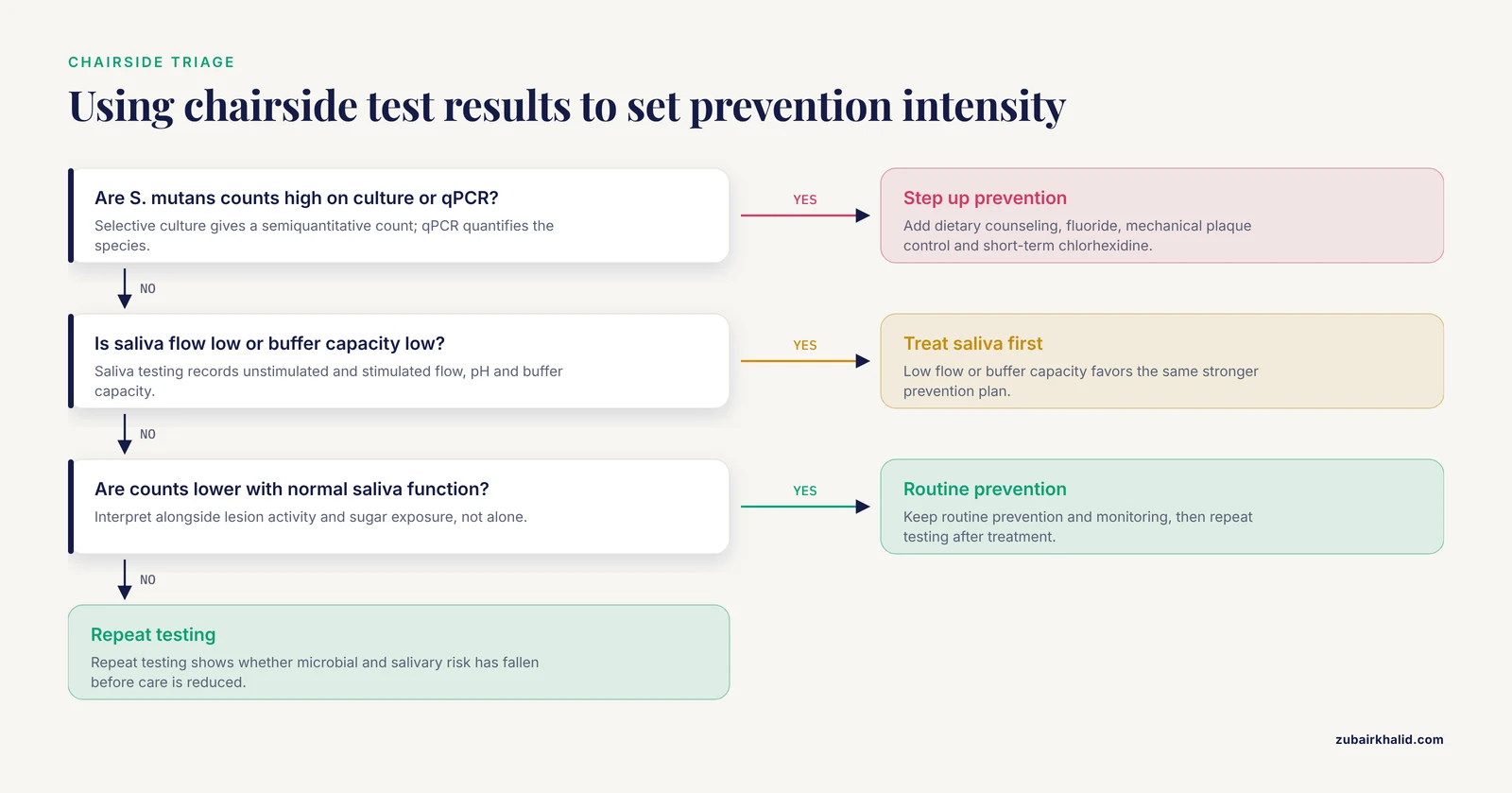
Caries risk assessment combines microbial load, saliva function, diet, and clinical lesions. Selective culture on Mitis Salivarius bacitracin agar gives a semiquantitative count of mutans streptococci; quantitative PCR can identify and quantify S. mutans specifically. Saliva testing records unstimulated and stimulated flow, pH, and buffer capacity. Chairside dip-slide tests estimate mutans streptococci and lactobacilli counts, while plaque sampling shows whether the cariogenic load is localized or widespread. These results are interpreted with lesion activity and sugar exposure, not as stand-alone diagnoses.
Use them to set prevention intensity. High S. mutans counts, low saliva flow, or low buffer capacity support dietary counseling, fluoride, mechanical plaque control, and short-term chlorhexidine use [1][3]. Lower counts with normal saliva function favor routine prevention and monitoring. Repeat testing after treatment shows whether microbial and salivary risk has fallen, guiding whether care can be stepped down.
Laboratory Models for Studying S. mutans Biofilms
In vitro models allow controlled study of S. mutans adhesion, matrix production, and antimicrobial susceptibility. The microtiter plate assay is the most common screening tool. Biofilms grow on polystyrene or glass wells, and total attached biomass is quantified by crystal violet staining [8]. This method is high-throughput but measures extracellular matrix and dead cells along with live bacteria, so it overestimates viable biofilm mass. Saliva-coated hydroxyapatite discs mimic the acquired enamel pellicle and tooth mineral surface, providing a more relevant substratum for adhesion and glucan-dependent biofilm assays [11]. Flow cell systems permit real-time observation of biofilm architecture under continuous medium flow, using confocal microscopy to assess thickness, channel formation, and matrix distribution. For viability, colony-forming unit (CFU) counts are often paired with crystal violet. CFU data require biofilm disruption and plating, and they underestimate cell numbers because S. mutans grows in chains and clumps, and some cells are viable but non-culturable. Interpreting either metric alone can therefore misrepresent biofilm behavior.
Related Guides
- Equine Strangles: Streptococcus equi subsp. equi Diagnostic Testing and Outbreak Management: Equine Streptococcus equi diagnostics and outbreak management comparison
- Streptococcus suis Serotype 2 in Pigs and Zoonotic Risk for Pork Workers: Meningitis and Diagnosis: Another Streptococcus species with zoonotic meningitis risk and diagnosis
- Streptococcus suis in Swine: Pathogenesis, Diagnostics, and Vaccine Strategies: Swine Streptococcus suis pathogenesis, diagnostics, and vaccine strategies
- Streptococcus zooepidemicus Bacterial Infection in Poultry: Etiology, Pathogenesis, and Diagnostic Approaches: Poultry Streptococcus zooepidemicus etiology, pathogenesis, and diagnostic approaches
- Streptococcus pyogenes (Group A Strep): Etiology and Clinical Relevance in Poultry: Group A Streptococcus etiology and clinical relevance in poultry
- Streptococcosis in Farmed Tilapia: Streptococcus agalactiae and Streptococcus iniae Pathogenesis, Rapid Diagnostic Tests, and Vaccine Development: Tilapia streptococcosis pathogenesis, rapid diagnostics, and vaccine development
- Tooth Resorption in Cats: Symptoms and Treatment: Feline tooth resorption symptoms and treatment for dental comparison
- Tooth Resorption in Dogs: Causes, Stages, and Symptoms: Canine tooth resorption causes, stages, and symptoms overview
- Cat Tooth Extraction: Recovery and Aftercare: Cat tooth extraction recovery guidance for advanced dental disease treatment
- Broken Tooth in Dogs: Symptoms, Types, and When It Hurts: Dog broken tooth signs and pain context for dental trauma
Frequently Asked Questions
What does Streptococcus mutans look like on Gram stain?
S. mutans appears as Gram-positive cocci arranged in pairs or chains. The cells are spherical to ovoid, approximately 0.5 to 0.75 µm in diameter, and retain crystal violet due to their thick peptidoglycan cell wall.
How does S. mutans cause tooth decay?
S. mutans adheres to tooth surfaces, produces glucosyltransferases that synthesize extracellular polysaccharides from sucrose, forms biofilms, and metabolizes dietary sugars into lactic acid [1]. The acid demineralizes enamel, leading to cavity formation [3].
What is the difference between S. mutans and S. sobrinus?
Both are cariogenic mutans streptococci, but S. mutans produces three glucosyltransferases (GtfB, GtfC, GtfD) and is more prevalent in human plaque [5][9]. S. sobrinus is less common but can also cause caries. S. mutans carries the comDE quorum-sensing system and produces mutacins that inhibit competitors [6][7].
How is S. mutans infection treated?
Treatment combines mechanical plaque removal, fluoride for remineralization, and antimicrobial agents such as chlorhexidine [1][3]. Emerging approaches include probiotics, postbiotics, and natural products that inhibit biofilm formation [1][2][4][12].
Can S. mutans be completely eliminated from the mouth?
No. The goal is to reduce S. mutans levels and restore microbial balance, not to eradicate the organism [4][12]. S. mutans is a normal member of the oral microbiota that becomes pathogenic when dietary sugars drive its overgrowth [12].
What dietary factors promote S. mutans growth?
Frequent consumption of sucrose and other fermentable carbohydrates promotes S. mutans growth and biofilm formation [1][11]. Sucrose is the preferred substrate for glucosyltransferase activity and extracellular polysaccharide synthesis [1][9][11].
Sources
- New strategies and mechanisms for targeting Streptococcus mutans biofilm formation to prevent dental caries: A review.
- Natural 2-phenyl chroman flavonoids against Streptococcus mutans: prospects of herbal remedies for dental caries.
- Oral hygiene agents at work: effects on Streptococcus mutans and caries risk.
- Probiotic Bifidobacterium animalis subsp. lactis BL-11 attenuates dental caries development in a Streptococcus mutans-induced rat model.
- Glucosyltransferase-modulated Streptococcus mutans adhesion to different surfaces involved in biofilm formation by atomic force microscopy.
- LiaS gene from two-component system is essential for caries pathogenicity in dual-species biofilms of Streptococcus mutans and Candida albicans.
- 3, 5-Di-tert-butylphenol combat against Streptococcus mutans by impeding acidogenicity, acidurance and biofilm formation.
- Inhibitory Effects of Ethyl Gallate on Streptococcus mutans Biofilm Formation by Optical Profilometry and Gene Expression Analysis.
- Chalcone derivatives against biofilm production of Streptococcus mutans by glucosyltransferase C inhibition.
- Antibacterial mechanism of areca nut essential oils against Streptococcus mutans by targeting the biofilm and the cell membrane.
- Raffinose Inhibits Streptococcus mutans Biofilm Formation by Targeting Glucosyltransferase.
- Postbiotics and phage synergy in precision oral microbiome engineering: systems biology strategies targeting Streptococcus mutans in dental caries.
- Effects of pH on the Properties of Membrane Vesicles Including Glucosyltransferase in Streptococcus mutans.
- The Streptococcus mutans collagen-binding protein Cnm enhances early biofilm formation with Candida albicans.
- Anti-biofilm Activities from Bergenia crassifolia Leaves against Streptococcus mutans.
- Anticariogenic activities of Libidibia ferrea, gallic acid and ethyl gallate against Streptococcus mutans in biofilm model.
- Sulfated vizantin inhibits biofilm maturation by Streptococcus mutans.
- Isolation and Characterisation of Probiotics for Antagonising the Cariogenic Bacterium Streptococcus mutans and Preventing Biofilm Formation.
- Influence of Surface Modification and Artificial Aging on Streptococcus mutans Biofilm Formation and Oral Cell Responses to Orthodontic PMMA: An In Vitro Study.
- Inhibitory effect of Bacillus velezensis on biofilm formation by Streptococcus mutans.