
Streptococcus zooepidemicus Bacterial Infection in Poultry: Etiology, Pathogenesis, and Diagnostic Approaches
Introduction
Streptococcus zooepidemicus (taxonomically classified as Streptococcus equi subsp. zooepidemicus) is a Lancefield group C beta-hemolytic streptococcus that causes acute septicemic disease in poultry, particularly in layer chickens and free-range flocks. Although historically considered an opportunistic pathogen associated with equine hosts, S. zooepidemicus has emerged as a significant cause of mortality and egg production losses in commercial poultry operations worldwide [1, 2]. The bacterium is capable of rapid horizontal transmission, often linked to environmental contamination or direct contact with carrier animals such as horses [2]. This article provides a detailed review of the Streptococcus zooepidemicus poultry bacterial infection, including its etiology, epidemiology, clinical manifestations, pathological findings, diagnostic methods, treatment options, and control strategies, with emphasis on peer-reviewed evidence from the past four decades.
Etiology and Taxonomy
S. zooepidemicus is a Gram-positive, catalase-negative, facultative anaerobic coccus that forms chains in liquid culture. It belongs to the pyogenic group of streptococci and expresses the Lancefield group C carbohydrate antigen. The organism is closely related to Streptococcus equi subsp. equi (the agent of strangles in horses) but differs in host range and virulence gene repertoire. In poultry, S. zooepidemicus produces a polysaccharide capsule that contributes to antiphagocytic properties and systemic dissemination. The bacterium secretes several exotoxins, including streptolysin O and streptokinase, which facilitate tissue invasion and fibrinolytic activity [3].
Epidemiology and Transmission
Host Range and Reservoir
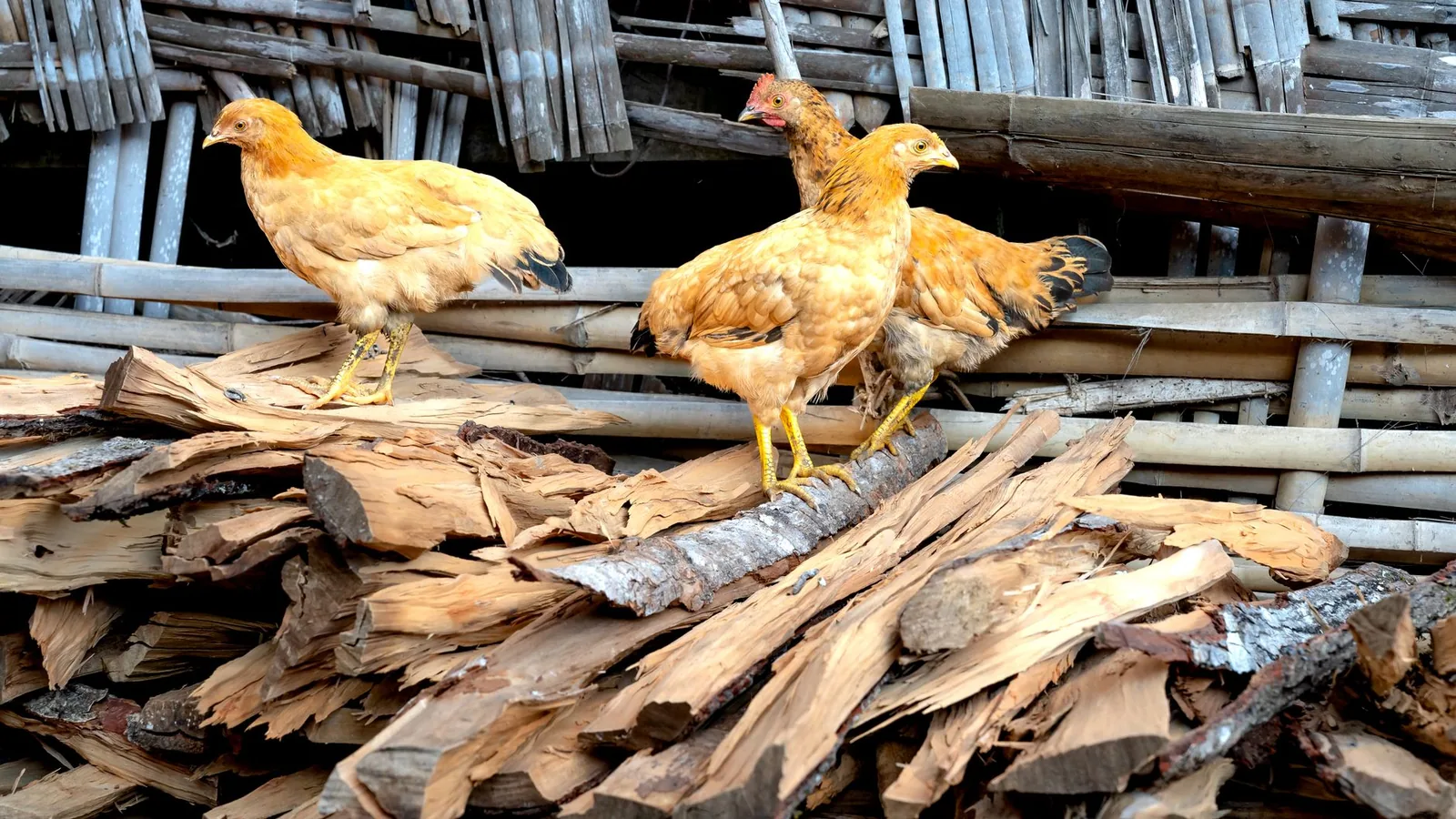
The primary reservoir of S. zooepidemicus is the equine upper respiratory tract, but the organism has been isolated from many mammals and birds. In poultry, outbreaks have been documented in layer flocks, broiler breeders, and free-range chickens [1, 2]. Turkeys, ducks, and other gallinaceous birds are also susceptible. A major outbreak in free-range chickens was directly linked to the presence of horses on the same farm, suggesting interspecies transmission via contaminated fomites or aerosolized respiratory secretions [2].
Transmission Dynamics
Horizontal transmission occurs through the fecal-oral route, as demonstrated by Garmyn et al. [1], who provided evidence of fecal shedding and subsequent ingestion of contaminated litter as a primary infection mechanism. The bacterium can survive for extended periods in organic material, including bedding, feed, and water. Vertical transmission has not been conclusively demonstrated, but the organism can colonize the oviduct and contaminate eggshells during passage. Co-infection with other respiratory pathogens, such as Ornithobacterium rhinotracheale, can exacerbate disease severity and complicate diagnosis [4].
Risk Factors
High stocking density, poor ventilation, concurrent immunosuppressive infections (e.g., infectious bursal disease), and stress from transport or molting predispose flocks to outbreaks. The introduction of new birds or contact with horses significantly increases the risk of introduction [2].
Clinical Signs
The incubation period in experimentally infected chickens ranges from 24 to 72 hours [5]. Clinical presentation varies from peracute death to subacute illness with the following signs:
- Sudden onset of depression, ruffled feathers, and anorexia.
- Cyanosis of the comb and wattles due to septicemic shock.
- Respiratory distress: dyspnea, rales, and nasal discharge.
- Diarrhea, often greenish or watery.
- Drop in egg production in laying hens, sometimes exceeding 30%.
- Neurologic signs (torticollis, ataxia) in a subset of cases, indicating meningoencephalitis.
- Increased mortality, which can reach 10-50% in untreated flocks [1, 5].
In chronic cases, birds may develop lameness due to fibrinous polyserositis or arthritis.
Pathology
Gross Lesions
Postmortem examination reveals lesions consistent with acute septicemia and fibrinous inflammation:
- Spleen: Splenomegaly with mottled appearance.
- Liver: Hepatomegaly, congestion, and focal necrosis.
- Heart: Pericarditis with fibrinous exudate; epicardial petechiae.
- Lungs: Congestion, edema, and fibrinous pneumonia.
- Serous membranes: Fibrinous peritonitis, airsacculitis, and perihepatitis.
- Oviduct: Salpingitis with caseous exudate in layers.
- Joints: Fibrinous arthritis in chronic cases.
Histopathology
Microscopic examination shows multifocal necrosis and heterophilic infiltration in the liver, spleen, and myocardium. Fibrin thrombi are frequently observed in small vessels, consistent with a disseminated intravascular coagulation (DIC)-like syndrome. Roy et al. [3] characterized the hemostatic response in infected layers using thromboelastography and prothrombin time, demonstrating a hypercoagulable state followed by consumptive coagulopathy. Fibrinogen levels rise acutely, contributing to the fibrinous exudates seen grossly.
Pathogenesis
After ingestion or inhalation, S. zooepidemicus adheres to mucosal epithelial cells via surface adhesins. The polysaccharide capsule inhibits phagocytosis, allowing the bacterium to enter the bloodstream and cause bacteremia. Systemic dissemination leads to colonization of the liver, spleen, heart, and serosal surfaces. The release of streptolysin O and other cytolysins damages host cell membranes, while streptokinase activates plasminogen, promoting fibrinolysis and tissue invasion. The resulting inflammatory cascade recruits heterophils and macrophages, but the bacterium's M-like proteins confer resistance to opsonophagocytosis. The host response includes a marked acute-phase reaction, with elevated ovotransferrin levels detectable by commercial ELISA kits [6].
Diagnostic Approaches
Clinical and Gross Pathology
A presumptive diagnosis is based on acute mortality, fibrinous polyserositis, and Gram-positive cocci in chains on impression smears from liver or spleen.
Bacteriological Culture
Definitive diagnosis requires isolation of S. zooepidemicus from affected tissues (liver, spleen, heart blood) on blood agar or selective streptococcal media. Colonies appear as small, gray, beta-hemolytic after 24-48 hours at 37°C in 5% CO2. The organism is catalase-negative and PYR (pyrrolidonyl arylamidase) positive. Lancefield group C antigen can be confirmed using commercial latex agglutination kits.
Molecular Detection
PCR assays targeting the sodA or 16S rRNA genes provide rapid species-level identification. Real-time PCR can quantify bacterial load in tissues or environmental samples. High-throughput sequencing of the gdh gene allows subspecies differentiation.
Serology
Commercial ELISA kits for chicken ovotransferrin (an acute-phase protein) have been evaluated as adjunctive diagnostic tools. Roy et al. [6] reported that ovotransferrin levels rise significantly in S. zooepidemicus-infected layers, but the response is not specific and overlaps with infections such as Gallibacterium anatis. Therefore, serology is best used in combination with culture or PCR.
Differential Diagnosis
The clinical and pathological presentation of S. zooepidemicus infection resembles other septicemic diseases of poultry:
| Disease | Key Differentiating Features |
|---|---|
| Fowl cholera (Pasteurella multocida) | Gram-negative bipolar rods; no beta-hemolysis on blood agar |
| Infectious Coryza (Avibacterium paragallinarum) | Swollen infraorbital sinuses; Gram-negative pleomorphic rods |
| Ornithobacterium rhinotracheale infection | Gram-negative rods; requires chocolate agar or CO2; co-infection common [4] |
| Escherichia coli colibacillosis | Gram-negative rods; lactose-fermenting on MacConkey; airsacculitis more prominent |
| Avian Cholera in waterfowl | Similar lesions but host species and Gram stain differentiate |
Diagnostic Workflow
The following Mermaid diagram outlines a recommended diagnostic algorithm for suspected S. zooepidemicus infection in poultry.
flowchart TD
 A[Acute mortality with fibrinous polyserositis] --> B[Impression smear from liver/spleen]
 B --> C{Gram-positive cocci in chains?}
 C -->|Yes| D[Culture on blood agar, 37°C, 5% CO2]
 C -->|No| E[Consider other septicemic pathogens]
 D --> F{Beta-hemolytic colonies?}
 F -->|Yes| G[Catalase negative, PYR positive]
 G --> H[Lancefield group C latex agglutination]
 H --> I[Confirm by PCR sodA or 16S rRNA]
 I --> J["Definitive diagnosis: S. zooepidemicus"]
 F -->|No| K[Re-evaluate culture conditions or consider other streptococci]
 E --> L[Perform culture and PCR for Pasteurella, E. coli, Ornithobacterium]
Treatment
Antimicrobial Therapy
S. zooepidemicus is generally susceptible to beta-lactam antibiotics, macrolides, and tetracyclines. However, antimicrobial susceptibility testing should be performed due to regional resistance patterns. In laying hens, withdrawal periods must be observed for egg safety. Commonly used agents include:
- Amoxicillin or ampicillin (water-soluble formulations) at 10-20 mg/kg body weight for 3-5 days.
- Oxytetracycline in feed or water at 20-40 mg/kg for 5-7 days.
- Erythromycin or tylosin at 10-25 mg/kg for 3-5 days.
Treatment is most effective when initiated early in the outbreak. In severe cases, parenteral administration of ceftiofur or florfenicol may be considered under veterinary supervision.
Supportive Care
Improving ventilation, reducing stocking density, and providing clean water and feed are essential. Removal of moribund birds and contaminated litter reduces bacterial load.
Control and Prevention
Biosecurity
Strict biosecurity measures are critical to prevent introduction and spread:
- Separate poultry operations from equine facilities. Horses should not share water sources or equipment with poultry.
- Implement all-in/all-out management with thorough cleaning and disinfection between flocks.
- Use footbaths with disinfectants (e.g., quaternary ammonium compounds, peroxygen compounds) at house entrances.
- Control rodents and wild birds that may act as mechanical vectors.
Vaccination
No commercial vaccine is currently available for S. zooepidemicus in poultry. Autogenous bacterins have been used in some outbreaks, but efficacy data are limited. Research on subunit vaccines targeting the capsule or M-like proteins is ongoing.
Monitoring
Regular clinical surveillance and postmortem examination of dead birds allow early detection. PCR-based environmental monitoring of litter and water can identify contamination before clinical signs appear.
Conclusion
Streptococcus zooepidemicus is an important cause of acute septicemic disease in poultry, particularly in layers and free-range flocks. The Streptococcus zooepidemicus poultry bacterial infection is characterized by high mortality, fibrinous polyserositis, and egg production losses. Transmission occurs via the fecal-oral route, and outbreaks are often linked to contact with horses or contaminated environments. Diagnosis relies on bacterial culture, PCR, and exclusion of other septicemic pathogens. Treatment with beta-lactam antibiotics is effective if initiated early, but control depends on robust biosecurity and separation from equine reservoirs. Further research is needed to develop vaccines and understand the molecular epidemiology of this emerging avian pathogen.
References
[1] Garmyn A, Van de Velde N, Braeckmans D, et al. An Outbreak Associated with Streptococcus equi Subsp. zooepidemicus in Layers: Evidence of Fecal Transmission. Avian Dis. 2020. URL: https://pubmed.ncbi.nlm.nih.gov/33205184/
[2] Bisgaard M, Bojesen AM, Petersen MR, et al. A major outbreak of Streptococcus equi subsp. zooepidemicus infections in free-range chickens is linked to horses. Avian Dis. 2012. URL: https://pubmed.ncbi.nlm.nih.gov/23050474/
[3] Roy K, Bertelsen MF, Pors SE, et al. Inflammation-induced haemostatic response in layer chickens infected with Streptococcus equi subsp. zooepidemicus as evaluated by fibrinogen, prothrombin time and thromboelastography. Avian Pathol. 2014. URL: https://pubmed.ncbi.nlm.nih.gov/25017320/
[4] Pan Q, Liu A, He C. Co-infection of Ornithobacterium rhinotracheale with Streptococcus zooepidemicus in chickens. Avian Dis. 2012. URL: https://pubmed.ncbi.nlm.nih.gov/23397838/
[5] Roy K, Bisgaard M, Kyvsgaard NC, et al. Pathogenicity of wild-type and small-colony variants of Streptococcus equi subsp. zooepidemicus in layer chickens. Avian Pathol. 2013. URL: https://pubmed.ncbi.nlm.nih.gov/23721084/
[6] Roy K, Kjelgaard-Hansen M, Pors SE, et al. Performance of a commercial Chicken-Ovo-transferrin-ELISA on the serum of brown layer chickens infected with Gallibacterium anatis and Streptococcus zooepidemicus. Avian Pathol. 2014. URL: https://pubmed.ncbi.nlm.nih.gov/24313352/
[7] Goren E, de Jong WA, van Eck JH. [Streptococcus zooepidemicus infection in laying hens (author's transl)]. Tijdschr Diergeneeskd. 1981. URL: https://pubmed.ncbi.nlm.nih.gov/7268749/ *** Disclaimer: This article is for educational and informational purposes only. It is not intended to substitute for professional veterinary advice, diagnosis, treatment, or regulatory guidance. Always consult a licensed veterinarian or qualified specialist regarding animal health, disease diagnosis, and therapeutic decisions.