Imaging Bacteria: Microscopy and Advanced Techniques
By Dr. Zubair Khalid, DVM, MS, PhD ·
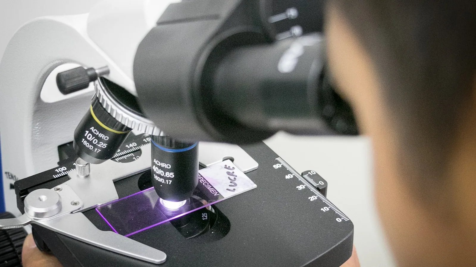
Imaging bacteria starts with a resolution problem. A typical rod-shaped bacterium measures roughly 1 to 5 micrometers long and about 0.5 to 1 micrometer wide, so a single cell sits close to the diffraction limit of a light microscope, where the smallest resolvable distance is about 200 nanometers. Seeing a bacterium is easy. Seeing its internal organization is not. That single constraint explains why bacterial imaging spans such a wide range of instruments, from a simple Gram stain on a glass slide to cryo-electron tomography that resolves individual macromolecular complexes inside a frozen cell [1].
This guide walks through the principal methods in current bench use. It covers what each technique actually resolves, whether it works on live or fixed cells, and where it fits in a laboratory workflow. It also flags the failure modes that produce false results, because most imaging errors in bacteriology come from sample handling rather than from the optics.
Why Bacterial Imaging Is Hard
Three physical facts shape every method below.
The first is size. Bacteria are small enough that many structures of interest, including ribosome arrangements, secretion systems, and membrane-associated complexes, fall below the resolution of conventional optical microscopy. This is the problem that motivated in-cell cryo-electron microscopy, which visualizes macromolecular complexes and the networks they form directly in their native cellular context [1].
The second is contrast. Bacteria are mostly water and have a refractive index close to that of their surroundings. In brightfield microscopy, an unstained cell is nearly invisible. Stains, phase contrast, or fluorescence labels are what create the signal.
The third is motion and fragility. Live bacteria swim, divide, and respond to light and heat. Fixing them preserves structure but can introduce artifacts. The choice between live and fixed imaging is therefore not cosmetic. It determines which questions the experiment can answer.
Brightfield Microscopy and the Gram Stain
Brightfield microscopy passes white light through a stained specimen and forms an image by absorption. It is the oldest and still the most widely used bacterial imaging method in routine laboratories, largely because of the Gram stain.
The Gram stain separates bacteria into two groups based on cell wall structure. Gram-positive cells retain crystal violet because their thick peptidoglycan layer traps the dye-iodine complex. Gram-negative cells lose crystal violet during alcohol decolorization and take up the pink counterstain safranin. The result, purple versus pink or red, is read directly under a brightfield microscope with a 100x oil immersion objective.
A related approach, morphology-defined classification from fluorescence microscopy of vaginal smears, illustrates how staining and imaging combine in practice. In a two-year real-world study of 4,492 patients, morphology-defined bacterial vaginosis was identified by the presence of clue cells or Gardnerella-like anaerobic bacteria on vaginal fluorescence microscopy, a definition explicitly distinct from Nugent scoring, Amsel criteria, or molecular profiling [2]. The point is that the imaging method and the diagnostic definition are tied together, and changing the imaging method changes what you can call a case.
Strengths and Limits of Brightfield
Brightfield is inexpensive, fast, and needs no specialized optics beyond a good objective. It cannot resolve anything below the diffraction limit, and it gives no information about viability, metabolic state, or molecular identity. A Gram stain tells you shape and staining class. It does not tell you which species you are looking at.
Phase Contrast and Darkfield
Phase contrast converts differences in refractive index into brightness differences, making live unstained bacteria visible. Darkfield illuminates the specimen from the side so that only scattered light reaches the objective, which is useful for thin or poorly staining organisms. Detection of thin spirochetes such as Leptospira benefits from methods that generate strong contrast against a clean background [3].
Fluorescence Microscopy
Fluorescence microscopy is the workhorse of modern bacterial imaging. A fluorophore absorbs light at one wavelength and emits at a longer wavelength. Filters separate excitation from emission, so the signal appears bright against a dark background. This gives high sensitivity and the ability to label specific molecules.
DNA Stains: DAPI and Its Relatives
DAPI (4',6-diamidino-2-phenylindole) binds AT-rich regions of double-stranded DNA and fluoresces blue under ultraviolet excitation. It is a fast way to count nucleoids, assess chromosome number per cell, and confirm that cells contain DNA. Because it stains all DNA, it does not distinguish bacterial DNA from host or environmental DNA, so it is used mainly for counting and localization rather than identification.
Fluorescence In Situ Hybridization (FISH)
FISH uses a fluorescently labeled nucleic acid probe that hybridizes to a complementary target sequence inside the cell, most commonly the 16S ribosomal RNA gene or its transcript. Because the probe sequence is chosen to match a specific taxonomic group, FISH identifies bacteria in situ while preserving their spatial context.
Two recent studies show FISH in action. In the ciliate Frontonia paramagna, FISH targeting the 16S rRNA gene, combined with V3-V4 high-throughput sequencing, consistently identified the bacterial symbiont Caedimonas localized within the host macronucleus, and transmission electron microscopy confirmed the intramacronuclear colonization [4]. In the giant kelp Macrocystis pyrifera, FISH imaging showed bacterial signal associated with host tissues across developmental stages, and the authors explicitly distinguished this signal from algal autofluorescence [5]. That distinction matters, because autofluorescence is the single most common source of false positives in environmental FISH.
Genetically Encoded Reporters: GFP and mRFP
Green fluorescent protein (GFP) and its variants are encoded directly in the genome, so a cell that expresses the reporter fluoresces without any external dye. This is powerful for live imaging of protein localization, promoter activity, and dynamic processes. Engineered strains that express fluorescent markers also enable automated detection of infected cells in high-throughput screens [6].
Reporter fusions go beyond simple labeling. An mRFP-GFP-LC3 tandem reporter measures autophagic flux because the GFP signal is quenched in acidic compartments while mRFP persists, so the ratio of the two signals reports whether a structure has progressed to an acidic stage. This reporter has been used to quantify Fusobacterium nucleatum-induced autophagy in colorectal cancer cells by immunofluorescence, with thin-section transmission electron microscopy used to confirm autophagosome ultrastructure [7].
Fluorescent Labels for Viability and Size
Chemically reactive dyes such as carboxyfluorescein succinimidyl ester (CFSE) covalently label cell proteins and persist through division, making them useful for tracking and for high-content imaging. A standardized CFSE-based method for Leptospira uses high-content fluorescence microscopy with the OPERA Phenix system and image analysis in ImageJ with the MicrobeJ plugin to automatically detect and measure individual bacteria, enabling reproducible size measurement across species and strains [3]. This is a good example of imaging bacteria as a quantitative measurement rather than just a picture.
Confocal Microscopy
A conventional fluorescence microscope collects light from the whole depth of the specimen, which produces out-of-focus blur. A confocal microscope uses a pinhole to reject light from outside the focal plane, so it builds an image from a thin optical section. Scanning the specimen in x, y, and z produces a three-dimensional reconstruction.
Confocal imaging is the standard for thick samples, biofilms, and tissue-associated bacteria. In the Helicobacter pylori saliva study, fluorescence-positive regions identified by confocal microscopy were then examined by field emission scanning electron microscopy, an approach known as correlative light and electron microscopy (CLEM) [8]. Confocal found the region of interest. Electron microscopy resolved the ultrastructure.
The resolution of confocal microscopy remains bounded by diffraction, so it improves contrast and sectioning rather than resolving power.
Super-Resolution Microscopy
Super-resolution methods break the diffraction limit by either localizing individual fluorophores or by using patterned illumination. Two families dominate bacterial imaging.
STORM and PALM
Stochastic optical reconstruction microscopy (STORM) and photoactivated localization microscopy (PALM) switch individual fluorophores on and off over many frames, localize each one's center precisely, and assemble the positions into a reconstruction. Resolution reaches roughly 20 to 50 nanometers. This is enough to resolve the arrangement of membrane proteins, the width of the bacterial cell envelope, and the spacing of molecular machines.
SIM
Structured illumination microscopy (SIM) projects a patterned light field onto the sample and extracts higher spatial frequencies from the resulting moiré patterns. SIM reaches roughly 100 nanometers, better than confocal but not as fine as STORM. Its advantage is speed and compatibility with live imaging, so it is often used for dynamic processes that STORM cannot capture.
Both methods demand careful sample preparation. Label density, fluorophore brightness, and drift control all determine whether the improved resolution is real.
Electron Microscopy
Electron microscopy replaces photons with electrons, whose much shorter wavelength pushes resolution below 1 nanometer. This is the only way to see bacterial ultrastructure at molecular detail.
Scanning Electron Microscopy (SEM)
SEM scans a focused electron beam across the surface of a specimen and collects secondary electrons. It produces detailed three-dimensional-looking images of surface topography. In materials and environmental work, SEM with quantitative image analysis characterized the nanofiber network of bacterial cellulose films, where optimized films showed a denser nanofiber structure that correlated with a 3.4-fold higher puncture strength [9]. SEM also confirmed the surface morphology of composite photocatalytic fiber membranes used for bacterial disinfection in wastewater [10].
SEM requires fixed, dehydrated, and usually metal-coated samples, so it images dead cells only.
Transmission Electron Microscopy (TEM)
TEM transmits electrons through an ultrathin section and forms an image from the electrons that scatter or pass through. It resolves internal structures, including membranes, ribosomes, flagella, and storage granules.
Several recent studies illustrate its range. TEM confirmed the formation of an iron nanoparticle-loaded bacterial cell factory, revealing rod-shaped cells with intact flagella and uniform spherical nanoparticles about 29.9 nanometers in diameter associated with the bacterial surface [11]. TEM showed preservation of mitochondrial ultrastructure in sepsis models, using a quantitative mitochondrial damage score to compare treated and untreated animals [12]. And in the kelp study, TEM revealed structures consistent with intracellular bacteria inside gametophyte cells [5].
TEM requires fixation, dehydration, embedding, and thin sectioning. Each step can introduce artifacts, and a structure that appears only after fixation should be treated with suspicion until it is confirmed another way.
Cryo-Electron Microscopy and Cryo-Electron Tomography
Cryo-EM avoids the chemical fixation and dehydration that cause the worst artifacts. The sample is plunge-frozen in vitreous ice, preserving near-native structure, and imaged at liquid nitrogen temperature. In-cell cryo-electron tomography (cryo-ET) tilts the specimen and reconstructs the three-dimensional density, which allows macromolecular complexes and the networks they form to be visualized in their native context [1]. In-cell single-particle analysis extends this to high-resolution structure determination of complexes inside intact cells.
These methods are transforming bacterial cell biology because they resolve structures that optical microscopy cannot, including subcellular organization that was once assumed to be a eukaryotic specialty [1]. Cryo-EM has also driven rapid progress in bacteriophage structural biology, where advances in specimen preparation, electron optics, and image processing have expanded what can be resolved in situ [13].
Correlative Light and Electron Microscopy
CLEM combines the molecular specificity of fluorescence with the resolution of electron microscopy. The workflow is straightforward in concept: find the feature of interest by light microscopy, then relocate the same region in the electron microscope. The H. pylori saliva study used exactly this logic, immunostaining samples with an anti-H. pylori antibody, identifying positive regions by confocal microscopy, and then examining those regions by FE-SEM [8]. Notably, salivary H. pylori appeared exclusively as coccoid or intermediate forms, with no spiral shapes observed, a morphological finding that light microscopy alone would not have resolved convincingly [8].
Method Comparison
| Method | Resolution limit | Live or fixed | Best use case |
|---|---|---|---|
| Brightfield (Gram stain) | ~200 nm | Fixed | Rapid classification by shape and cell wall type |
| Phase contrast / darkfield | ~200 nm | Live | Motile or poorly staining organisms |
| Fluorescence (DAPI, FISH, GFP) | ~200 nm | Either | Molecular identification and live reporter imaging |
| Confocal | ~200 nm (improved contrast) | Either | Optical sectioning of biofilms and thick samples |
| SIM | ~100 nm | Live-favorable | Fast super-resolution of dynamic processes |
| STORM / PALM | 20 to 50 nm | Mostly fixed | Molecular arrangement at the nanoscale |
| SEM | < 1 nm (surface) | Fixed | Surface topography and biofilm architecture |
| TEM | < 1 nm (internal) | Fixed | Internal ultrastructure and organelle morphology |
| Cryo-EM / cryo-ET | < 1 nm | Frozen-hydrated | Native-state macromolecular complexes in cells |
| CLEM | < 1 nm with molecular context | Fixed | Linking a specific molecule to an ultrastructural feature |
Common Mistakes and Limitations
Gram Stain Variability
Gram staining is not perfectly reproducible. Results shift with the age of the culture, because older cells lose cell wall integrity and can decolorize unpredictably. Over-decolorization turns Gram-positive cells pink. Under-decolorization leaves Gram-negative cells purple. Both errors are common with inexperienced technique or old reagents. The standard safeguard is to run a known Gram-positive and a known Gram-negative control on the same slide batch. As the bacterial vaginosis study shows, the imaging definition used also changes what counts as a positive finding, and microscopy-based definitions are not interchangeable with molecular or scoring-based ones [2].
Autofluorescence
Many biological samples fluoresce on their own. Chlorophyll, lignin, and various algal pigments emit across the green and red channels, which overlaps with common fluorophores. In the kelp study, the authors had to distinguish bacterial FISH signal from algal autofluorescence explicitly [5]. Environmental and plant-associated samples are the worst offenders. Practical controls include unstained samples imaged with identical settings, single-color channels to check for bleed-through, and if possible a probe with a different emission wavelength.
Fixation and Preparation Artifacts in EM
Chemical fixation cross-links proteins and can collapse or distort membranes. Dehydration removes water and shrinks the specimen. Embedding medium can extract lipids. The result is that a TEM image may not reflect the living cell. Cryo-EM avoids these steps but introduces its own challenges, including ice thickness, beam damage, and the difficulty of finding rare structures [1]. A reported structure should be treated as provisional until confirmed by an independent method or by cryo-ET in native context.
Misreading Resolution Claims
Resolution is not the same as detection. A method may detect a single fluorophore with high sensitivity while resolving structures only at a much coarser scale. Super-resolution numbers quoted in the literature describe the best-case localization precision under optimal labeling, not a guaranteed performance on every sample.
Ignoring the Live-versus-Fixed Question
A fixation protocol that preserves one structure can destroy another. GFP reporters work only in live or gently fixed cells, since fixation can quench or mislocalize the fluorophore. Calcium imaging with genetically encoded indicators must be done live, because the signal is transient [6]. Matching the method to the biological question is the first decision, not the last.
What Imaging Can and Cannot Tell You
Imaging bacteria answers questions about location, shape, number, and arrangement. It can tell you that a symbiont sits inside a host nucleus [4], that a nanoparticle binds the bacterial surface [11], that a bacterium in saliva has a coccoid form [8], or that an antiviral compound reduces fluorescence in infected cells [14].
It cannot, by itself, confirm viability, quantify gene expression without a calibrated reporter, or prove causality. Those require complementary methods. Sequencing paired with FISH is a common and effective combination, as the Frontonia and kelp studies both demonstrate [4][5].
Individual samples still require expert interpretation, and a veterinarian or clinical microbiologist should be consulted for any diagnostic decision.
Frequently Asked Questions
What is the resolution limit of a light microscope when imaging bacteria?
About 200 nanometers. This is set by the diffraction of visible light and means a typical bacterium is visible but its internal structures are not resolvable by brightfield or conventional fluorescence microscopy.
Can you see live bacteria under a microscope?
Yes. Phase contrast, darkfield, and fluorescence microscopy all work on live cells. Genetically encoded reporters such as GFP are designed for live imaging. Fixing is required only for methods such as Gram staining and electron microscopy.
What does DAPI stain in bacteria?
DAPI binds AT-rich double-stranded DNA and fluoresces blue. It labels the bacterial nucleoid and is used to count cells and assess chromosome number, but it does not distinguish bacterial DNA from other DNA in the sample.
How does FISH identify bacteria?
FISH uses a fluorescent probe that hybridizes to a specific nucleic acid sequence, usually 16S rRNA. Because the probe sequence is taxon-specific, the fluorescence identifies the organism while preserving its position in the sample.
What is the difference between SEM and TEM?
SEM images the surface by collecting secondary electrons and produces topographic views. TEM transmits electrons through an ultrathin section and reveals internal structures. Both resolve below 1 nanometer but require fixed, dehydrated samples.
Why does cryo-EM give better preservation than conventional TEM?
Cryo-EM freezes the sample in vitreous ice without chemical fixation or dehydration, so membranes and macromolecular complexes stay close to their native state. Conventional TEM preparation can shrink, distort, or extract cellular material.
What causes autofluorescence in bacterial imaging?
Pigments such as chlorophyll and various metabolic cofactors emit light without any added label. This signal overlaps common fluorophore channels and can be mistaken for specific staining, especially in environmental or plant-associated samples.
Is super-resolution microscopy better than confocal for bacteria?
It depends on the question. Super-resolution reaches 20 to 50 nanometers and resolves molecular arrangements, but confocal offers faster imaging and better performance on thick samples. Super-resolution is often used for structure, confocal for context.
Related Articles
- Reptile Diagnostic Imaging: Radiography and Advanced Techniques
- Advanced Imaging: CT, MRI, and Scintigraphy
- Equine Diagnostic Imaging: Radiography, Ultrasound, and Advanced Modalities
- Veterinary Imaging Careers: Radiography, Ultrasound, and Advanced Modalities
- Resolution in Microscopy: Limits, Factors, and Enhancement Techniques
- Advanced Imaging in Exotic Companion Mammals
- CLSM Microscopy: Confocal Laser Scanning Basics
Sources
- Advancing bacterial cell biology with in-cell cryo-electron microscopy.
- Morphology-defined bacterial vaginosis and HPV-related cervical screening abnormalities: a two-year real-world study with histopathologic correlation.
- Bacterial Size Analysis Using High-Content Imaging: Application to Leptospira Strains.
- Detection of endocytobionts inhabiting the macronucleus of Frontonia paramagna (Ciliophora, Peniculida).
- Experimental evidence for early bacterial inheritance in the giant kelp, Macrocystis pyrifera.
- High-throughput quantitation of pathogen-induced calcium signals captured through live-cell fluorescence microscopy.
- Fluorescence and Electron Microscopy of Fusobacterium nucleatum-Induced Autophagy.
- Correlative Light and Electron Microscopy Visualization of Helicobacter pylori in Human Saliva.
- High-yield biosynthesis and quantitative structure-property relationships of bacterial cellulose films.
- Electrospinning Preparation of Silk Fibroin/Titanium-Based Photocatalytic Fiber Membrane for Bacteria Disinfection in Wastewater.
- TEM analysis of iron nanoparticle-loaded endophytic bacteria mitigating salinity-induced root stress effects in maize.
- Xiangdan Injection attenuates experimental sepsis-induced myocardial injury by suppressing inflammation and preserving mitochondrial ultrastructure.
- Overview and future potential in structural studies of bacteriophages by cryogenic electron microscopy.
- Quantitative Fluorescence Imaging of Alphavirus Infection for Antiviral Screenings.