Bacterial Spores (Endospores): Structure and Staining
By Dr. Zubair Khalid, DVM, MS, PhD ·
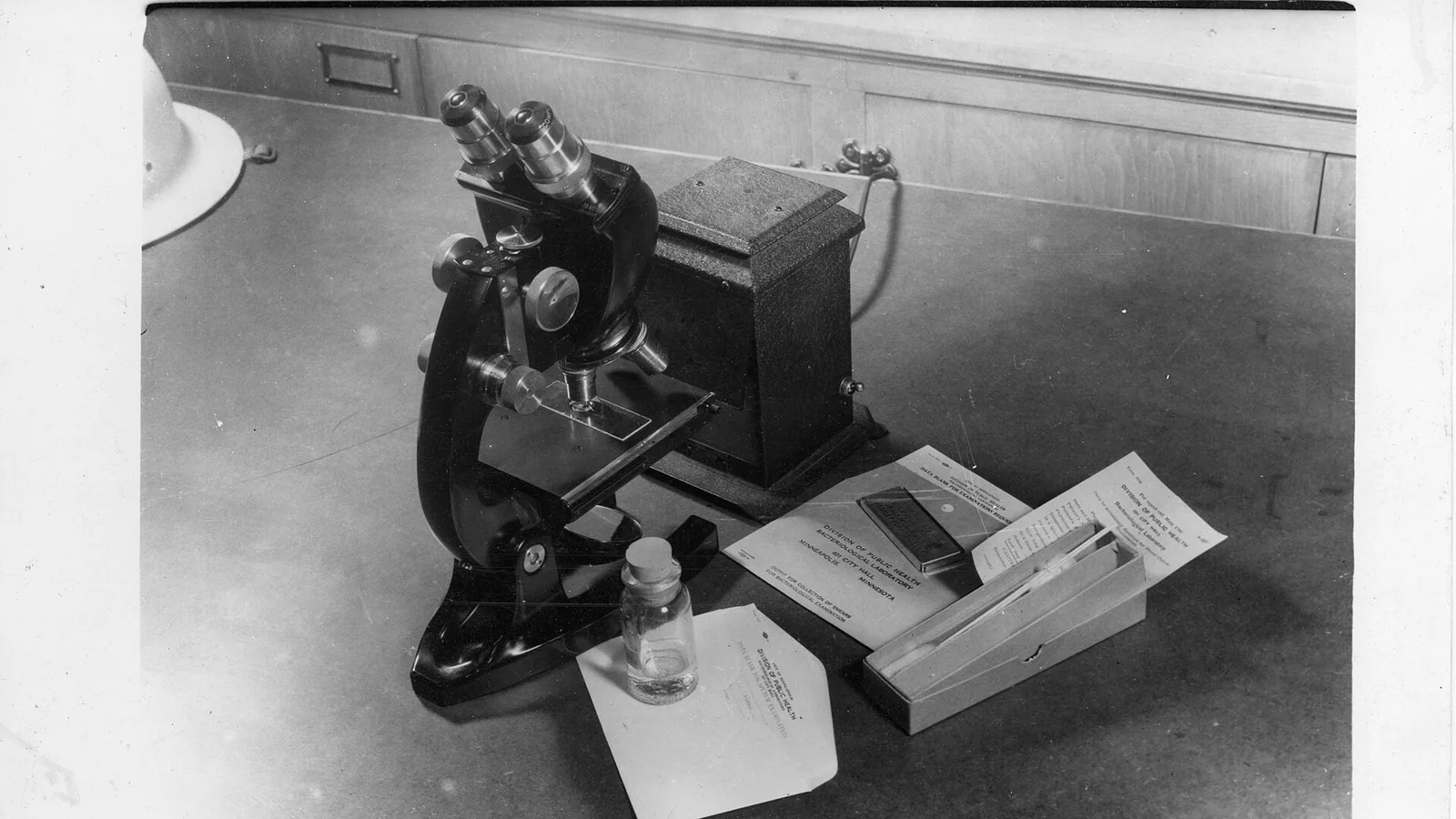
A bacterial spore, also called an endospore, is a dormant, dehydrated, and highly resistant cell form that develops inside a small group of Gram-positive bacteria, usually one spore per parent cell. It is a survival structure, not a reproductive one, and it is built from the inside out so that a core of DNA and ribosomes sits behind four to six protective layers.
That architecture explains almost everything a laboratory does with spores. A spore can survive boiling, drying, ultraviolet light, alcohol, and many detergents that destroy the same bacterium when it is growing. It can wait in dust, soil, a food-processing line, or a hospital room for years, then germinate within minutes once conditions improve. For the diagnostician, the spore is a target that must be stained, quantified, and often broken open before it can be identified or killed. Understanding the structure, the staining chemistry, and the germination triggers is the difference between a valid sterility test and a false pass.
Bacterial Spore vs Endospore: Clearing Up the Terminology
The two words are used interchangeably in most laboratory writing, and for practical purposes in bacteriology they mean the same thing. "Endospore" is the more precise term because it names the location. The structure develops inside the bacterial cell, and the finished spore is released when the mother cell lyses. A few other bacterial structures are labeled spores but are not endospores, and keeping them straight prevents a real diagnostic error.
Vegetative cell. The growing, metabolically active form of the bacterium. It divides by binary fission, takes up nutrients, and is the form that a routine Gram stain or culture plate normally shows. Vegetative cells are comparatively fragile. Spores are what remains when the vegetative cell converts itself into a survival package.
Arthroconidia and conidia of actinomycetes. Streptomyces and related genera produce external, bead-like cells by fragmentation of filaments. These are reproductive conidia, not endospores. They are more heat tolerant than the parent hyphae but far less resistant than a true endospore.
Cysts. Azotobacter and some other Gram-negative genera form cysts that resist drying. Cysts are not endospores and do not show the same core-cortex-coat layering.
Exospores. A small number of budding or filamentous bacteria make spores by budding at the cell surface. In bacteriology the term "exospore" means an externally formed spore, so it is the opposite of an endospore.
Fungal spores. Molds and yeasts produce reproductive spores in enormous numbers. Fungal spores are larger on average, do not stain with malachite green, and are killed by many agents that a bacterial endospore survives. A spore stain that shows green oval bodies inside a red bacterial cell is showing an endospore. Fungal spores are a different problem entirely, usually handled with lactophenol cotton blue or calcofluor white.
| Term | What it is | Typical examples | Key diagnostic point |
|---|---|---|---|
| Endospore | Dormant survival form built inside a bacterial cell | Bacillus, Clostridium, Paenibacillus, Sporosarcina | One per cell, green with malachite green, extremely resistant |
| Vegetative cell | Growing, dividing bacterial form | Any actively multiplying bacterium | Red or pink with the Schaeffer-Fulton counterstain |
| Arthroconidia | External reproductive fragments of filaments | Streptomyces spp. | Reproductive, moderate resistance, not layered like an endospore |
| Cyst | Resting cell of some Gram-negatives | Azotobacter | Different envelope, different resistance profile |
| Fungal spore | Reproductive propagule of molds and yeasts | Aspergillus, Penicillium | Not stained or identified by bacterial spore methods |
The practical rule is simple. If the structure is inside a rod-shaped bacterial cell, is refractile under phase contrast, stains green with Schaeffer-Fulton, and survives a heat shock that kills the surrounding cells, it is an endospore.
Why Bacterial Spores Matter
Spores are the reason several of the most serious bacterial diseases cannot be eliminated by ordinary hygiene. Clostridium difficile spores survive alcohol-based hand rubs and persist on surfaces, and germination in the gut after antibiotic disruption is the first step in C. difficile infection [1]. Bacillus anthracis spores are the agent of inhalational anthrax and the reason environmental screening programs exist, with detection methods designed around the spore biomarker dipicolinic acid [2]. Clostridium perfringens and Bacillus cereus spores pass through cooking, survive cooling, and germinate in food to produce toxin [3], [4].
In food manufacturing, spores set the practical limit of thermal processing. Alicyclobacillus acidoterrestris spores survive pasteurization temperatures in fruit juice and spoil the product without producing gas [5]. Bacillus cereus spores in milk are the target of rapid detection platforms built on dipicolinic acid recognition [4]. Paenibacillus spores survive processing and cause spoilage in dairy and other foods, and their detection is complicated by the fact that viable spores and vegetative cells respond differently to the same assay [6], [7].
In the laboratory, spores define sterilization validation. Biological indicators for steam, dry heat, ethylene oxide, and hydrogen peroxide cycles are prepared spores of defined resistance, and a sterility assurance level is only as trustworthy as the spore preparation used to test it.
Bacterial Spore Structure
The spore is best understood as a set of concentric layers, each with a distinct biochemical job. From the inside out, the layers are the core, the inner membrane, the germ cell wall, the cortex, the outer membrane (present in some species), the coat, and, in some species, the exosporium. Not every species has every layer. Bacillus subtilis lacks a true exosporium. Bacillus anthracis and Bacillus cereus have a prominent one. Turicibacter sanguinis, a strict anaerobe from the mammalian gut, builds a canonical core-cortex-coat arrangement but adds a dual-layered outer envelope and shows two distinct electron-dense coat morphologies, which shows how much structural variation exists outside the classical Bacillus and Clostridium models [8].
The Core
The core is the spore equivalent of cytoplasm. It holds the chromosome, ribosomes, tRNA, and the enzymes needed for the first steps of outgrowth. Two features make it unusual. First, the core is heavily dehydrated relative to a growing cell, which shuts down enzyme activity and protects DNA and proteins from heat denaturation. Second, the core accumulates large amounts of dipicolinic acid (DPA, pyridine-2,6-dicarboxylic acid), mostly as the calcium chelate Ca-DPA. DPA typically makes up a substantial fraction of spore dry weight and is a defining biochemical marker for endospores, which is why detection platforms target it [2], [4].
The core also contains small acid-soluble proteins (SASPs), a family of low-molecular-weight proteins that bind DNA. In Clostridium perfringens, the Ssp4 variants bind cooperatively to DNA and show preferential binding to GC-rich regions, with significantly longer residence time when DPA is present [3]. That binding saturates the chromosome, changes its conformation, and contributes directly to UV and heat resistance.
The Inner Membrane
The inner membrane is the permeability barrier of the spore. In a dormant spore it is unusually compressed and has low lipid mobility, and its biophysical state controls whether germinants can reach their receptors. Cardiolipin content in the inner membrane influences germination efficiency and the size of the superdormant fraction under high hydrostatic pressure, and cortex cross-linking changes how much compressive force the membrane experiences [9]. The inner membrane is also the site where the SpoVA channel releases DPA during germination [8].
The Germ Cell Wall and Cortex
Between the inner membrane and the coat lies the cortex, a thick layer of peptidoglycan that is structurally related to the vegetative cell wall but chemically distinct. Much of its muramic acid is present in the delta-lactam form, and the glycan chains are lightly cross-linked, which makes the cortex flexible and allows it to act as a spring. The cortex keeps the dehydrated core at a lower osmotic potential than the surrounding medium and holds the spore in its dormant state. The enzymes that build it, including the N-acetylmuramic acid deacetylase PdaA and the amidase CwlD, are not cosmetic. Disruption of pdaA in Bacillus cereus ATCC 14579 produces spores with strongly increased UV-C resistance but reduced heat resistance and lower viable spore yield, and subsequent heat stress selects for attenuated CwlD variants that restore viability and heat resistance [10]. Cortex composition is therefore a tunable resistance trait, not a fixed scaffold.
The Coat
The coat is a proteinaceous shell assembled on the outer surface of the developing spore. In Bacillus subtilis it consists of dozens of proteins organized into a lamellar inner coat and a more electron-dense outer coat, and its assembly depends on a defined morphogenetic sequence. SpoIVA, an ATPase, polymerizes into static filaments at the forespore surface and forms the basement layer on which coat proteins assemble, and SpoIVA also contributes to efficient engulfment of the forespore during sporulation [11]. The molecular chaperone DnaK is required for proper inner coat assembly, and its deletion mislocalizes the morphogenetic protein SafA, reduces accumulation of full-length SafA, alters the transcription of spoIIID and sigK, and ultimately impairs coat integrity and function [12].
The coat is the main reason spore staining is difficult. It is a dense, cross-linked, largely impermeable multilayer, and many fluorescent probes simply cannot cross it without harsh permeabilization, which kills the spore [13]. That barrier is also the reason spores resist lysozyme, many proteases, and a wide range of chemical disinfectants.
The Exosporium
The exosporium is a loosely attached outermost layer found in B. anthracis, B. cereus, and some Clostridium species, but absent in B. subtilis. It is rich in glycoproteins and often shows hair-like projections. It contributes to surface adhesion and host interaction and provides a further diffusion barrier. Its presence or absence is a species-level structural marker, not a universal spore feature.
| Layer (inside to outside) | Main composition | Principal function |
|---|---|---|
| Core | DNA, ribosomes, Ca-DPA, SASPs, low water | Stores the genome in a protected, inactive state |
| Inner membrane | Compressed lipid bilayer, cardiolipin | Permeability barrier and germinant receptor environment |
| Germ cell wall | Peptidoglycan | Becomes the wall of the outgrowing cell |
| Cortex | Loosely cross-linked peptidoglycan with muramic lactam | Maintains dehydration and dormancy, buffers pressure |
| Outer membrane | Lipid bilayer, species dependent | Additional permeability barrier |
| Coat | Cross-linked protein layers | Chemical and enzymatic resistance, physical shield |
| Exosporium | Glycoprotein, species dependent | Adhesion and extra diffusion barrier |
Bacterial Spore Formation: Sporulation Step by Step
Sporulation is a programmed developmental pathway triggered by nutrient limitation, most often starvation for carbon or nitrogen. It is not a stress response that any cell can enter at any moment. It requires a committed cell, an intact chromosome, and roughly several hours of coordinated gene expression. The classical model comes from Bacillus subtilis and is divided into stages.
Stage 0, initiation. The cell integrates starvation signals through a phosphorelay that activates the response regulator Spo0A by phosphorylation. Spo0A is the master switch, and its activity level determines whether the cell commits. Turicibacter sanguinis uses a hybrid regulatory architecture that combines Clostridial-type Spo0A initiation with Bacillus-like late-stage sigma factor control, which shows the pathway has been rewired across lineages even though the broad stages are conserved [8].
Stage I, axial filament. The chromosome replicates and condenses into an elongated axial filament running the length of the cell.
Stage II, asymmetric septation. The cell divides asymmetrically near one pole rather than at the midpoint. This creates two compartments with different sizes and different transcriptional programs, the larger mother cell and the smaller forespore. The asymmetry is what makes sporulation possible, since the two cells then signal to each other and divide labor.
Stage III, engulfment. The mother cell membrane migrates around the forespore and pinches it off, so the forespore ends up surrounded by two membranes derived from the mother cell. SpoIVA polymerizes at the forespore surface during this step and serves as the basement layer for coat assembly, and mutants that cannot hydrolyze ATP or form filaments show engulfment defects such as septal membrane bulges [11].
Stage IV, cortex synthesis. Peptidoglycan is deposited between the two membranes around the forespore to form the cortex. The cross-linking pattern and the presence of muramic lactam are set here, and the enzymes that set them determine later resistance properties [10].
Stage V, coat assembly. Coat proteins synthesized in the mother cell are deposited on the forespore surface in a defined order, with DnaK and other quality-control factors supporting correct folding and localization [12].
Stage VI, maturation. The spore acquires its full resistance. DPA is accumulated as Ca-DPA, SASPs are synthesized and saturate the chromosome, the core dehydrates, and the inner membrane compacts.
Stage VII, lysis and release. The mother cell lyses and the mature spore is released into the environment as a free, dormant particle.
The result is a cell that has, in effect, converted itself into a seed. It has no measurable metabolism, no growth, and no susceptibility to antibiotics that act on active pathways, which is why beta-lactams and aminoglycosides do not sterilize a spore population.
Bacterial Spore Germination
Germination is the reverse program, and it is much faster than sporulation. It is triggered when specific small molecules called germinants are recognized by receptors in the inner membrane. Different species respond to different germinants, which is one reason spore behavior is species specific. Bacillus species commonly respond to single amino acids such as L-alanine or to purine nucleosides. Clostridium difficile responds to a combination of taurocholate and glycine, and calcium enhances the response [1]. Alicyclobacillus acidoterrestris responds to L-valine, and 40 mM L-valine was identified as an effective germinant in a juice-spoilage inactivation study [5].
Germination proceeds in stages.
Activation. A dormant spore is often held in a state in which it will not germinate even in the presence of germinant. A sublethal heat shock, aging, or chemical treatment can activate it and make it responsive. This is why heat shock at about 80 degrees Celsius for 10 minutes is both a selection step in culture and a standard measure of spore heat resistance [14].
Stage I, DPA release. Once germinant binds its receptor, the SpoVA channel opens in the inner membrane and Ca-DPA is released from the core into the surrounding medium [8]. This is the earliest measurable event and the most useful for assay design. Terbium chloride is commonly added to germination buffers because Tb3+ complexes with released DPA and the resulting fluorescence can be tracked in real time, although the terbium concentration must be titrated carefully because terbium itself can influence germination in some media [1].
Stage II, cortex hydrolysis and core hydration. Cortex-lytic enzymes, including CwlJ and SleB in Bacillus and CwlJ- and SleM-type hydrolases in other lineages, degrade the cortex [8]. With the cortex removed, water enters the core, the core expands, and the SASPs are degraded by specific proteases. The spore loses its refractility and its resistance at the same time. Morphological work on Bacillus cereus and Bacillus subtilis shows the process in detail. Within 15 to 30 minutes of germination induction, spherical structures containing material with the same elemental ratio as the spore appear on the surface, suggesting leakage of cellular material. By 360 minutes, outgrowing cells are elongated with hemispherical structures at the poles, and discoid structures with a central hole of 20 to 30 nanometers are visible. These structures were shown by confocal microscopy with fluorescently labeled anti-spore antibodies to originate from the spore coat, and they break down after detaching from the cells [15].
Outgrowth. The hydrated cell resumes metabolism, synthesizes new RNA and protein, and emerges from the remains of the coat as a vegetative cell. In a food or clinical sample, outgrowth is what produces visible colonies, toxin, or disease.
Germination Is Heterogeneous
A population of genetically identical spores does not germinate in synchrony. Under 150 MPa of isostatic high pressure at 37 degrees Celsius, Bacillus subtilis spores separated by flow cytometry into four subpopulations: heat-resistant and mostly cultivable superdormant spores that remained dormant, heat-sensitive and cultivable germinated spores, heat-sensitive and partially cultivable germinated spores, and membrane-compromised cells with barely detectable cultivability [14]. Similar heterogeneity appears after very high pressure treatment at 550 MPa and 60 degrees Celsius, where the post-treatment temperature and time strongly determine how many spores progress to full germination and how many become membrane-damaged [16]. Superdormant spores are the practical problem in sterilization, because they can survive a treatment that inactivates the rest of the population [9].
Germination also happens inside hosts. Donor spore populations transplanted into mice germinated in the recipient intestine, and a two-step labeling strategy with fluorescein isothiocyanate on the donor spores and a cyanine-tagged D-amino acid in the recipient allowed germinated donor spores to be identified by their dual fluorescence and then classified by cell sorting, 16S rDNA sequencing, and fluorescence in situ hybridization [17]. The gut is a germination chamber as well as a transmission route.
Bacterial Spore Resistance: Why Spores Survive
Resistance is not one property. It is a set of independent mechanisms that each cover a different threat, and a change that improves one can worsen another.
Dehydration. The low water content of the core stops most heat-induced protein denaturation and Maillard chemistry. A hydrated cell cannot tolerate the same temperature as a dehydrated core.
Ca-DPA. Dipicolinic acid chelates calcium and stabilizes DNA against heat and oxidative damage, and its release is the trigger for the transition back to a hydrated state.
SASPs. Alpha and beta type small acid-soluble proteins bind the chromosome, change its conformation, and physically protect it from UV-induced pyrimidine dimer formation and from oxidative damage. The C. perfringens Ssp4 work shows that DNA binding is cooperative, is preferential for GC-rich regions, and is strengthened when DPA is present, and that a single aspartic acid residue at position 36 stabilizes the protein structure without changing DNA affinity [3].
Cortex and coat. The cortex buffers external pressure and osmotic stress, and the coat blocks chemical and enzymatic attack. The two layers interact. In B. subtilis exposed to high hydrostatic pressure, coat defects appear to alleviate cortex compression on the inner membrane, which reduces germination efficiency at 200 MPa but enhances it at 500 MPa, expanding the superdormant subpopulation and increasing resistance at 200 MPa while diminishing it at 500 MPa. Increased cortex cross-linking combined with core expansion modifies inner membrane properties and strongly promotes germination at both pressures, lowering resistance. Reduced cardiolipin in the inner membrane increases germination efficiency at 500 MPa [9].
DNA repair. During outgrowth, spore repair enzymes fix damage accumulated during dormancy. A spore that survives UV is not necessarily undamaged, it is a spore that can repair the damage once it rehydrates.
The practical consequence is that resistance claims must specify the stress. A B. cereus variant selected for enhanced UV-C resistance through a pdaA phase variation pays for it with reduced heat resistance and lower spore yield, and subsequent heat stress selects for CwlD variants that restore heat resistance while retaining some UV advantage [10]. There is no single "most resistant spore."
Bacterial Spore Examples
Endospore formation is concentrated in the phylum Firmicutes, particularly the orders Bacillales and Clostridiales, but the trait is broader than the classical textbook list.
| Organism | Spore relevance | Notable feature |
|---|---|---|
| Bacillus anthracis | Inhalational and cutaneous anthrax | Exosporium present, environmental persistence, DPA-based detection [2] |
| Bacillus cereus | Foodborne illness, spoilage | Cortex variation alters UV-C and heat resistance [10], DPA detection in milk [4] |
| Bacillus subtilis | Model organism, biological indicators | Best-characterized coat and sporulation genetics [9], [11], [12] |
| Clostridium difficile | Antibiotic-associated colitis | Germinates in response to taurocholate and glycine, calcium enhances [1] |
| Clostridium perfringens | Foodborne gastroenteritis, gas gangrene | Ssp4 SASP variants protect DNA [3] |
| Clostridium botulinum | Botulism | Heat-resistant spores in low-oxygen foods |
| Clostridium tetani | Tetanus | Terminal spores give the characteristic drumstick appearance |
| Alicyclobacillus acidoterrestris | Juice spoilage | L-Valine germinant plus pulsed electric field inactivation [5] |
| Paenibacillus spp. | Dairy and food spoilage | Purification method affects measured viability [7] |
| Sporosarcina spp. | Environmental and food isolates | Spore germination tracked by flow cytometry [6] |
| Turicibacter sanguinis | Gut commensal | Noncanonical coat architecture and hybrid regulatory control [8] |
The list matters for bench work because the species determines the germinant, the antibody, the PCR target, and the disinfectant susceptibility. A method validated on B. subtilis is not automatically valid for C. difficile.
Bacterial Spore Stain: Principles and Bench Methods
A spore stain is a differential stain that uses the impermeability of the spore coat to separate spores from vegetative cells in a single field. The principle is straightforward. The primary dye is forced into the spore with heat or prolonged contact, the smear is then washed so that dye leaves the vegetative cells but stays trapped in the spore, and a contrasting counterstain colors everything else.
The Schaeffer-Fulton Method
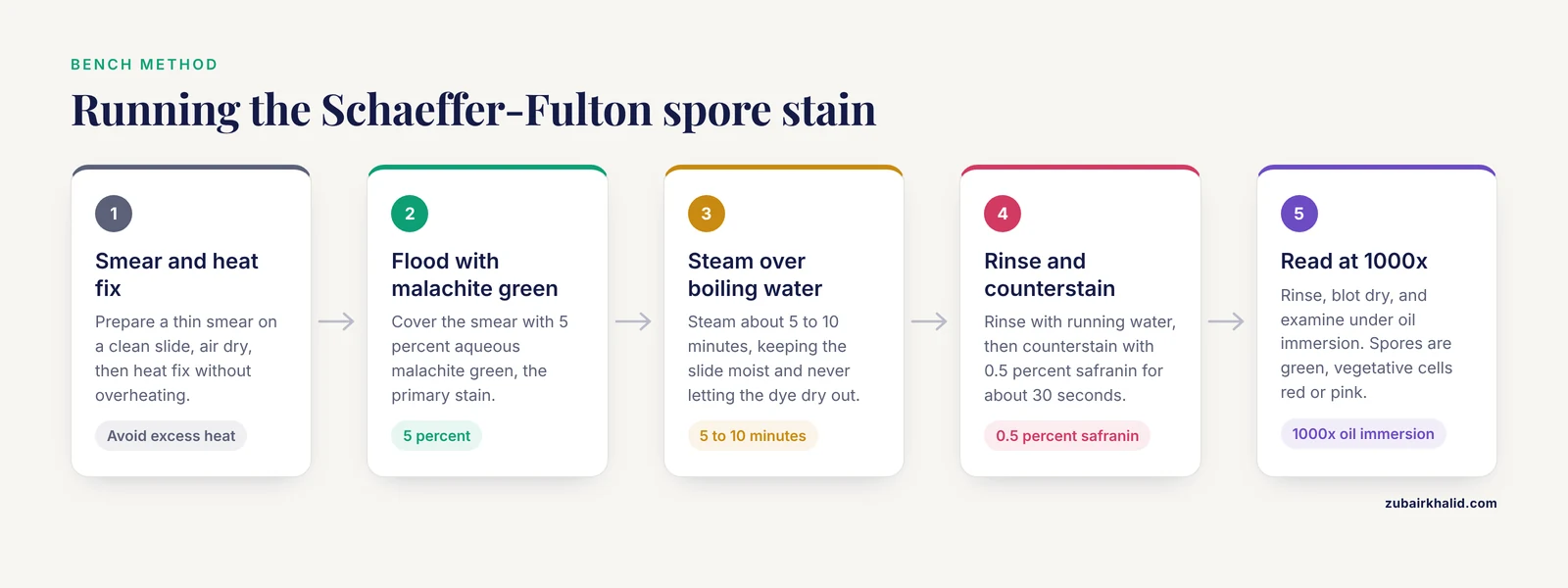
Schaeffer-Fulton is the standard endospore stain in most teaching and clinical laboratories, and it remains the reference method against which newer approaches are compared [18]. The bench sequence is:
- Prepare a thin smear on a clean slide, air dry, and heat fix. Do not overheat, because excessive heat damages spore structure.
- Flood the smear with 5 percent aqueous malachite green, the primary stain.
- Steam the slide over a boiling water bath for about 5 to 10 minutes, keeping the slide moist and never letting the dye dry out. The heat drives malachite green through the coat.
- Rinse thoroughly with running water to remove excess dye.
- Counterstain with 0.5 percent safranin for about 30 seconds.
- Rinse, blot dry, and examine under oil immersion at 1000x.
The result is unambiguous. Spores appear green, and vegetative cells, including the mother cell around a developing spore, appear red or pink. Free spores and spores still inside a sporangium both stain green.
The limitation is not the chemistry but the handling. Schaeffer-Fulton is sample-destructive and demands significant operator involvement, which rules out live imaging of the same spore over time. Newer lanthanide-beta-diketonate complexes stain instantaneously with minimal processing and leave the core hollow and nonfluorescent in dormant spores, then enter the core and become brightly fluorescent on germination, allowing live imaging of single spores by low-cost microscopy including in milk [18]. The trade-off is that the classical stain gives a permanent, inexpensive, unambiguous morphological answer while the fluorescent method gives dynamic information.
Why Ordinary Stains Fail
A Gram stain of a sporulating culture is misleading. Spores do not take up crystal violet reliably, so they appear as unstained refractile ovals, and the culture may read as Gram variable or Gram negative. A culture of Bacillus that looks Gram negative is usually a culture that has largely converted to spores. The spore stain exists because the Gram stain cannot do this job.
Alternative Staining and Imaging Approaches
Dipicolinic acid probes. Because DPA is unique to spores, DPA-reactive chemistries act as indirect spore stains. A polychromatic fluorescent nanoprobe made of aminoclay-coated silicon nanoparticles functionalized with europium ions responds selectively to DPA and detects as few as 0.3 x 10^5 CFU per milliliter of Bacillus subtilis spores within 10 seconds, and it also supports visual readout for screening B. anthracis simulants [2]. Molecularly imprinted polymers targeting DPA, read by a thermal method, achieved an imprinting factor of 2.62 at 100 micromolar DPA and limits of detection of 0.20 micromolar in aqueous solution and 0.060 micromolar in spiked ultrahigh-temperature milk [4].
Zwitterionic fluorophores. Diaminodicyanoquinodimethane dyes penetrate the endospore system because of their dipolar structure and fluoresce strongly in their rigid or aggregated state. Nanomolar amounts of the BT2 and BHADQ derivatives give effective endospore staining and allow selective imaging of spores in the presence of vegetative bacteria without forcing germination [13].
Metabolic dyes. 5-Cyano-2,3-ditolyl-2H-tetrazolium chloride (CTC) is reduced to a fluorescent formazan by metabolically active cells, so it stains germinating spores but not dormant ones. Flow cytometry with CTC distinguished vegetative cells from spores and quantified germination in Paenibacillus, Sporosarcina, and Clostridium in food samples, and CTC staining also verified the absence of residual vegetative cells in purified Paenibacillus spore preparations [6], [7].
Raman and isotope labeling. Spores grown in 30 percent deuterium oxide broth reach steady-state deuterium labeling within about 48 hours and give Raman peaks at 2200 and 2300 per centimeter in the cell silent region. Those peaks decrease rapidly on germination in nondeuterated medium, and inactivated spores do not lose them, so the signal can indicate viability [19].
Viability dyes with flow cytometry. SYTO16 reports germination and propidium iodide reports membrane damage, and the combination resolves multiple physiological states in a single run [16], [14].
Antibody-based imaging. Fluorescence-labeled anti-spore antibodies have been used to show that coat-derived structures persist during outgrowth [15].
Detecting and Testing Spores in Practice
Heat shock selection. A sample is held at about 80 degrees Celsius for 10 minutes and then cultured. Vegetative cells die and spores survive, which both selects for spore formers and provides a rough heat resistance measure [14].
Spore purification. Crude sporulating cultures contain vegetative cells, cell debris, and germinating spores. Two common purification routes are lysozyme treatment and density gradient centrifugation with HistDenz. In four Paenibacillus strains, lysozyme treatment reduced spore viability in some strains, while HistDenz purification more reliably preserved germination potential, and spores produced on solid medium outperformed those produced in liquid medium for some strains [7]. Purification quality directly changes the answer a viability assay gives.
Phase contrast microscopy. Dormant spores are phase bright and refractile. Germinating spores lose refractility as the core hydrates, so a simple wet mount tracks germination in real time.
Cultural enumeration. Plate counts after heat shock give a spore count, but they only count spores that can germinate and grow under the chosen conditions, which undercounts superdormant and injured spores.
Flow cytometry. Complete absence of metabolically active vegetative cells can be verified by CTC staining combined with flow cytometry, and this is faster than waiting for colonies [6], [7]. Flow cytometry also resolves germinated, superdormant, and membrane-compromised subpopulations after pressure treatment [16], [14].
Chemical and biomarker assays. DPA measurement by terbium fluorescence, nanoprobe fluorescence, or imprinted polymer thermal sensing provides a rapid indirect spore signal and works in complex matrices such as milk [1], [2], [4].
Structural imaging. Scanning and transmission electron microscopy show layer architecture, coat detachment, and outgrowth morphology that no stain can show at the same resolution [15].
Molecular identification. Nucleic acid methods identify the species but do not by themselves prove that the detected DNA came from a viable spore, so molecular results are usually paired with a viability or germination assay.
Disinfection and inactivation testing. Sporicidal testing is done on purified, characterized spore suspensions because resistance depends on the purification and sporulation conditions used to make them [7]. Peracetic acid at 0.2 percent inhibited spore germination in a flow cytometry assay, shown by a decreased count of metabolically active spores [6]. Germinant pretreatment lowers resistance. A. acidoterrestris spores incubated for 4 hours with 40 mM L-valine and then treated with 210 seconds of 0.18 kV per centimeter pulsed electric field combined with ohmic heating dropped by 1.73 log units, compared with 0.54 log units in the nongerminant control, and the treated spores showed structural disruption with leakage of nucleic acids and proteins by scanning electron and confocal microscopy [5].
Clinical and Food Safety Relevance
In clinical microbiology, spores explain persistence and relapse. C. difficile spores survive on surfaces and on hands, resist alcohol, and are killed reliably only by sporicidal agents such as chlorine-based disinfectants and peracetic acid. B. anthracis requires specific environmental sampling and confirmatory identification. C. perfringens and B. cereus cause foodborne illness when spores survive cooking and germinate during slow cooling.
In food microbiology, spores set process targets. A thermal process validated against vegetative cells fails if spores are present. B. cereus spores in milk and dairy products survive ultrahigh-temperature treatment in some cases and are detected through DPA chemistries [4]. Juice spoilage by A. acidoterrestris is a pasteurization-survivor problem that requires either higher heat or a germination-then-inactivation strategy [5]. Paenibacillus contamination in dairy powders is a recurring spoilage issue, and reliable detection depends on properly purified and characterized spore preparations [6], [7].
In biosecurity and environmental monitoring, spore detection is a first-line capability, and rapid DPA-based screening supports presumptive identification before culture confirmation [2]. In space exploration, distinguishing dormant from germinated spores is a planetary-protection requirement, which is one reason live-imaging methods have been developed [18].
Quick Review
- A bacterial spore (endospore) is a dormant, dehydrated survival form built inside one bacterial cell, not a reproductive spore.
- Structure runs from core, inner membrane, germ cell wall, cortex, outer membrane, coat, and, in some species, exosporium.
- The core carries DNA, ribosomes, Ca-DPA, and SASPs, and its low water content is the basis of heat resistance.
- Sporulation is a staged developmental program triggered by nutrient starvation and controlled by Spo0A and compartment-specific sigma factors.
- Germination starts with germinant recognition and DPA release, continues with cortex hydrolysis and core hydration, and ends with outgrowth.
- Schaeffer-Fulton staining uses malachite green with steam and safranin counterstain, giving green spores and red vegetative cells.
- Resistance is stress-specific. Coat, cortex, membrane, and SASP changes can trade heat resistance against UV or pressure resistance.
Common Mistakes and Limitations
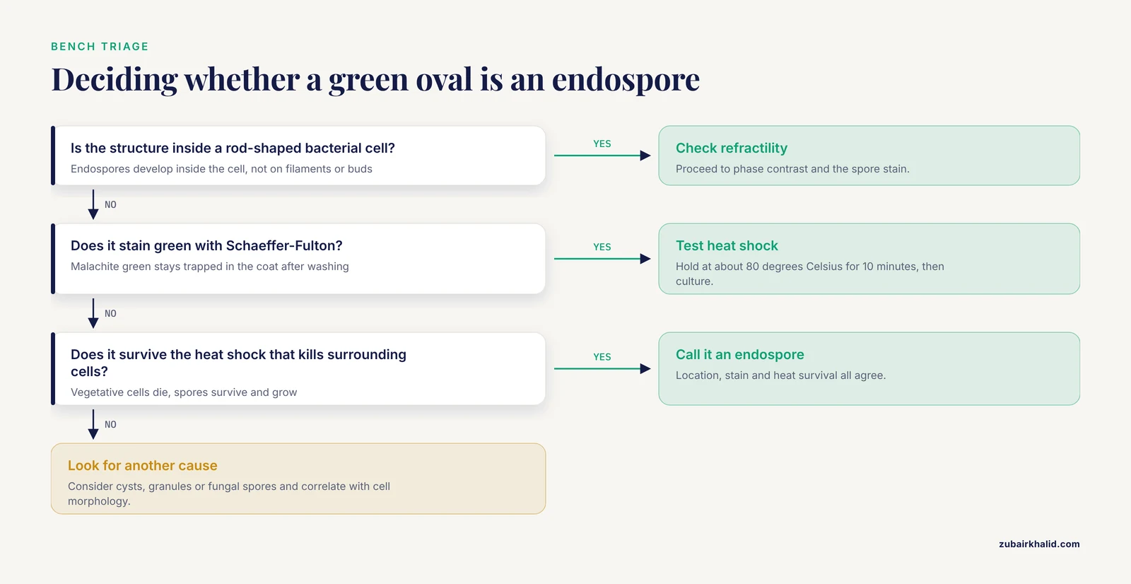
Reading a Gram stain as a spore stain. Spores appear unstained and refractile on Gram stain, and a heavily sporulated culture can read as Gram negative. Confirm with Schaeffer-Fulton or phase contrast before calling a culture contaminated or misidentified.
Assuming a green oval is always an endospore. Calcium and polyphosphate granules can take up dye, and fungal spores in a mixed specimen do not stain the way bacterial endospores do. Correlate with the cell morphology and the culture.
Overheating during Schaeffer-Fulton. Excess heat or a slide allowed to dry during steaming distorts spore morphology and can rupture the coat, producing false negatives or atypical shapes.
Confusing dormancy with death. A dormant spore is viable. A negative culture from a spore-containing sample may mean the germination conditions were wrong, not that the spores were absent or dead.
Ignoring superdormant subpopulations. A single germination condition does not germinate every spore in a population. Pressure-treated B. subtilis populations contain superdormant spores that remain heat resistant and cultivable after the rest of the population germinates [14], and superdormant fractions change with coat, cortex, and membrane alterations [9]. Inactivation studies that report an average log reduction can hide a resistant tail.
Extrapolating resistance across stresses. Enhanced UV-C resistance in a B. cereus pdaA variant came with reduced heat resistance and lower spore yield [10]. Quoting a single resistance figure without naming the stress and the strain overstates what is known.
Treating a purified spore preparation as uniform. Purification by lysozyme treatment reduced viability in some Paenibacillus strains, whereas HistDenz gradient purification better preserved germination potential, and the sporulation medium changed growth performance [7]. A viability comparison between two spore preparations may be measuring preparation quality rather than biology.
Ignoring matrix effects in detection. DPA-based sensing behaves differently in buffer, in milk, and in juice [4], [2]. A limit of detection measured in water is not a limit of detection in food.
Misreading a single positive assay as proof of viability. DPA release, CTC reduction, and SYTO16 uptake measure different things. Pair a viability or germination marker with a structural or identity marker before drawing a conclusion.
Individual clinical or food-safety decisions need a qualified microbiologist or veterinarian who can interpret results against the sample, the method, and the organism.
Related Guides
- Negative Controls in Bacterial Staining: Why They Matter and How to Use Them: Negative controls help validate spore stain interpretation
- Alternative Counterstains for Gram Staining: Alternative counterstains may improve spore stain contrast
- Gram Stain Reagent Contamination vs. True Bacterial Morphology: Explains reagent contamination versus real bacterial morphology in stains
- The Impact of Smear Thickness on Artifact Formation: Smear thickness affects artifact formation during differential staining
- Gram Staining Anaerobic Broth Cultures: Special Considerations: Anaerobic culture staining is relevant for Clostridium spore-formers
- Gram Staining Colonies from Agar Plates: Colony staining guidance supports bench work with Bacillus and Clostridium isolates
Frequently Asked Questions
What is the difference between a bacterial spore and an endospore?
They are the same structure. "Endospore" is the more precise term because the spore develops inside the bacterial cell, while "spore" is the general word used in most laboratory practice.
Why do spores stain green with the Schaeffer-Fulton method?
Malachite green is driven through the impermeable spore coat by steam and remains trapped inside after washing. The safranin counterstain cannot displace it, so spores stay green while vegetative cells take up the red counterstain.
Are bacterial spores alive?
A dormant spore is viable but metabolically inactive. It has no measurable growth or respiration until germination begins, and it can be cultured after germination under suitable conditions.
How long can bacterial spores survive?
Survival depends on the species, the stress, and the environment. Spores are built for long-term persistence in soil, dust, and dried food residues, and some species remain viable after years of storage.
Do all bacteria produce spores?
No. Endospore formation is limited to a subset of Gram-positive bacteria, mainly within the Firmicutes, including Bacillus, Clostridium, Paenibacillus, Sporosarcina, and related genera. Most clinically important Gram-negative bacteria do not form endospores.
What kills bacterial spores?
Sporicidal conditions include moist heat under pressure (autoclaving), dry heat, hydrogen peroxide vapor, peracetic acid, glutaraldehyde, and high concentrations of chlorine. Alcohols and many quaternary ammonium compounds do not reliably kill spores. Germinant pretreatment followed by a physical treatment can also reduce resistance by forcing spores to germinate before inactivation [5].
Sources
- Terbium chloride influences Clostridium difficile spore germination.
- A Dual-Mode Fluorescent Nanoprobe for the Detection and Visual Screening of Pathogenic Bacterial Spores.
- Structural and dynamic basis of Ssp4-mediated DNA protection in foodborne bacterial spores.
- Molecularly Imprinted Polymers as an Indirect Sensing Approach for Bacillus cereus Detection.
- Enhancing Spore Inactivation: Low-Intensity Pulsed Electric Field Combined with Ohmic Heating and Germinant Pretreatment.
- Analysis of vegetative cells and spore germination in food using flow cytometry.
- Optimization of Spore Purification and Quality Assessment in Paenibacillus Species.
- Distinctive spore architecture and developmental biology of Turicibacter sanguinis reveal unexpected diversity among gut spore formers.
- New insights into the structure-based mechanism of Bacillus subtilis spore resistance to high hydrostatic pressure.
- Cortex modulation shapes UV-C and heat resistance properties of Bacillus cereus endospores.
- SpoIVA contributes to efficient engulfment through a cytoskeletal-like mechanism during Bacillus subtilis sporulation.
- Chaperone protein DnaK-mediated regulation of spore coat assembly in Bacillus subtilis.
- Zwitterionic small molecule based fluorophores for efficient and selective imaging of bacterial endospores.
- Flow Cytometry Combined With Single Cell Sorting to Study Heterogeneous Germination of Bacillus Spores Under High Pressure.
- Morphological Changes in Spores during Germination in Bacillus cereus and Bacillus subtilis.
- Post-high-pressure temperature and time - Overlooked parameters in high pressure treatment of bacterial spores.
- Visualizing germination of microbiota endospores in the mammalian gut.
- Elucidating Bacterial Spore Dynamics through Lanthanide-Enhanced Live Imaging.
- Assessing CaDPA levels, metabolic activity, and spore detection through deuterium labeling.