AlphaFold Sequence Length Limits: How to Prepare Long Proteins
By Dr. Zubair Khalid, DVM, MS, PhD ·
The most common cause of an AlphaFold sequence length limit failure is not a hard cutoff in the software. It is memory. AlphaFold's internal pair representation grows with the square of the residue count, so a protein twice as long needs roughly four times the memory, and a protein three times as long needs roughly nine times as much. On a consumer GPU this means a 700 residue protein predicts in minutes while a 2,500 residue protein crashes with an out of memory error before the first recycle finishes.
The first thing to check is the exact residue count of your sequence, followed by a search of the AlphaFold Database. Many long proteins already have a full-length predicted structure waiting for you, and downloading an existing model takes seconds where a new prediction takes hours. This article explains why the different length ceilings exist, gives you a short diagnostic sequence to identify which ceiling you are hitting, and then walks through practical fixes from the easiest to the most involved, including domain trimming, chain splitting, MSA subsampling, and alternative predictors.
What the AlphaFold Sequence Length Limit Actually Is
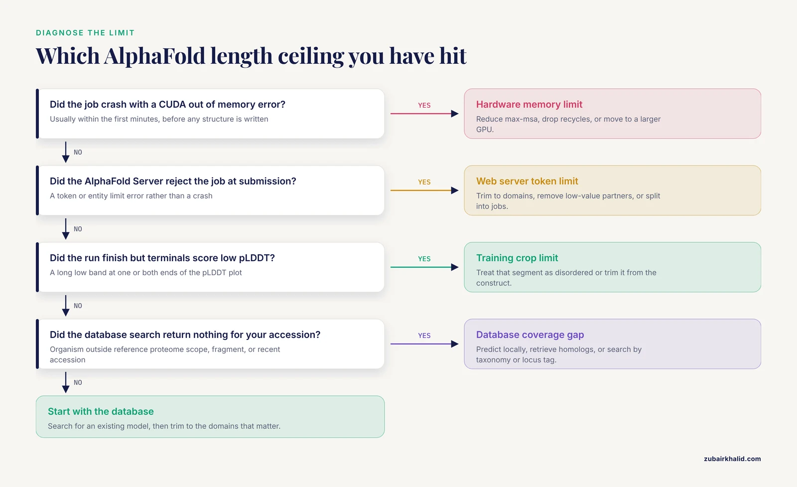
People mean at least four different things when they talk about AlphaFold sequence length limits, and the fix depends entirely on which one you have hit.
Hardware memory limits. AlphaFold 2 and its reimplementations such as ColabFold hold a pair representation with one entry per residue pair, plus a single-sequence representation, on the accelerator. The pair representation and the triangle updates that operate on it dominate memory use. Practical ceilings reported by the community sit around 1,000 to 1,400 residues on a free 16 GB T4 GPU in Google Colab, with higher ceilings on A100 cards with 40 GB or 80 GB of memory. These are working limits, not documented guarantees, and they shift with recycle count, model type, and MSA depth.
Web server token limits. The AlphaFold Server operated by Google DeepMind and Isomorphic Labs does not limit you by residues alone. It converts every entity in your job, including each chain, ligand, ion, modification, and copy, into tokens, and enforces a total token budget per job along with caps on the number of distinct entities. A long protein chain consumes a large share of that budget by itself, so adding ligands or interaction partners can push a sequence that fit alone over the limit. The exact caps are published in the AlphaFold Server FAQ and have been raised over the life of the service, so check the current values before planning a submission.
Training crop limits. AlphaFold 2 was trained on randomly cropped windows of roughly 384 residues for single chains, and similar crops apply to complex models. Inference can run on much longer sequences, but accuracy on regions far outside the training crop size depends heavily on the quality of the multiple sequence alignment and the presence of templates. In practice this shows up as degraded accuracy in terminal segments rather than an outright crash. Benchmarking of AlphaFold 3 found that it captured overall backbone features well for most proteins tested, but small misfolding of N- and C-terminal segments was common and often did not affect the biological interpretation [1].
Database coverage limits. The AlphaFold Database contains over 200 million predicted structures spanning UniProt reference proteomes. Coverage is broad, but if your protein is from an unindexed organism, is a short fragment, or falls outside the database's organism and accession scope, no precomputed model exists and you must predict it yourself. Tools built to retrieve models by accession handle the common cases. The AlphaFoldDB Structure Extractor, for example, accepts NCBI Taxonomy IDs, RefSeq accessions, locus tags, and UniProt or AlphaFold accessions, and returns structures in PDB, mmCIF, bCIF, or PAE JSON formats with an ID mapping file, which removes most friction from bulk retrieval [2].
What Causes the AlphaFold Sequence Length Limit
Understanding the mechanism behind each ceiling tells you which fixes can work and which cannot.
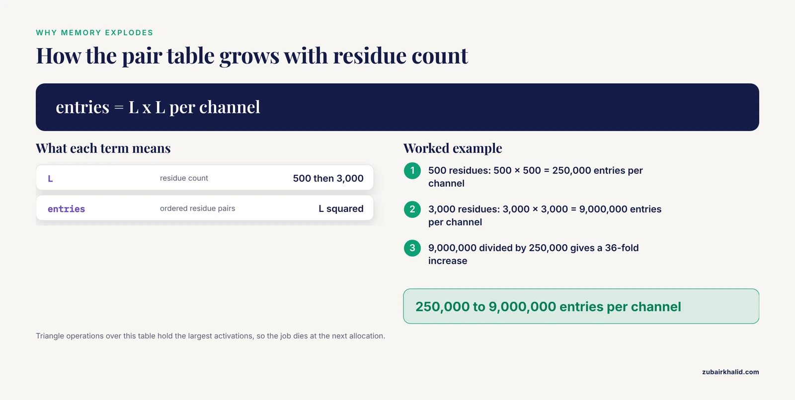
Quadratic and super-quadratic memory growth. The core of AlphaFold is the pair representation, a tensor with one vector per ordered pair of residues. A 500 residue protein needs a pair table with 250,000 entries per channel. A 3,000 residue protein needs 9,000,000 entries per channel, a 36-fold increase. On top of that, the triangle multiplication and triangle update operations iterate over rows and columns of this table, and their intermediate activations are the largest consumers of memory. Backpropagation must retain these activations, so memory during training-style inference exceeds the memory of a forward pass alone. This is why the failure is usually abrupt. A sequence that runs at 90 percent of available memory does not run 10 percent slower, it dies at the next allocation.
MSA depth interacts with length. The multiple sequence alignment (MSA) is the set of evolutionarily related sequences that AlphaFold aligns against your query. Deeper MSAs sharpen inference but consume memory in proportion to the number of aligned sequences times the query length. A long query with a 50,000 sequence MSA uses far more memory than the same query with a 5,000 sequence MSA. Reducing MSA depth is therefore one of the levers you can pull when a long protein barely exceeds your memory.
Recycling multiplies the cost. AlphaFold refines its prediction by feeding the output structure back through the network, a process called recycling. Each recycle is a full pass through the network. More recycles usually improve accuracy on structured regions, but they increase runtime roughly linearly and can be the difference between fitting and not fitting when memory is tight, because intermediate states are held between passes depending on the implementation.
Disordered regions waste budget. Long proteins frequently contain intrinsically disordered regions (IDRs), segments that never adopt a single stable structure. AlphaFold will happily spend memory and tokens modeling these regions, and the per-residue confidence score, pLDDT, will report low values there. Work on alpha-synuclein, an intrinsically disordered protein central to Parkinson's disease research, shows why this matters. That study combined sequence-level embeddings with AlphaFold coordinates filtered by pLDTT, down-weighting unreliable structural regions, precisely because structure-only representations perform poorly in disordered segments [3]. Every residue you spend on a region that will be flagged low confidence anyway is a residue you could have allocated to a domain that matters.
Score distortion from long, loosely packed sequences. Length does not only cause crashes. The inter-chain predicted TM score, ipTM, aggregates alignment quality over entire chains, and its mathematical formulation means that adding disordered or irrelevant residues to a chain changes the score even when the modeled interaction itself is unchanged. Dunbrack analyzed this problem and showed that trimming full-length constructs to the interacting domains changes ipTM substantially, and proposed corrections that restrict the score to residue pairs with good predicted aligned error [4]. If you model long constructs and interpret ipTM, you need to know that the score is length sensitive.
First Checks Before You Change Anything
Work through this sequence in order. Most readers stop at step 2 or 3.
- Count the residues exactly. Open the FASTA file and count. A quick terminal check for a single-sequence file is
tail -n +2 query.fasta | tr -d '\n' | wc -c. Everything downstream depends on whether you are facing 900 residues or 3,900. - Search the AlphaFold Database. Paste the UniProt accession at alphafold.ebi.ac.uk or query by sequence. The database covers UniProt reference proteomes and includes many full-length models longer than local GPUs can handle. If a model exists, download it along with the PAE file and inspect pLDDT before deciding you need a custom prediction. For batches, the Structure Extractor web server and API accept RefSeq, UniProt, Ensembl-style locus tags, and taxonomy IDs and return coordinate files plus the per-residue confidence data [2].
- Annotate domains and disorder. Run the sequence through InterPro or Pfam to find domain boundaries, and through a disorder predictor such as IUPred or the disorder annotations in UniProt. Mark linker regions. This map is the input for every fix described below.
- Check your MSA. If you already ran a job, look at the MSA depth in the output. If you have not run one yet, a shallow MSA on a long protein predicts both a crash and poor accuracy, because accuracy beyond the training crop size depends on deep, clean alignments.
- Reproduce the failure cheaply. Run the shortest construct that interests you, such as one domain, before committing hours to a full-length job. A domain-only run that produces high pLDDT tells you the pipeline works and gives you a baseline for judging the full-length result.
Troubleshooting Table
| Symptom | Likely cause | How to confirm | Fix |
|---|---|---|---|
| CUDA out of memory error during feature extraction or the first forward pass | Pair representation plus deep MSA exceed GPU memory | Error appears within the first minutes, before any structure is written | Reduce -max-msa in ColabFold, drop recycles, or move to a larger GPU or local install |
| Run dies partway through recycles on a job that started fine | Activations from repeated passes accumulate or a later, larger allocation fails | Log shows progress through early recycles then OOM | Reduce recycle count, reduce MSA depth, or split the sequence |
| AlphaFold Server rejects the job outright | Total token budget exceeded by the chain plus partners, ligands, or copies | Server reports a token or entity limit error at submission | Trim to domains, remove low-value partners, or split into separate jobs |
| Prediction completes but a 300 residue terminal segment has pLDDT below 50 | Intrinsically disordered region or an MSA too shallow to support distant residues | pLDDT plot shows a long low band at one or both ends | Treat that segment as disordered, model it separately if needed, or trim it from the construct |
| ipTM or interface score changes when you shorten a construct | Score formulation aggregates over the whole chain including disordered residues | Recompute the score on trimmed constructs, compare with PAE at the interface | Judge the interface by PAE and local pLDDT, not by the full-chain score alone [4] |
| No model exists in the AlphaFold Database for your protein | Organism outside reference proteome scope, fragment, or recent accession | Database search returns nothing for the accession | Predict locally or through a server, retrieve homologs' models instead, or use a retrieval tool by taxonomy or locus tag [2] |
| Model of a split two-domain construct shows no clear interface | Domains were split at a wrong boundary or genuinely do not form a stable interface | PAE shows high error between the two chains | Re-examine domain boundaries against Pfam and structures of homologs, consider modeling the linker |
| Full-length model looks inflated or stretched compared with expected compactness | Disordered regions modeled as extended chains fill space | Superimpose on domain-only models or known structures | Use the domain models for biology, treat the full-length model as one conformational hypothesis |
How to Fix the AlphaFold Sequence Length Limit
These solutions run from the easiest and most common to the most involved. Apply the first one that resolves your case.
Check the AlphaFold Database before predicting anything
The cheapest fix is to not run a prediction at all. The AlphaFold Database holds predicted structures for UniProt reference proteomes, and for many long multidomain proteins the full-length model already exists. Download the mmCIF or PDB file plus the predicted aligned error (PAE) file, which encodes the expected positional error between every pair of residues. For programmatic or bulk work, the Structure Extractor handles batches of up to thousands of accessions and reports unmapped identifiers separately, so you can trace exactly which inputs produced structures [2]. A precomputed model also tells you something no fresh run can: whether a different MSA and a different random seed produced a stable, well-ordered fold.
Trim to domains and model fragments
The highest-yield fix for a long protein is to stop modeling the whole thing. Split the sequence into folded domains using Pfam or InterPro boundaries, and predict each domain separately. Predictions on single domains of 100 to 400 residues run fast, fit on modest hardware, and usually return high pLDDT across the fold.
The gains here are documented. A study of domain-domain interface types curated from the 3did resource generated a reference set of verified domain-domain interfaces, identified a high-confidence subset by logistic regression, and then applied fragment-guided AlphaFold modeling to thousands of predicted human domain-domain interactions. Guided fragment modeling produced confident models for a large share of predicted interfaces that confident full-length models had missed [5]. The lesson generalizes: restricting the model to the residues that matter improves both the odds of success and the interpretation of the scores.
Boundary choice matters more than most people expect. In a study of acyl carrier protein (ACP) domains from modular polyketide synthases, two segments prepared with existing domain boundaries behaved completely differently. One adopted the expected fold, while the other, despite 49 percent sequence identity to the first, failed to fold. Secondary structure prediction revealed an overlooked N-terminal helix, H0, that was a rigid structural component of the domain and a thermodynamic stabilizer. Constructs lacking H0 were substantially less stable [6]. If your split cuts through a structural element that happens to belong to the neighboring domain, your fragment prediction will fail and the failure will look mysterious. Before splitting, check secondary structure predictions and structural homologs, not just Pfam span coordinates.
Trimming also changes your scores, and you should plan for that. Because ipTM aggregates over entire chains, trimming disordered or accessory residues from a construct can raise or lower the reported score without changing the modeled interface geometry [4]. Set your expectations using the interface PAE block for the residues you care about.
Split a long protein into pieces and model the assembly
When the biology requires the full-length protein, model it in pieces and assemble the pieces. Two practical variants exist.
Consecutive fragments with overlap. Split the chain into overlapping windows of, say, 400 residues with 40 residue overlaps. Model each window, then superimpose the overlapping segments to build a composite structure. This works best for proteins with repeating or modular architecture. Judge each overlap by the local pLDDT and the PAE between the shared residues.
Domain assembly from separate predictions. Model each domain separately, then model domain pairs if the biology suggests they contact each other. AlphaFold is far more likely to produce a confident domain-domain model when the construct contains just the two interacting fragments than when it contains two full-length proteins with long disordered tails. The interface-type reference work mentioned above formalized exactly this strategy and demonstrated its success rate on human interaction data [5].
Multidomain bacterial proteins illustrate both the power and the subtlety of this approach. Histidine kinases are multidomain two-component signaling proteins with sensor, dimerization, and catalytic domains, and AlphaFold models captured whether the kinase dimerizes in a cis or trans phosphorylation mechanism. Notably, the mechanism traced to a difference in the lengths of the helices surrounding the DHp loop, and very small changes in those helices could switch the mechanism [7]. Two implications follow. First, domain-resolved modeling can capture functionally decisive geometry. Second, results can be sensitive to construct boundaries at the residue level, so test at least two boundary choices when the biology depends on the answer.
Reduce memory demand on the job you already have
If your sequence is only modestly over your hardware limit, three settings can pull it back under.
Subsample the MSA. In ColabFold, the -max-msa flag controls how many sequences and extra sequences enter the model, with default values on the order of tens of thousands for the deepest settings. Dropping to a shallow setting such as -max-msa 16:32 cuts MSA memory sharply. The tradeoff is accuracy, particularly for proteins with few homologs, so compare the pLDDT profile against a deeper run on a shorter construct before trusting the shallow result.
Reduce recycles. The default three recycles in ColabFold can be reduced to one or zero for a memory-constrained first pass. Accuracy on structured domains usually survives, interface quality suffers more. Use reduced recycles to establish that a construct fits, then reserve recycles for the final runs.
Disable or slim optional outputs. Turning off Amber relaxation and saving only the top-ranked model reduces disk and some processing overhead, though the dominant memory cost is in the network itself rather than post-processing.
Move to bigger hardware or a local install
When tuning fails, change the hardware rather than the science. Options in rough order of cost and effort.
- Paid cloud GPU sessions. A 40 GB A100 handles sequences roughly two to three times longer than a 16 GB T4 in the same pipeline. Colab and equivalent cloud providers offer these as paid sessions. This is often the fastest path for a one-off 2,500 residue protein.
- Local AlphaFold installation. Running the open-source AlphaFold or a reimplementation such as OpenFold on an institutional multi-GPU machine removes session time limits and, importantly, keeps your sequence data on your own infrastructure. Installation is nontrivial, requiring a database of reference genomes and MSAs, so budget a day for setup the first time.
- AlphaFold Server for complex jobs. The AlphaFold Server handles protein-protein, protein-nucleic acid, and protein-ligand modeling through a browser, with the token budget described earlier. It is the right tool when your question involves partners and ligands rather than a bare monomer, and when your sequence fits the published caps. For DNA-bound complexes, a systematic comparison of AlphaFold 3, RoseTTAFold2NA, and Chai-1 on the competence protein ComP found that several distinct protein-DNA binding modes were robustly modeled across platforms, which suggests that running a second tool is a useful cross-check for server-scale jobs [8].
A data safety note applies here. Public web servers, including AlphaFold Server and free notebook services, receive your sequence in plaintext over the network and store it under the provider's policies. If your target sequence is unpublished, proprietary, or part of a restricted project, run predictions on local hardware instead, or model a published homolog to protect the confidential sequence.
Use alternative predictors and modeling strategies
AlphaFold is not the only option for a long protein, and sometimes it is not even the best one.
Template-based and comparative modeling. When close experimental structures of homologous domains exist, classical homology modeling can assemble a full-length model that respects known disordered regions. A full-length model of coagulation factor XII built from four structural templates covering more than 70 percent of the sequence, with loop modeling for the connectors, produced a more compact and realistic disordered-region geometry than the corresponding AlphaFold prediction, while the ordered regions agreed closely with the AF model [9]. If your long protein contains well-characterized domains, this route deserves consideration.
Other deep learning predictors. Chai-1, RoseTTAFold2, and related systems have different memory profiles and different strengths. Running a borderline-length protein through a second predictor is standard practice, and disagreement between predictors is itself diagnostic information. The ComP study cited above used three platforms in parallel and treated agreement across platforms as the criterion for a robust model [8].
Structure-guided assembly from AlphaFold pairs. For protein complexes involving a long chain, curating likely domain-domain interfaces before prediction raises success rates. The interface-type reference set built from 3did, with 2,411 of 5,724 candidate interface types rated high confidence, was applied to tens of thousands of human protein interactions to prioritize AlphaFold modeling, and the guided models succeeded where unguided full-length models often did not [5].
Handle disorder and post-translational context deliberately
Long proteins are rich in disorder and in post-translational modification sites, and both interact with length limits.
For disorder, extract the pLDDT track from any AlphaFold model or prediction and treat the standard bands as guidance. Residues above 90 are very confidently placed, 70 to 90 are confident, 50 to 70 are low confidence, and below 50 the model is effectively stating that no single structure exists, which is how AlphaFold flags disorder. pLDDT is an imperfect error predictor. A homology-controlled benchmark of AlphaFold Database models against experimental structures across 231 chains found that low pLDDT regions carried a contact error rate of 0.0089 versus 0.0024 above the pLDDT 70 threshold, a 3.70-fold enrichment, but the discrimination overall was modest with an AUC of 0.672, and pLDDT below 70 captured only 46 percent of all contact errors [10]. In other words, low pLDDT marks risk, not proven error, and high pLDDT does not guarantee correctness.
For modified residues, note that AlphaFold 3 accepts certain modifications as inputs and that their predicted geometry carries caveats. A curated rotamer library for common post-translational modifications, built from RCSB PDB structures, improved the prediction of experimental PTM geometries in folded proteins relative to existing methods including AlphaFold 3 [11]. If a specific phosphorylated or acetylated sidechain is central to your question, validate its predicted geometry against the library or against experimental structures rather than accepting the model at face value.
A Short Alphafold Tutorial: Preparing a 2,500 Residue Protein
This worked sequence covers the most common real-world case, a long multidomain protein that exceeds a free GPU but contains clear folded domains.
Step 1. Confirm the length.
tail -n +2 query.fasta | tr -d '\n' | wc -cSuppose the output is 2,512. That sequence will not fit a free 16 GB GPU at default settings.
Step 2. Look for an existing model. Search the UniProt accession on the AlphaFold Database site. If a model exists, download the coordinates and PAE file. Plot pLDDT. Domains above 70 are likely usable as is. If no model exists, continue.
Step 3. Map the domains. Run InterProScan or query InterPro with the sequence. Note Pfam boundaries and any signal peptides or transmembrane helices from tools such as Phobius or TMHMM. Mark low-complexity and coiled-coil regions, since these often lack a single structure and waste modeling budget.
Step 4. Design constructs. A typical plan for a five-domain 2,500 residue protein:
- Domain 1, residues 1-260, with 10 residues of linker on each side if the boundary is uncertain.
- Domain 2, residues 280-610.
- Domain 3, residues 630-1,050.
- Domain 4, residues 1,070-1,700.
- Domain 5, residues 1,720-2,512.
Include 10 to 20 residues beyond the annotated boundary on each side when a neighboring region might contribute structural elements, for the reasons demonstrated by the H0 helix in ACP domains [6]. Where two adjacent domains are known or suspected to interact, also prepare a construct containing both.
Step 5. Run the predictions. With ColabFold installed locally or in a notebook session:
colabfold_batch domain1.fasta out_d1/ --num-recycle 3
colabfold_batch d2_d3_pair.fasta out_d2d3/ --num-recycle 3
ColabFold searches sequence databases and generates MSAs automatically through its integrated MMseqs2 pipeline. If you have a precomputed deep MSA, supply it instead, since a better MSA usually improves long-protein accuracy.
Step 6. Inspect results. For each output, check the top-ranked model's pLDDT profile and the PAE matrix. A well-ordered domain shows a dark diagonal block with low PAE inside the domain. If you predicted a pair construct, look for a dark off-diagonal block at the interface. High PAE between domains means AlphaFold found no confident relative placement, which is a real biological signal, not merely a failure.
Step 7. Assemble. Superimpose domains with any structural alignment tool, respecting any pair models that define relative orientations. For complexes with many partners, the AlphaFold Server or a local multimer pipeline can then model the assembly using shorter, domain-only inputs, which keeps you under the token budget.
Step 8. Cross-check borderline calls. For any interface or conformation that matters to your conclusions, run a second predictor such as Chai-1 or RoseTTAFold2 on the same constructs and require agreement, following the practice used in the ComP work where two of six binding modes were robustly modeled across platforms [8]. Similar cross-validation with sequence and genome context analysis strengthened complex-level conclusions in the archaeal polymerase study, where AlphaFold models combined with genome context established a heteropentameric composition with high confidence scores [12].
Interpreting Confidence on Long Proteins
Long predictions demand stricter reading habits than short ones.
Read PAE, not just pLDDT. The pLDDT track scores each residue locally. The PAE matrix scores expected error between every pair of residues and is the only view that tells you whether two domains were placed confidently relative to each other. A protein can show brilliant per-residue scores with a PAE matrix that reveals the domains are floating freely relative to one another. In that case, each domain model is trustworthy, and the full-length conformation is one sample from a possible ensemble, not a determined structure.
Expect terminal softness. Benchmarks of AlphaFold 3 found small misfolding at N- and C-terminal segments was common across the protein series tested, often without affecting the biological interpretation [1]. For long proteins, where terminals are frequently disordered linkers anyway, this reinforces the practice of separating structured-domain conclusions from full-chain conformational claims.
Do not over-trust high pLDDT. The contact-error benchmark discussed above quantified the limits of pLDDT as an error detector, with a per-pair contact error rate of 0.0036 across the benchmark and weak discrimination by pLDDT [10]. For long proteins, where the sheer number of residue pairs multiplies the chance of a wrong long-range contact, spot-check any contact that carries argumentative weight against homologous experimental structures when they exist.
Watch the linker problem. Low-confidence linkers between confidently modeled domains can adopt arbitrary extended conformations, and downstream analyses that assume compactness, such as binding site searches or docking, will inherit that arbitrariness. Sequence and structure mining tools that scan the AlphaFold Database for active site geometries, such as ActSeek, work on the database's per-residue coordinate models, and their results on flexible regions inherit the same uncertainty [13]. When you mine or model long proteins, restrict geometry-dependent conclusions to regions with high pLDDT and low PAE to the rest of the fold.
Common Mistakes and Limitations
These are the errors this article's authors and colleagues see most often.
Modeling the full length when only one domain matters. Every unnecessary residue raises cost and lowers interpretability. If your hypothesis concerns a catalytic domain, model the domain, and model the domain with its binding partner, not the entire 2,500 residue parent.
Splitting at Pfam coordinates without checking structure. Pfam boundaries are based on multiple sequence alignments and can miss structural elements that belong with the domain, as the H0 helix case demonstrated with a fragment that failed to fold outright [6]. Check secondary structure predictions and neighboring homolog structures before finalizing cuts.
Reading ipTM as an interaction probability without considering construct length. The score changes when you trim, independent of whether the interaction is real, because of how it aggregates over whole chains [4]. Use interface PAE for geometry and reserve interaction confidence questions for constructs designed to answer them.
Assuming the model conformation is the conformation. AlphaFold returns a plausible structure, and for a long protein with flexible inter-domain linkers, many conformations are consistent with the same confidence profile. Molecular dynamics and experimental restraints, not the model alone, resolve which conformation exists under your conditions. The factor XII study showed a hand-built full-length model with compact disordered regions coexisting with an AlphaFold model whose ordered parts matched closely but whose disordered parts did not [9].
Ignoring the MSA. A long protein with a shallow or redundant MSA will predict poorly no matter how much memory you throw at it. If homologs are scarce, invest in expanding the database search or consider whether templates can carry the prediction instead.
Confusing database absence with biological absence. If the AlphaFold Database lacks your protein, that reflects indexing scope, not protein properties. Retrieval tools that accept taxonomy IDs and locus tags recover many cases that an accession search misses [2].
Single-run conclusions. MSA depth, seeds, and tool version all shift results. For decisions that matter, predict the same construct under at least two settings or two tools and require concordance, as done across three platforms for the ComP-DNA system [8].
Prevention Practices
A few habits prevent most length-limit problems before they start.
Build the length and domain map into project planning. The moment a new protein enters a project, record its residue count, Pfam architecture, and disorder profile. This five-minute annotation determines whether you predict full length, in fragments, or not at all.
Default to the database. Make searching the AlphaFold Database the first step of any structure question, and retrieve models in bulk with a tool that maps your identifiers cleanly and reports unmapped inputs [2]. Reserve live prediction for proteins the database lacks or for complexes the database does not cover.
Keep constructs minimal and justified. A construct file that documents why each residue range was included makes results reproducible and makes score interpretation honest, particularly given the length sensitivity of aggregated scores [4].
Standardize the confidence review. Adopt a fixed rule for what you will call a confident model, such as mean pLDDT above a stated threshold within a domain and a dark diagonal PAE block, and apply it identically across a project. The weak but real relationship between pLDDT and error is manageable when applied consistently [10].
Version and archive your inputs. Save the exact FASTA, MSA files, and command lines with every prediction. ColabFold writes an args.json of settings with each run. Archived inputs turn a mysterious result into a repeatable one.
Frequently Asked Questions
What is the maximum sequence length AlphaFold can handle?
There is no single number. AlphaFold 2 on a free 16 GB GPU typically handles around 1,000 to 1,400 residues, larger GPUs extend that range, and the AlphaFold Server enforces a total token budget across all entities in a job rather than a fixed residue cap. Accuracy also degrades gracefully for regions far longer than the training crop size, so the practical and quality ceilings are separate limits.
Why does AlphaFold run out of memory on long proteins?
Memory grows with the square of sequence length because the model keeps a pair representation with an entry for every residue pair and runs triangle operations over that table. Doubling the length roughly quadruples the memory, and backpropagation-time activations push the requirement higher still.
Does the AlphaFold Database have models for very long proteins?
Usually yes for proteins in UniProt reference proteomes. The database contains over 200 million full-length predicted structures, including many longer than local GPUs can handle, and bulk retrieval tools accept taxonomy IDs, RefSeq accessions, and locus tags in addition to UniProt accessions [2].
Should I split a long protein into domains before prediction?
Almost always, if your question concerns specific domains or interfaces. Fragment-guided modeling produced confident models for a large fraction of human domain-domain interactions that confident full-length models missed, and domain-only runs are cheaper, faster, and easier to interpret [5]. Just verify split boundaries against secondary structure so you do not cut through an element that belongs with the domain [6].
Does trimming residues change AlphaFold confidence scores?
Yes, and that is expected behavior rather than a bug. The ipTM score aggregates over entire chains, so adding or removing disordered or accessory residues shifts the score even when the modeled interface is unchanged [4]. Judge geometry by interface PAE and local pLDDT instead.
Can AlphaFold predict intrinsically disordered regions?
It predicts them as low pLDDT regions, which is an honest statement that no single structure exists rather than a prediction failure. For disordered proteins such as alpha-synuclein, structure-aware representations need to be filtered by per-residue confidence to avoid overweighting unreliable coordinates [3].
How does the length limit differ between AlphaFold 2 and AlphaFold 3?
AlphaFold 3 handles multi-entity jobs with a token-based budget and generally improves accuracy, including on protein-nucleic acid and protein-ligand systems, while AlphaFold 2 monomer workflows are constrained mainly by accelerator memory. Both share the underlying quadratic growth in pair representation, so neither escapes the length problem, and benchmarks of AF3 still report common terminal-segment misfolding on longer constructs [1].
What is the first thing to try when a sequence exceeds the limit?
Search the AlphaFold Database for an existing model, then trim the sequence to the domains that matter for your question. Between those two steps, most length-limit problems disappear without new hardware.
Sources
- Benchmarking Google DeepMind’s AlphaFold 3 Performance for Protein 3D-Structure Prediction
- AlphaFold Database Structure Extractor: a web server and API to download AlphaFold structures using common protein accessions
- A Fusion-based Sequence-Structure Protein Encoder of α-synuclein under Structural Uncertainty
- Rēs ipSAE loquunt: What’s wrong with AlphaFold’s ipTM score and how to fix it
- Generation of a high confidence set of domain-domain interface types to guide protein complex structure predictions by AlphaFold.
- Modular type I polyketide synthase acyl carrier protein domains share a common N-terminally extended fold
- Phosphorylation Mechanism Switching in Histidine Kinases Is a Tool for Fast Protein Evolution: Insights From AlphaFold Models
- Two different and robustly modeled DNA binding modes of Competence Protein ComP - systematic modeling with AlphaFold 3, RoseTTAFold2NA, Chai-1 and re-docking in HADDOCK
- Modeling and dynamical analysis of the full-length structure of factor XII with zinc.
- Where Does AlphaFold Make Contact Errors? A Homology-Controlled Characterization of pLDDT, Contact Error, and the Limits of Contact Refinement
- A curated rotamer library for common post-translational modifications of proteins
- Common themes in architecture and interactions of prokaryotic PolB2 and Pol V mutasomes inferred from in silico studies
- ActSeek: fast and accurate search algorithm of active sites in alphafold database