
Feline Bartonellosis: Clinical Manifestations and Diagnostic Challenges in Cats
Introduction
Feline bartonellosis is a vector-borne bacterial disease caused by species of the genus Bartonella, primarily Bartonella henselae. The domestic cat (Felis catus) serves as the principal reservoir host for this pathogen, which is transmitted among cats by arthropod vectors, most notably the cat flea (Ctenocephalides felis). Although many infected cats remain subclinical, a subset develops a spectrum of clinical signs ranging from mild febrile illness to severe systemic inflammation. The diagnostic workup of feline bartonellosis is complicated by the fastidious nature of the bacterium, the intermittent bacteremia it produces, and the imperfect sensitivity and specificity of available serological and molecular assays. This article provides an exhaustive review of the clinical manifestations of feline bartonellosis and the diagnostic challenges encountered in veterinary practice, with a focus on B. henselae and its relevance to comparative host-range biology.
Etiology and Taxonomy
The genus Bartonella comprises Gram-negative, facultative intracellular, hemotropic bacteria belonging to the class Alphaproteobacteria. More than 20 species have been described, of which at least eight have been isolated from or detected in cats: B. henselae, B. clarridgeiae, B. koehlerae, B. quintana, B. bovis, B. vinsonii subsp. berkhoffii, B. elizabethae, and B. grahamii [1, 2]. B. henselae is the most prevalent and clinically relevant species in cats and is the primary etiological agent of cat-scratch disease in humans, a zoonosis that underscores the importance of feline bartonellosis from a One Health perspective. However, this review confines its discussion to the feline host except where direct comparative host-range parallels are drawn.
B. henselae is a small (0.3-0.5 × 1.0-2.0 μm), curved rod that exhibits a tropism for erythrocytes and endothelial cells. The bacterium possesses a type IV secretion system (VirB/VirD4) that mediates the injection of effector proteins into host cells, subverting immune responses and promoting intracellular survival [3]. The lipopolysaccharide of Bartonella is atypical, with reduced endotoxic activity, which may contribute to the chronic, often subclinical nature of infection [4].
Epidemiology and Vector-Borne Transmission
The global seroprevalence of B. henselae in cats varies widely, ranging from 10% to 80% depending on geographic region, climate, and population demographics [5, 6]. Risk factors for seropositivity include outdoor access, multi-cat households, and infestation with fleas. Young cats (<1 year of age) are more likely to be bacteremic than older cats, suggesting that age-related immunity develops after initial exposure [7].
Transmission occurs primarily through the inoculation of infected flea feces into the skin or mucous membranes. C. felis ingests B. henselae during a blood meal from a bacteremic cat; the bacteria replicate within the flea gut and are shed in feces for weeks [8]. Cats become infected when they groom and inoculate contaminated flea feces into bite wounds or abraded skin. Direct cat-to-cat transmission via biting has also been documented, though flea-borne transmission is considered the dominant route [9]. Ixodid ticks have been implicated as potential vectors for some Bartonella species, but their role in feline bartonellosis remains less well defined [10].
Clinical Manifestations
The clinical spectrum of feline bartonellosis is broad and nonspecific. Many infected cats are asymptomatic, particularly those that are adult and immunocompetent. When clinical signs do occur, they are often associated with bacteremia and the host inflammatory response.
Acute and Subacute Signs
Fever is the most commonly reported clinical sign, often exceeding 39.5°C and lasting 2-4 days [11]. Lethargy, anorexia, and lymphadenomegaly (particularly of the mandibular, prescapular, and popliteal lymph nodes) are frequently observed. Lymph node enlargement may be painful on palpation and can persist for weeks [12].
Ocular Manifestations
Ocular disease is a well-recognized manifestation of feline bartonellosis. Uveitis (anterior or panuveitis) has been associated with B. henselae infection in cats, with some studies reporting seropositivity rates of 50-70% in cats with idiopathic uveitis [13, 14]. The proposed mechanism involves immune-mediated inflammation triggered by bacterial antigens within the uveal tract. Chorioretinitis, retinal detachment, and optic neuritis have also been described [15].
Oral and Respiratory Signs
Gingivitis and stomatitis, particularly lymphocytic-plasmacytic stomatitis, have been linked to Bartonella infection. In one study, cats with chronic gingivostomatitis were significantly more likely to be seropositive for B. henselae than healthy controls [16]. Sneezing, nasal discharge, and conjunctivitis may occur but are less specific.
Cardiac and Vascular Involvement
Myocarditis and endocarditis are rare but serious complications of feline bartonellosis. B. henselae has been isolated from the aortic and mitral valves of cats with vegetative endocarditis [17]. The pathogenesis involves bacterial adherence to valvular endothelium via the VirB/VirD4 secretion system, leading to platelet aggregation and vegetation formation [18].
Neurologic Signs
Neurologic abnormalities, including seizures, ataxia, and behavioral changes, have been reported in cats with bartonellosis, though definitive causal evidence is limited. Meningitis and meningoencephalitis have been documented in experimental infections [19].
Reproductive Disorders
Abortion, stillbirth, and neonatal mortality have been associated with B. henselae infection in queens, although the evidence is largely circumstantial [20].
Coinfections
Cats infected with B. henselae are frequently coinfected with other vector-borne pathogens, including Mycoplasma haemofelis, [Anaplasma phagocytophilum](/knowledge/bacteria/Equine Granulocytic Anaplasmosis/anaplasma-phagocytophilum-equine-granulocytic-anaplasmosis-tick), and Ehrlichia species. Coinfection may exacerbate clinical signs and complicate diagnostic interpretation [21]. For a discussion of similar diagnostic challenges in other vector-borne diseases, see the article on Anaplasma phagocytophilum in Livestock and Companion Animals: Diagnostics and Tick-Borne Epidemiology.
Diagnostic Challenges
The diagnosis of feline bartonellosis is fraught with difficulties due to the biology of the organism and the limitations of current testing modalities.
Direct Detection Methods
Blood Culture
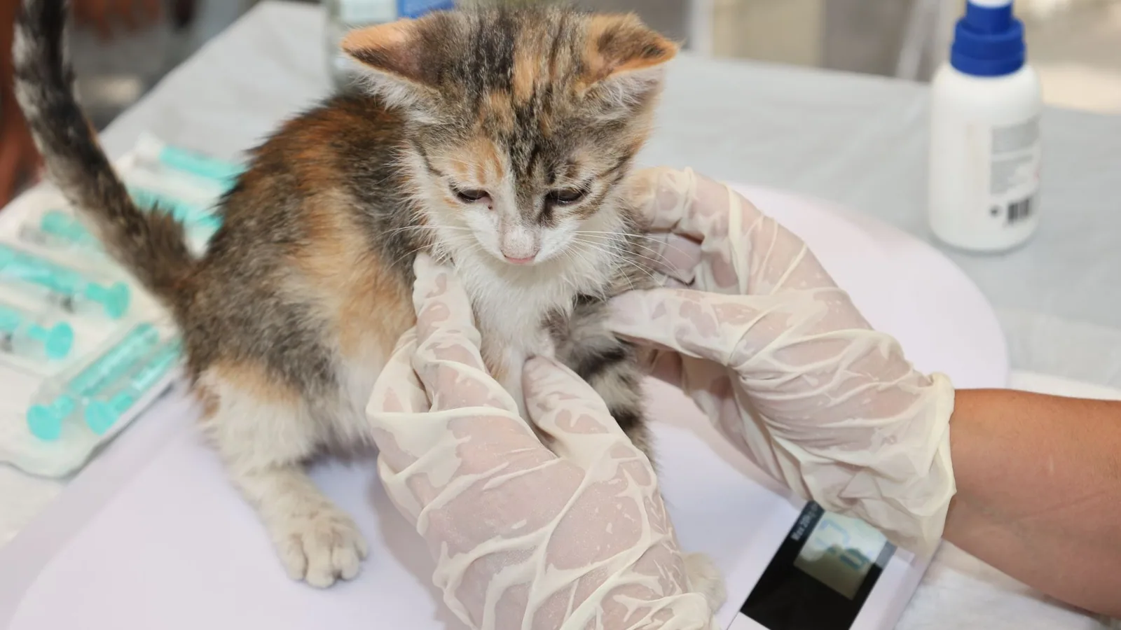
B. henselae is a fastidious, slow-growing bacterium that requires specialized culture conditions. Isolation is performed using blood agar or chocolate agar incubated at 35-37°C in 5% CO₂ for up to 4-6 weeks [22]. The sensitivity of blood culture is low, estimated at 30-50%, because bacteremia is often intermittent and of low magnitude [23]. Lysis-centrifugation techniques can improve yield but are labor-intensive and not routinely available in commercial diagnostic laboratories.
Polymerase Chain Reaction (PCR)
PCR assays targeting the 16S rRNA gene, the citrate synthase gene (gltA), or the riboflavin synthase gene (ribC) are widely used for the detection of Bartonella DNA in blood, tissue, and flea specimens [24, 25]. Real-time PCR (qPCR) offers higher sensitivity and quantification of bacterial load. However, PCR sensitivity is also affected by the intermittent nature of bacteremia. A single negative PCR result does not rule out infection; serial sampling over several weeks is recommended [26].
The specificity of PCR is generally high, but false positives can occur due to contamination or the presence of non-viable organisms. Additionally, PCR cannot distinguish between active infection and past exposure, as DNA may persist in tissues after bacterial clearance [27].
Immunohistochemistry and In Situ Hybridization
These techniques can detect Bartonella antigens or nucleic acids in formalin-fixed, paraffin-embedded tissues, such as lymph node or heart valve biopsies. They are useful for confirming the etiology of lesions but are not practical for routine screening [28].
Serological Methods
Indirect Fluorescent Antibody (IFA) Assay
IFA is the most commonly used serological test for feline bartonellosis. It detects IgG antibodies against B. henselae whole-cell antigens. A fourfold rise in titer between acute and convalescent samples is considered indicative of active infection [29]. However, IFA has several limitations:
- Cross-reactivity among Bartonella species and with other bacteria (e.g., Coxiella burnetii, Chlamydia felis) can produce false positives [30].
- Antibody titers may remain elevated for months after bacterial clearance, making it difficult to distinguish current from past infection [31].
- Seronegative bacteremic cats are well documented, particularly in the early stages of infection or in immunocompromised individuals [32].
Enzyme-Linked Immunosorbent Assay (ELISA)
ELISA-based serological tests, including those for Feline Leukemia Virus p27 antigen detection, have been adapted for Bartonella antibody detection. Commercial ELISA kits for B. henselae IgG and IgM are available but suffer from similar limitations as IFA, with variable sensitivity and specificity [33]. The use of recombinant antigens (e.g., 17-kDa protein, Pap31) has improved specificity in some assays but has not been widely validated in cats [34].
Comparative Performance of PCR and Serology
The following table summarizes the key characteristics of PCR and serology for the diagnosis of feline bartonellosis.
| Feature | PCR | Serology (IFA/ELISA) |
|---|---|---|
| Target | Bacterial DNA | Host antibodies (IgG, IgM) |
| Sensitivity (per sample) | 40-70% (single blood draw) | 60-80% (single titer) |
| Specificity | >95% (with proper controls) | 70-90% (cross-reactivity) |
| Distinguishes active vs. past infection | No (DNA persistence) | No (antibody persistence) |
| Time to result | 1-2 days | 1-3 days |
| Sample type | Whole blood (EDTA), tissue, flea | Serum or plasma |
| Effect of intermittent bacteremia | False negatives | Not affected |
| Effect of immunosuppression | False negatives | False negatives (low antibody) |
Diagnostic Algorithm
Given the limitations of individual tests, a diagnostic algorithm combining PCR and serology with clinical assessment is recommended. The following Mermaid diagram illustrates a decision tree for the diagnostic workup of a cat with suspected bartonellosis.
flowchart TD
 A[Cat with clinical signs suggestive of bartonellosis] --> B{Perform blood PCR and serology}
 B --> C[PCR positive, serology positive]
 C --> D[High probability of active infection]
 D --> E[Consider treatment and flea control]
 B --> F[PCR positive, serology negative]
 F --> G[Possible early infection or immunosuppression]
 G --> H[Repeat serology in 2-4 weeks]
 H --> I[Seroconversion?]
 I -->|Yes| D
 I -->|No| J[Consider non-infectious causes]
 B --> K[PCR negative, serology positive]
 K --> L[Past exposure or chronic antibody persistence]
 L --> M[Assess clinical signs and rule out other causes]
 M --> N[If signs persist, consider repeat PCR or tissue biopsy]
 B --> O[PCR negative, serology negative]
 O --> P[Low probability of bartonellosis]
 P --> Q[Investigate alternative diagnoses]
This algorithm emphasizes the need for serial testing and integration of clinical findings. For a similar approach to diagnostic algorithms in other feline infectious diseases, refer to the article on Feline Upper Respiratory Tract Infection Complex: Multiplex PCR Panel Interpretation and Treatment Algorithms.
Treatment
The treatment of feline bartonellosis remains controversial. Many cats clear bacteremia spontaneously without antimicrobial therapy. Antibiotics are generally reserved for cats with clinical signs or for those that are bacteremic and living in households with immunocompromised humans.
Doxycycline (10 mg/kg orally every 12-24 hours for 4-6 weeks) is the most commonly recommended antimicrobial. Azithromycin (10 mg/kg orally every 24 hours for 7 days, then every 48 hours for 2-3 weeks) is an alternative, though resistance has been reported [35]. Fluoroquinolones (e.g., enrofloxacin) have variable efficacy and are not considered first-line [36]. Rifampin may be used in combination with doxycycline for refractory cases [37].
Treatment failure is common, and bacteremia may recur after cessation of therapy. Concurrent flea control is essential to prevent reinfection. For a discussion of antimicrobial resistance in related bacterial pathogens, see the article on Antimicrobial Resistance in Livestock-Associated Staphylococcus aureus: Genomic Epidemiology and One Health Implications.
Conclusion
Feline bartonellosis is a complex, vector-borne disease with many clinical manifestations. The diagnosis is challenging due to the intermittent bacteremia, the fastidious nature of Bartonella, and the limitations of both PCR and serology. A combination of molecular and serological testing, interpreted in the context of clinical signs and risk factors, offers the best approach for accurate diagnosis. Ongoing research into improved diagnostic antigens and the development of point-of-care molecular assays may enhance our ability to detect and manage this infection in cats.
References
[1] Breitschwerdt EB, Kordick DL. Bartonella infection in animals: carriership, reservoir potential, pathogenicity, and zoonotic potential for human infection. Clin Microbiol Rev. 2000;13(3):428-438.
[2] Chomel BB, Boulouis HJ, Maruyama S, Breitschwerdt EB. Bartonella spp. in pets and effect on human health. Emerg Infect Dis. 2006;12(3):389-394.
[3] Schmid MC, Schulein R, Dehio M, et al. The VirB type IV secretion system of Bartonella henselae mediates invasion, proinflammatory activation and antiapoptotic protection of endothelial cells. Mol Microbiol. 2004;52(1):81-92.
[4] Minnick MF, Battisti JM. Pestilence, persistence and pathogenicity: infection strategies of Bartonella. Future Microbiol. 2009;4(6):743-758.
[5] Guptill L, Wu CC, HogenEsch H, et al. Prevalence of Bartonella henselae antibodies in pet cats throughout regions of North America. J Infect Dis. 1997;176(5):1140-1145.
[6] Maruyama S, Kabeya H, Nakao R, et al. Seroprevalence of Bartonella henselae, Toxoplasma gondii, FIV and FeLV infections in domestic cats in Japan. Microbiol Immunol. 2003;47(2):147-153.
[7] Abbott RC, Chomel BB, Kasten RW, et al. Experimental and natural infection with Bartonella henselae in domestic cats. Comp Immunol Microbiol Infect Dis. 1997;20(1):41-51.
[8] Higgins JA, Radulovic S, Jaworski DC, Azad AF. Acquisition of the cat scratch disease agent Bartonella henselae by cat fleas (Siphonaptera: Pulicidae). J Med Entomol. 1996;33(3):490-495.
[9] Chomel BB, Kasten RW, Floyd-Hawkins K, et al. Experimental transmission of Bartonella henselae by the cat flea. J Clin Microbiol. 1996;34(8):1952-1956.
[10] Billeter SA, Levy MG, Chomel BB, Breitschwerdt EB. Vector transmission of Bartonella species with emphasis on the potential for tick transmission. Med Vet Entomol. 2008;22(1):1-15.
[11] Kordick DL, Breitschwerdt EB. Relapsing bacteremia after blood transmission of Bartonella henselae to cats. Am J Vet Res. 1997;58(5):492-497.
[12] Guptill L. Feline bartonellosis. Vet Clin North Am Small Anim Pract. 2010;40(6):1073-1090.
[13] Lappin MR, Black JC. Bartonella spp. infection as a cause of uveitis in cats. Vet Ophthalmol. 1999;2(1):47-52.
[14] Ketring KL, Zuckerman EE, Hardy WD. Bartonella: a new etiological agent of feline ocular disease. J Am Anim Hosp Assoc. 2004;40(1):6-12.
[15] Powell CC, Lappin MR. Clinical ocular bartonellosis in cats. J Feline Med Surg. 2001;3(4):199-204.
[16] Dowers KL, Hawley JR, Brewer MM, et al. Association of Bartonella species, feline calicivirus, and feline herpesvirus 1 infection with gingivostomatitis in cats. J Feline Med Surg. 2010;12(4):314-321.
[17] Chomel BB, Kasten RW, Williams C, et al. Bartonella endocarditis: a pathology shared by animal reservoirs and patients. Ann N Y Acad Sci. 2009;1166:120-126.
[18] Varanat M, Broadhurst J, Linder KE, et al. Identification of Bartonella henselae in the heart of a cat with endocarditis. J Vet Diagn Invest. 2012;24(4):773-777.
[19] Breitschwerdt EB, Hegarty BC, Maggi RG, et al. Bartonella species as a potential cause of meningoencephalitis in cats. J Feline Med Surg. 2007;9(5):390-398.
[20] Guptill L, Slater LN, Wu CC, et al. Experimental infection of young specific pathogen-free cats with Bartonella henselae. J Infect Dis. 1997;176(1):206-216.
[21] Lappin MR, Breitschwerdt EB, Brewer M, et al. Prevalence of Bartonella species, Mycoplasma species, Ehrlichia species, and [Anaplasma phagocytophilum](/knowledge/bacteria/Equine Granulocytic Anaplasmosis/anaplasma-phagocytophilum-equine-granulocytic-anaplasmosis-tick) DNA in the blood of cats and their fleas in the United States. J Feline Med Surg. 2006;8(2):85-92.
[22] Brenner SA, Rooney JA, Manzewitsch P, Regnery RL. Isolation of Bartonella (Rochalimaea) henselae: effects of methods of blood collection and handling. J Clin Microbiol. 1997;35(3):544-547.
[23] Kordick DL, Wilson KH, Sexton DJ, et al. Prolonged bacteremia in cats experimentally infected with Bartonella henselae. J Clin Microbiol. 1995;33(12):3245-3251.
[24] Relman DA, Loutit JS, Schmidt TM, et al. The agent of bacillary angiomatosis. An approach to the identification of uncultured pathogens. N Engl J Med. 1990;323(23):1573-1580.
[25] Norman AF, Regnery RL, Jameson P, et al. Differentiation of Bartonella-like isolates at the species level by PCR-restriction fragment length polymorphism in the citrate synthase gene. J Clin Microbiol. 1995;33(7):1797-1803.
[26] Jensen WA, Fall MZ, Rooney J, et al. Rapid identification and differentiation of Bartonella species using a single-step PCR assay. J Clin Microbiol. 2000;38(5):1717-1722.
[27] Duncan AW, Maggi RG, Breitschwerdt EB. A combined approach for the enhanced detection and isolation of Bartonella species in dog blood samples: pre-enrichment liquid culture followed by PCR and subculture onto agar plates. J Microbiol Methods. 2007;69(2):273-281.
[28] Varanat M, Maggi RG, Linder KE, Breitschwerdt EB. Molecular prevalence of Bartonella, Babesia, and Ehrlichia species in dogs from the southeastern United States. J Vet Intern Med. 2011;25(3):488-494.
[29] Regnery RL, Olson JG, Perkins BA, Bibb W. Serological response to Rochalimaea henselae antigen in suspected cat-scratch disease. Lancet. 1992;339(8807):1443-1445.
[30] La Scola B, Raoult D. Serological cross-reactions between Bartonella quintana, Bartonella henselae, and Coxiella burnetii. J Clin Microbiol. 1996;34(9):2270-2274.
[31] Kordick DL, Breitschwerdt EB. Persistent infection of pets within a household with three Bartonella species. Emerg Infect Dis. 1998;4(2):325-328.
[32] Breitschwerdt EB, Maggi RG, Chomel BB, Lappin MR. Bartonellosis: an emerging infectious disease of zoonotic importance to animals and human beings. J Vet Emerg Crit Care. 2010;20(1):8-30.
[33] Lappin MR, Breitschwerdt EB, Brewer M, et al. Use of serologic tests to predict outcome of experimental Bartonella henselae infection in cats. Am J Vet Res. 2002;63(8):1139-1144.
[34] Anderson B, Sims K, Regnery R, et al. Detection of Rochalimaea henselae DNA in specimens from cat scratch disease patients by PCR. J Clin Microbiol. 1994;32(4):942-948.
[35] Greene CE, McDermott M, Jameson PH, et al. Bartonella henselae infection in cats: evaluation of treatment with doxycycline and azithromycin. J Vet Intern Med. 2002;16(3):306-312.
[36] Ketring KL, Zuckerman EE, Hardy WD. Treatment of Bartonella infection in cats. J Am Vet Med Assoc. 2005;226(4):555-559.
[37] Breitschwerdt EB, Linder KL, Day MJ, et al. Koch's postulates and the pathogenesis of comparative infectious disease causation associated with Bartonella species. J Comp Pathol. 2013;148(2-3):115-125.
[38] Chomel BB, Boulouis HJ, Breitschwerdt EB. Cat scratch disease and other zoonotic Bartonella infections. J Am Vet Med Assoc. 2004;224(8):1270-1279.
[39] Guptill L, Wu CC, HogenEsch H, et al. Humoral and cellular immune responses of cats experimentally infected with Bartonella henselae. Am J Vet Res. 1998;59(10):1285-1290.
[40] Karem KL, Dubie KA, Anderson B, et al. Characterization of Bartonella henselae-specific immunity in BALB/c mice. Immunology. 2000;99(3):385-392.
[41] Maggi RG, Duncan AW, Breitschwerdt EB. Novel chemically modified liquid medium that will support the growth of seven Bartonella species. J Clin Microbiol. 2005;43(6):2651-2655.
[42] Maruyama S, Nakamura Y, Kabeya H, et al. Prevalence of Bartonella henselae and Bartonella clarridgeiae in domestic cats from Thailand. J Vet Med Sci. 2001;63(4):443-447.
[43] Minnick MF, Smitherman LS, Samuels DS. Mitogenic effect of Bartonella bacilliformis on human vascular endothelial cells and involvement of GroEL. Infect Immun. 2003;71(12):6933-6942.
[44] Resto-Ruiz S, Burgess A, Anderson BE. The role of the host immune response in pathogenesis of Bartonella infections. Curr Opin Microbiol. 2003;6(1):43-48.
[45] Rolain JM, Franc M, Davoust B, Raoult D. Molecular detection of Bartonella quintana, B. koehlerae, B. henselae, B. clarridgeiae, Rickettsia felis, and Wolbachia pipientis in cat fleas, France. Emerg Infect Dis. 2003;9(3):338-342.
[46] Sander A, Posselt M, Oberle K, Bredt W. Seroprevalence of antibodies to Bartonella henselae in patients with cat scratch disease and in healthy controls: evaluation and comparison of two commercial serological tests. Clin Diagn Lab Immunol. 1998;5(4):486-490.
Disclaimer: This article is for educational and informational purposes only. It is not intended to substitute for professional veterinary advice, diagnosis, treatment, or regulatory guidance. Always consult a licensed veterinarian or qualified specialist regarding animal health, disease diagnosis, and therapeutic decisions.