
Bartonella henselae: Cat Scratch Disease, Clinical Presentation, Zoonosis, and Diagnostic Approaches
Etiology and Taxonomy
Bartonella henselae is a fastidious, Gram-negative, facultative intracellular bacillus belonging to the family Bartonellaceae within the order Rhizobiales. The organism is a hemotropic bacterium that exhibits tropism for erythrocytes and endothelial cells in its feline reservoir host. B. henselae is the primary etiologic agent of cat scratch disease (CSD), a zoonotic bacterial infection transmitted from cats to humans. The bacterium is characterized by its ability to adhere to and invade erythrocytes, a process mediated by type IV secretion systems and autotransporter adhesins such as BadA (Bartonella adhesin A) and the recently characterized BafA variants [1]. Strain-level differences in BafA have been associated with differential vasoproliferative traits, which may influence clinical manifestations in both reservoir and incidental hosts [1].
Epidemiology and Zoonotic Transmission
Bartonella henselae cat scratch disease clinical zoonosis is a globally distributed infection. The domestic cat (Felis catus) serves as the primary reservoir host, with bacteremia rates in feline populations ranging from 5% to 40% depending on geographic region, flea infestation prevalence, and cat age. Young kittens, particularly those under one year of age, demonstrate higher rates of bacteremia and are more likely to transmit the organism to humans. The cat flea (Ctenocephalides felis) is the principal arthropod vector responsible for horizontal transmission among cats. Flea feces containing viable B. henselae organisms are inoculated into feline skin through scratching or biting, establishing infection in the reservoir host.
Transmission to humans occurs through scratches, bites, or licks from an infected cat, particularly when flea feces contaminate claws or oral mucosa. Direct inoculation of B. henselae into human skin or mucous membranes initiates the zoonotic infection. The organism does not require an arthropod vector for transmission from cats to humans, although flea feces serve as the environmental source of infectious bacteria on feline pelage and claws.
Clinical Presentation in Cats
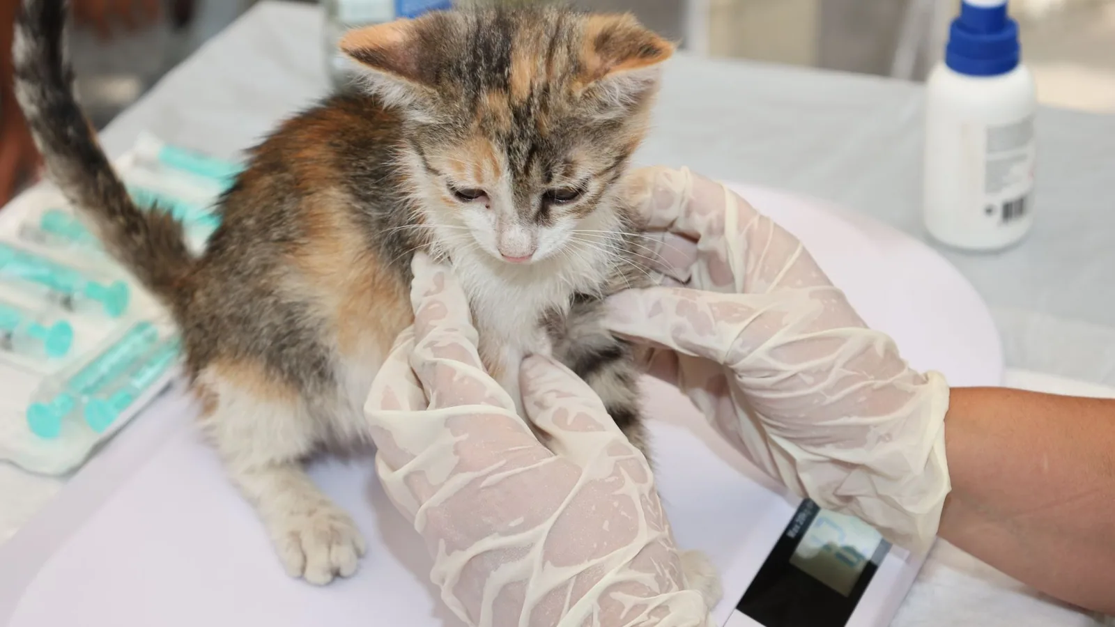
Reservoir Host Infection
In immunocompetent cats, B. henselae infection is typically subclinical or associated with mild, self-limiting signs. Experimental and natural infection studies indicate that cats may develop transient fever, lethargy, and mild lymphadenopathy. The bacterium establishes a long-term intracrythrocytic bacteremia that can persist for months to years, allowing the cat to serve as a continuous source of infection for fleas and, subsequently, for humans.
Feline Disease Manifestations
Although most infected cats remain asymptomatic, B. henselae has been associated with several clinical syndromes in felines:
- Feline endocarditis: B. henselae is a recognized cause of blood culture-negative endocarditis in cats, presenting with valvular vegetative lesions, fever, and cardiac murmurs.
- Feline uveitis: The organism has been implicated in cases of anterior uveitis and chorioretinitis in cats, although definitive causation remains debated.
- Feline lymphadenitis: Regional lymphadenopathy, particularly involving the submandibular, prescapular, or popliteal lymph nodes, may occur following flea exposure.
- Feline gingivitis and stomatitis: Some studies have identified B. henselae DNA in oral tissues of cats with chronic gingivostomatitis, though the etiologic role remains unclear.
Clinical Presentation in Humans: Cat Scratch Disease
Typical CSD
The classic presentation of CSD in immunocompetent individuals begins with a primary inoculation lesion at the site of a cat scratch or bite. This lesion appears as a papule, pustule, or vesicle 3 to 10 days after inoculation and persists for 1 to 3 weeks. Regional lymphadenopathy develops 1 to 3 weeks later, most commonly involving the axillary, epitrochlear, cervical, or inguinal lymph nodes [2, 3]. The lymph nodes are typically tender, erythematous, and may suppurate, forming abscesses that require drainage [4]. Lymphadenopathy resolves spontaneously over 2 to 4 months in most cases, although suppurative nodes may persist longer [4, 5].
Atypical CSD
Atypical manifestations occur in 5% to 20% of CSD cases and involve extranodal sites. These presentations are more common in immunocompromised individuals but can occur in immunocompetent patients as well.
Neurologic involvement: Encephalitis is a well-documented neurologic complication of CSD, presenting with headache, seizures, altered mental status, and focal neurologic deficits [6]. Cranial nerve neuropathies, including facial nerve palsy and optic neuritis, have been reported [7]. Peripheral neurological involvement, such as radiculitis and mononeuritis multiplex, has been described in immunocompetent adolescents [8]. Meningitis with bone marrow hemophagocytosis has been documented in pediatric cases [9].
Ocular manifestations: Parinaud oculoglandular syndrome, characterized by unilateral conjunctivitis and ipsilateral preauricular lymphadenopathy, is a classic ocular presentation [10, 11, 47]. Neuroretinitis, presenting with optic disc edema and macular star formation, is a well-recognized complication [12, 13, 14, 15, 16, 17]. Chorioretinitis, branch retinal artery occlusion, and serous retinal detachment have also been reported [15, 16, 17].
Hepatosplenic disease: Disseminated infection can produce granulomatous lesions in the liver and spleen, presenting with fever, abdominal pain, and hepatosplenomegaly [18, 19]. Hepatic abscesses and osteomyelitis have been documented in pediatric patients [19].
Musculoskeletal involvement: Osteomyelitis, spondylodiscitis, and septic arthritis are rare but documented complications [20, 21, 19]. Bone and joint infections may require prolonged antimicrobial therapy and surgical intervention.
Cardiovascular involvement: B. henselae is a recognized cause of blood culture-negative infective endocarditis, particularly in patients with preexisting valvular disease [22, 23, 24, 45]. Endocarditis may be complicated by immune complex glomerulonephritis, presenting with hematuria, proteinuria, and renal dysfunction [22, 23]. Discordant ANCA positivity has been reported in association with B. henselae endocarditis-associated glomerulonephritis [22].
Multisystemic and thrombo-inflammatory syndromes: Severe disseminated infection can produce a multisystemic thrombo-inflammatory syndrome characterized by fever, coagulopathy, and multiorgan dysfunction [25]. Pleural and extradural spinal involvement has been reported in systemic bartonellosis [26].
Unusual presentations: CSD has been reported to mimic neoplastic disease, presenting with persistent lymphadenopathy that raises concern for lymphoma [27, 46]. Co-infection with other pathogens, such as Epstein-Barr virus (infectious mononucleosis) and Sporothrix brasiliensis, has been documented [28, 47]. Pneumonia with empyema is a rare pulmonary manifestation.
Pathology and Pathogenesis
Histopathology
The characteristic histopathologic lesion of CSD is a granulomatous lymphadenitis. Early lesions demonstrate lymphoid hyperplasia with follicular hyperplasia and paracortical expansion. As the lesion progresses, necrotizing granulomas with central microabscesses surrounded by palisading epithelioid histiocytes develop. The Warthin-Starry silver stain reveals clusters of small, pleomorphic bacilli within the necrotic centers and within macrophages.
Molecular Pathogenesis
B. henselae employs multiple virulence mechanisms to establish infection and evade host immune responses. The type IV secretion system (VirB/VirD4) translocates effector proteins into host cells, modulating cytoskeletal dynamics, apoptosis, and inflammatory signaling. The Bartonella adhesin A (BadA) mediates adherence to endothelial cells and erythrocytes, while the autotransporter BafA promotes vasoproliferation through activation of the vascular endothelial growth factor (VEGF) receptor pathway [1]. Strain-specific variations in BafA may account for differences in vasoproliferative potential and clinical outcome [1].
The organism survives within erythrocytes by inhibiting phagolysosomal fusion and resisting oxidative killing. Intracellular persistence within erythrocytes allows the bacterium to evade antibody-mediated clearance and maintain prolonged bacteremia in the feline reservoir host.
Diagnostic Approaches
Serologic Testing
Serology remains the most widely used diagnostic modality for CSD. Indirect immunofluorescence assays (IFA) and enzyme-linked immunosorbent assays (ELISA) detect antibodies against B. henselae. Chemiluminescence immunoassays, such as the VirClia IgG test, have demonstrated utility for serologic diagnosis [29, 30]. A four-fold rise in IgG titer between acute and convalescent sera, or a single elevated IgM titer, supports active infection. However, serologic cross-reactivity occurs among Bartonella species and with Coxiella burnetii, necessitating confirmatory testing [30]. Seronegative cases, particularly in immunocompromised patients, have been documented [18].
Molecular Diagnostics
Polymerase chain reaction (PCR) assays targeting the 16S rRNA gene, the citrate synthase gene (gltA), or the riboflavin synthase gene (ribC) provide species-level identification of B. henselae. Real-time PCR offers rapid detection with high sensitivity and specificity. Metagenomic next-generation sequencing (mNGS) has emerged as a powerful tool for diagnosing atypical CSD, particularly in cases with negative serology or unusual clinical presentations [31, 20]. mNGS can detect B. henselae DNA in blood, lymph node aspirates, cerebrospinal fluid, and tissue biopsies, providing definitive etiologic diagnosis [31, 20].
Culture
B. henselae is a fastidious organism requiring specialized culture conditions. Isolation is performed on chocolate agar or blood agar supplemented with 5% defibrinated sheep blood, incubated at 35-37 degrees Celsius in 5% carbon dioxide for 2 to 6 weeks. The organism produces small, adherent, pitting colonies after prolonged incubation. Culture sensitivity is low, particularly in clinical specimens, and is not recommended as a primary diagnostic method.
Histopathology and Cytology
Lymph node biopsy or fine-needle aspiration cytology may demonstrate granulomatous inflammation with necrotizing microabscesses. Warthin-Starry silver staining reveals intracellular bacilli within macrophages and within necrotic centers. Immunohistochemistry using monoclonal antibodies against B. henselae can confirm the presence of the organism in tissue sections.
Diagnostic Algorithm
The following Mermaid diagram illustrates a diagnostic decision tree for suspected CSD:
flowchart TD
 A[Patient with lymphadenopathy and cat exposure] --> B{"Serology: IFA or ELISA"}
 B -->|Positive IgM or 4-fold IgG rise| C[Probable CSD]
 B -->|Negative or equivocal| D{Clinical suspicion high?}
 D -->|Yes| E[Perform PCR on lymph node aspirate or blood]
 D -->|No| F[Consider alternative diagnoses]
 E -->|Positive| G[Confirmed CSD]
 E -->|Negative| H[Consider mNGS or biopsy]
 H -->|Positive| G
 H -->|Negative| F
 C --> I{Atypical features?}
 I -->|Yes| J["Imaging: ultrasound, CT, MRI"]
 I -->|No| K[Supportive care]
 J --> L[Evaluate for hepatosplenic, neurologic, ocular, or cardiac involvement]
 L --> M["Targeted diagnostics: echocardiography, lumbar puncture, ophthalmologic exam"]
Treatment and Management
Feline Treatment
Treatment of B. henselae infection in cats is controversial. Antimicrobial therapy does not reliably eliminate bacteremia, and treated cats often become re-infected following flea exposure. Doxycycline (10 mg/kg orally every 12 hours for 14 days) has been shown to reduce bacteremia transiently but does not achieve consistent clearance. Azithromycin and enrofloxacin have also been used with variable success. Strict flea control using veterinary-approved adulticides and insect growth regulators is the most effective strategy for preventing infection and re-infection in cats.
Human Treatment
Typical CSD: In immunocompetent patients with mild to moderate lymphadenopathy, treatment is primarily supportive. Lymphadenopathy resolves spontaneously over 2 to 4 months. Analgesics and warm compresses may provide symptomatic relief. Suppurative lymph nodes may require needle aspiration for pain relief.
Atypical or severe CSD: Antimicrobial therapy is indicated for patients with severe lymphadenopathy, hepatosplenic disease, endocarditis, neurologic involvement, or ocular disease. Azithromycin (500 mg orally on day 1, then 250 mg daily for 5 days) is the preferred agent for uncomplicated CSD. Doxycycline (100 mg orally every 12 hours) combined with rifampin (300 mg orally every 12 hours) is recommended for neuroretinitis, encephalitis, and endocarditis. Treatment duration ranges from 2 to 6 weeks depending on the severity and site of infection. Endocarditis may require 6 weeks or more of combination therapy, often with gentamicin added for the initial 2 weeks.
Control and Prevention
Flea Control
Comprehensive flea control is the cornerstone of CSD prevention in cats. Year-round use of veterinary-approved flea adulticides (e.g., fipronil, imidacloprid, selamectin) combined with insect growth regulators (e.g., lufenuron, methoprene) reduces flea populations and interrupts transmission of B. henselae among cats. Environmental flea control, including vacuuming and treatment of indoor and outdoor environments, is essential for reducing flea burdens.
Cat Management
Declawing is not recommended as a preventive measure. Owners should be educated about the risks of rough play with cats, particularly kittens, and should avoid allowing cats to lick open wounds or mucous membranes. Prompt wound cleansing with soap and water following cat scratches or bites reduces the risk of bacterial inoculation.
Public Health Education
Veterinarians play a critical role in educating cat owners about the zoonotic potential of B. henselae. Immunocompromised individuals, including those with HIV/AIDS, organ transplant recipients, and patients receiving immunosuppressive therapy (e.g., abatacept for rheumatoid arthritis), should be advised of the increased risk of severe CSD [32, 33, 34, 44]. Adopting adult cats rather than kittens, maintaining strict flea control, and avoiding rough play are recommended risk-reduction strategies.
Conclusion
Bartonella henselae cat scratch disease clinical zoonosis represents a significant veterinary and public health concern. The bacterium is maintained in feline reservoir populations through flea-borne transmission and is transmitted to humans through direct contact with infected cats. Clinical manifestations range from self-limiting lymphadenopathy to severe systemic disease involving the nervous system, eyes, heart, liver, spleen, and bones. Diagnosis relies on serology, PCR, and increasingly on metagenomic sequencing for atypical presentations. Effective control requires rigorous flea management in cats and public education regarding zoonotic risk. Continued research into strain-specific virulence factors, such as BafA variants, may further elucidate the mechanisms underlying the diverse clinical presentations of this important zoonotic pathogen.
References
[1] Kondo Y, Suzuki M, Sato S, et al. Differential vasoproliferative traits of Bartonella henselae strains associated with autotransporter BafA variants. Microbiol Spectr. 2025. URL: https://pubmed.ncbi.nlm.nih.gov/39611834/
[2] Sabir S, Daley SF, Huang B. Cat Scratch Disease. PubMed. 2026. URL: https://pubmed.ncbi.nlm.nih.gov/29489252/
[3] Öcal-Demir S, Kahraman K, Bozbeyoğlu G, et al. Clinical Presentation of Cat Scratch Disease in Pediatric Patients-A Single-Center Study. Turk Arch Pediatr. 2024. URL: https://pubmed.ncbi.nlm.nih.gov/39540783/
[4] Inoue S, Sumii S, Araki K, et al. Treatment-Resistant Femoral Lymph Node Abscess Caused by Cat Scratch Disease: A Case Confirmed by Polymerase Chain Reaction. Cureus. 2025. URL: https://pubmed.ncbi.nlm.nih.gov/41583232/
[5] Shoewu OA, Oscanoa C, Baker AH, et al. Persistent Postauricular Swelling in a Pediatric Patient: Unraveling the Cat Scratch Disease. Cureus. 2025. URL: https://pubmed.ncbi.nlm.nih.gov/40861698/
[6] Yakubovsky M, Kadar L, Katchman E, et al. Cat scratch disease encephalitis: New insights into rate, clinical features, and long-term outcomes. Int J Infect Dis. 2026. URL: https://pubmed.ncbi.nlm.nih.gov/41544843/
[7] Yakubovsky M, Kosman A, Kadar L, et al. Cranial nerve neuropathies: a rare manifestation of cat scratch disease. BMC Infect Dis. 2026. URL: https://pubmed.ncbi.nlm.nih.gov/41580648/
[8] Arabeyre C, Lheritier S, Rached A, et al. Peripheral neurological involvement due to Bartonella henselae in an immunocompetent adolescent: a case report and literature review. Diagn Microbiol Infect Dis. 2026. URL: https://pubmed.ncbi.nlm.nih.gov/42127721/
[9] DU WQ, Liu LJ, Zhang L, et al. [A case of Bartonella henselae meningitis characterized by bone marrow hemophagocytosis]. Zhongguo Dang Dai Er Ke Za Zhi. 2026. URL: https://pubmed.ncbi.nlm.nih.gov/42130363/
[10] Mummaneni NS, Villatoro GA, Yen KG, et al. Conservative Management of Eyelid Necrotizing Fasciitis in a Pediatric Patient With Concurrent Parinaud's Oculoglandular Syndrome. Ophthalmic Plast Reconstr Surg. 2025. URL: https://pubmed.ncbi.nlm.nih.gov/41022040/
[11] Abd Rahim NN, Ami M, Zainal Abidin Z. Parinaud Oculoglandular Syndrome Among Female Adults in Malaysia: A Case Series. Cureus. 2024. URL: https://pubmed.ncbi.nlm.nih.gov/39286673/
[12] Rajan RS, Kuganasan S, Ali NS, et al. Clinical and imaging characteristics of neuroretinitis secondary to cat scratch disease from tertiary centers in Malaysia: a retrospective study. J Ophthalmic Inflamm Infect. 2025. URL: https://pubmed.ncbi.nlm.nih.gov/41273610/
[13] Nicula CA, Lăpuște AI, Lăpușan AI. Neuroretinitis and chorioretinitis in a cat-scratched young boy: a case report. Rom J Ophthalmol. 2025. URL: https://pubmed.ncbi.nlm.nih.gov/40330972/
[14] Shi J, Cheema MR. Clinical Characteristics and Visual Outcomes of Cat Scratch Disease. Korean J Ophthalmol. 2025. URL: https://pubmed.ncbi.nlm.nih.gov/40235103/
[15] Zhang W, Chen C, Song Y, et al. A CASE OF BARTONELLA NEURORETINITIS PRESENTING WITH CONCOMITANT FROSTED BRANCH ANGIITIS ON LONG-TERM FOLLOWUP. Retin Cases Brief Rep. 2026. URL: https://pubmed.ncbi.nlm.nih.gov/39933158/
[16] Skrehot HC, Alsoudi AF, Nolan RP, et al. Atypical Haemorrhagic Presentation of Neuro-Retinitis and Serous Retinal Detachment Secondary to Cat-Scratch Disease. Neuroophthalmology. 2024. URL: https://pubmed.ncbi.nlm.nih.gov/39156225/
[17] Jafari S, Micieli JA. Cat Scratch Disease Presenting with Right Branch Retinal Artery Occlusion and Left Neuroretinitis. Case Rep Ophthalmol. 2024. URL: https://pubmed.ncbi.nlm.nih.gov/39144650/
[18] Osawa H, Shibutani K, Mori N. Suspected Hepatosplenic Cat Scratch Disease with No Major Symptoms and Negative Serology: A Case Report. Am J Case Rep. 2025. URL: https://pubmed.ncbi.nlm.nih.gov/41160532/
[19] Antonson M, Klingemann L, Neemann K. Bartonella henselae Hepatic Abscesses and Associated Osteomyelitis in a Pediatric Patient. Case Rep Infect Dis. 2024. URL: https://pubmed.ncbi.nlm.nih.gov/39135548/
[20] Wu X, Yin Y, Guo Y, et al. A case of atypical cat scratch disease with bone and joint infection diagnosed through clinical metagenomics. IDCases. 2026. URL: https://pubmed.ncbi.nlm.nih.gov/41551358/
[21] El Assaad H, Schumann E, Klemann C, et al. Presumed Bartonella-Associated Spondylodiscitis in a 3-Year-Old Child: A Case Report and Review of the Literature. Children (Basel). 2025. URL: https://pubmed.ncbi.nlm.nih.gov/40426827/
[22] Yow BLH, Kittaka RC, Yeung CSJ. Discordant ANCA Positivity in Immune Complex Glomerulonephritis Secondary to Bartonella henselae Prosthetic Valve Endocarditis. Nephrology (Carlton). 2026. URL: https://pubmed.ncbi.nlm.nih.gov/42168832/
[23] Bae J, Ipaye A, Pocock J, et al. Subacute Bartonella endocarditis with glomerulonephritis: a diagnostic and therapeutic challenge in blood culture-negative infective endocarditis. Access Microbiol. 2025. URL: https://pubmed.ncbi.nlm.nih.gov/41427080/
[24] Ghozzia M, Azaiez F, Trabelsi M, et al. Navigating diagnostic challenges in Bartonella-induced infective endocarditis: a case report. J Med Case Rep. 2025. URL: https://pubmed.ncbi.nlm.nih.gov/40016798/
[25] Gobbo G, Zacchia M, Nalesso F, et al. Bartonella henselae infection associated with a multisystemic thrombo-inflammatory syndrome in an immunocompetent adult. Infection. 2026. URL: https://pubmed.ncbi.nlm.nih.gov/41986819/
[26] Kamiya Rodas IA, Bustos AC. Pleural and extradural spinal involvement in a patient with systemic bartonellosis. Arch Argent Pediatr. 2025. URL: https://pubmed.ncbi.nlm.nih.gov/40693648/
[27] Smith E, Lawless R, Hoellein A. Cat-Scratch Disease Mimicking Neoplastic Etiology in a Complex Clinical Presentation: A Case Report. Cureus. 2024. URL: https://pubmed.ncbi.nlm.nih.gov/39280565/
[28] Huang W, Yuan J, He H. Case Report: Co-infection with cat-scratch disease and infectious mononucleosis: a rare clinical entity. Front Pediatr. 2025. URL: https://pubmed.ncbi.nlm.nih.gov/41378194/
[29] Pollani C, Floch P, Grouteau G, et al. Real-life assessment of the VirClia IgG chemiluminescence test for the diagnosis of Bartonella henselae infection. Diagn Microbiol Infect Dis. 2026. URL: https://pubmed.ncbi.nlm.nih.gov/41242076/
[30] Tortosa-Carreres J, Lloret-Sos C, Sahuquillo-Arce JM, et al. VirClia chemiluminescent assays as a tool for detecting Bartonella Henselae and Coxiella burnetii infections. Eur J Clin Microbiol Infect Dis. 2025. URL: https://pubmed.ncbi.nlm.nih.gov/40802230/
[31] Lai SY, Chang L, Duan JX, et al. Clinical and epidemiological characteristics of cat scratch disease in children from southwestern China: a retrospective analysis of mNGS-confirmed cases. Front Public Health. 2025. URL: https://pubmed.ncbi.nlm.nih.gov/41641055/
[32] Saito H, Takahashi Y, Tsuge S, et al. Disseminated cat-scratch disease during abatacept therapy for rheumatoid arthritis in an older patient: A case report and review of the literature. J Infect Chemother. 2025. URL: https://pubmed.ncbi.nlm.nih.gov/40379021/
[33] Doğan B, Güney İ, Ömeroğlu E. Unexplained Fever in a Hemodialysis Patient Possibly due to Cat Scratch Disease Opportunistic Infection. Hemodial Int. 2025. URL: https://pubmed.ncbi.nlm.nih.gov/40044409/
[34] Rošić Despalatović B, Bratanić A, Božić D, et al. A case of generalized cat scratch disease in a patient with ulcerative colitis on immunosuppressive therapy. Ther Adv Infect Dis. 2024. URL: https://pubmed.ncbi.nlm.nih.gov/39524988/
[35] Dhillon T, Pereira N. Uncovering Latent Tuberculosis Masquerading as Bartonella henselae Infection in a 12-Year-Old Patient. Cureus. 2025. URL: https://pubmed.ncbi.nlm.nih.gov/40949059/
[36] Kucarz TJ, Souza CA, Hatanaka SA, et al. Multi systemic compromise due to Bartonella henselae in a child. Rev Inst Med Trop Sao Paulo. 2025. URL: https://pubmed.ncbi.nlm.nih.gov/40699062/
[37] Zabari N, Abdul Hadi FS, Seman Z, et al. Seropositivity of Bartonella henselae Among Suspected Cat Scratch Disease Patients in Malaysia: A 5-Year Retrospective Study (2015-2019). Trop Biomed. 2025. URL: https://pubmed.ncbi.nlm.nih.gov/40618362/
[38] Endeladze M, Zhamutashvili M, Gognadze T, et al. CASE REPORT OF CAT SCRATCH DISEASE (BARTONELLA). Georgian Med News. 2025. URL: https://pubmed.ncbi.nlm.nih.gov/40305794/
[39] Yin QL, Liu YQ, Zhang HM, et al. Cat scratch disease in children with nocturnal fever: A case report. World J Clin Cases. 2024. URL: https://pubmed.ncbi.nlm.nih.gov/39687640/
[40] Martínez Lindado MA, Praino ML, Caratozzolo A, et al. Cat Scratch Disease in Pediatrics: Who Has Systemic Involvement? Pediatr Infect Dis J. 2025. URL: https://pubmed.ncbi.nlm.nih.gov/39230263/
[41] Nguyen M, Singh S, Sam B, et al. Three-Month History of Lymphadenopathy Caused by Bartonella henselae in a 13-Year-Old Following a Dog Scratch. Cureus. 2024. URL: https://pubmed.ncbi.nlm.nih.gov/39229389/
Disclaimer: This article is for educational and informational purposes only. It is not intended to substitute for professional veterinary advice, diagnosis, treatment, or regulatory guidance. Always consult a licensed veterinarian or qualified specialist regarding animal health, disease diagnosis, and therapeutic decisions.