In Vitro vs In Vivo: Key Differences Explained
By Dr. Zubair Khalid, DVM, MS, PhD ·
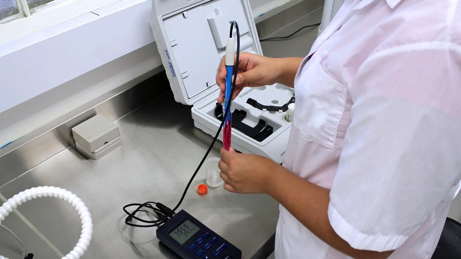
In vitro means the experiment happens outside a living organism, in a dish, tube, or engineered device, while in vivo means it happens inside a whole living animal or human. The rule of thumb is simple: use in vitro when you need to isolate a mechanism and run many conditions cheaply, and use in vivo when the question depends on the whole body, such as absorption, distribution, metabolism, excretion, or immune response.
The two are not competitors. They answer different questions, and most drug programs need both. A cell assay can tell you whether a compound kills a parasite. Only a living animal can tell you whether the drug reaches the parasite after being swallowed, survives liver metabolism, and avoids poisoning the host.
What "In Vitro" Actually Means
In vitro is Latin for "in glass." In practice it covers any biological system removed from a whole organism:
- Immortalized cell lines grown in flasks
- Primary cells freshly isolated from tissue
- Tissue slices and explants
- Isolated enzymes and subcellular fractions such as liver microsomes
- Three-dimensional spheroids, organoids, and organ-on-chip devices
The defining feature is that the system has no circulatory system, no immune surveillance, no endocrine signaling from distant organs, and no excretion route. Whatever you add to the medium is what the cells see. That is both the strength and the weakness of the approach.
A well-designed in vitro study can hold pH, temperature, oxygen, glucose, and compound concentration constant in a way no living animal allows. That control is why in vitro work dominates early discovery. A 2026 review of 3D spheroid models in breast cancer makes the point directly: conventional two-dimensional culture fails to capture the structural organization, cellular diversity, and microenvironmental gradients of real tumors, which limits its predictive value [1]. The same review notes that newer 3D systems, including hanging drop cultures, hydrogels, microfluidics, patient-derived organoids, and bioprinting, recover some of that complexity [1].
Complexity in a dish is not the same as complexity in a body. A bioprinted gastrointestinal stromal tumor model showed higher metabolic activity, better long-term survival, and upregulated tumor-associated genes compared with 2D monolayers, and cells from those constructs behaved differently again after transplantation into animals [2]. Each step up in complexity changed the biology.
What "In Vivo" Actually Means
In vivo is Latin for "within the living." The experiment runs inside an intact organism, whether that is a mouse, rat, zebrafish embryo, dog, pig, or human volunteer. The system brings everything the dish lacks:
- Absorption from the gut, lung, or skin
- Distribution through blood and into tissues
- Metabolism by liver enzymes
- Excretion by kidney or bile
- Immune and inflammatory responses
- Hormonal and neural signaling
- Behavior, appetite, and body weight effects
Those four processes, absorption, distribution, metabolism, and excretion, are collectively called pharmacokinetics, often abbreviated ADME. No isolated cell system performs ADME. A hepatocyte culture can metabolize a compound, but it cannot tell you the fraction of an oral dose that reaches the bloodstream. That requires a whole organism with a gut and a portal circulation.
In vivo work is also where toxicity shows up as a whole-animal event. The classic abnormal toxicity test used for veterinary autogenous vaccine release is an in vivo assay, and a 2026 study compared it against two in vitro alternatives, a fish embryo acute toxicity test and an L929 cytotoxicity assay, to see whether the animal test could be replaced [3]. The MTS cytotoxicity assay tracked the in vivo pass or negative outcome more closely than the embryo test, but the comparison itself shows why the in vivo reference exists: it captures the integrated response that single-cell assays approximate at best [3].
In Vitro vs In Vivo: Side-by-Side Comparison
| Criterion | In Vitro | In Vivo |
|---|---|---|
| System | Isolated cells, tissues, enzymes, or organ-on-chip | Whole living animal or human |
| Cost per data point | Low to moderate, reagents and plasticware dominate | High, includes housing, husbandry, veterinary care, and personnel |
| Control of variables | High, medium composition and dose are set exactly | Lower, subject to inter-animal variability and compensation |
| Throughput | High, hundreds to thousands of wells per week | Low, limited by animal numbers and ethics review |
| ADME captured | No | Yes |
| Immune and endocrine context | Absent unless co-cultured | Intact |
| Ethical and regulatory burden | Lower, still requires biosafety oversight | High, requires animal ethics approval and welfare oversight |
| Translational relevance | Mechanistic, often poor at predicting whole-body outcomes | Higher for integrated outcomes, imperfect across species |
| Best use | Mechanism, screening, dose-finding, assay development | Efficacy, safety, pharmacokinetics, confirmation |
Read that table as a division of labor, not a ranking. In vitro wins on control and throughput. In vivo wins on physiological realism. Neither wins on everything.
Where In Vitro Excels
Mechanism and causality
If you want to know whether a compound inhibits a specific enzyme, you purify or express the enzyme and measure kinetics. Adding a whole animal introduces dozens of confounders. A 2026 study on the antidiabetic potential of Crinum amoenum bulb extract ran α-amylase inhibition assays alongside molecular docking, molecular dynamics simulations, and ADMET prediction, then confirmed hypoglycemic effects in Wistar rats [4]. The enzyme assay isolated the mechanism. The rat study tested whether it mattered in a living system.
Throughput and cost
Multi-well plates let you test a compound across a concentration series in a single afternoon. Scaling that to animals is impossible on the same timeline. This is why virtually every drug discovery funnel starts with in vitro screening and narrows before any animal is used.
Human-relevant systems
Species differences are a real problem in animal work. A 2026 review of placental models states plainly that in vivo animal models and 2D cultures have not been sufficiently representative of whole human tissue, partly because of interspecies differences in placental physiology [5]. Three-dimensional human organoids and placenta-on-chip models were developed specifically to close that gap [5]. Kidney-on-chip and iPSC-derived organoid models serve a similar purpose, recapitulating fluid shear stress and mechanical strain that static culture cannot reproduce [6].
Precise, tunable environments
Mechanically active biomaterials let researchers dial matrix stiffness and viscoelasticity to study how physical cues drive stem cell differentiation [7]. You cannot dial stiffness in a living animal. A systems biology framework called LIV2TRANS was built to map microphysiological systems onto in vivo data and identify which experimental conditions, such as TGFβ signaling, most improve translatability for a liver disease model [8]. That work only makes sense because in vitro conditions are adjustable in the first place.
Where In Vivo Excels
Pharmacokinetics
This is the sharpest dividing line. In vitro systems do not absorb, distribute, metabolize, and excrete a drug as a coordinated whole. Skin physiologically based pharmacokinetic models exist precisely because percutaneous absorption, local tissue exposure, and systemic plasma concentration are whole-body phenomena that require mechanistic modeling to predict from in vitro diffusion data [9]. The term for that bridge is in vitro to in vivo extrapolation, or IVIVE [9].
Integrated efficacy
A drug can kill every cell in a dish and fail in a mouse because it never reaches the target tissue, is destroyed by liver enzymes, or triggers an immune response that clears it. A 2026 study of an engineered oncolytic adenovirus illustrates the layering. The virus was tested in syngeneic and humanized mouse models, with molecular analysis by Western blot, immunofluorescence, and qPCR, and immunological outcomes assessed by flow cytometry and in vitro co-culture assays [10]. The in vitro co-culture explained the immune mechanism. The mouse models showed tumor control and synergy with anti-PD-1 and CAR-T therapy [10]. Neither alone would have been convincing.
Toxicity in context
Local neurotoxicity assessment for extractables and leachables from drug container systems is a regulatory requirement, and animal testing remains the main approach for high-risk routes such as intrathecal or epidural administration [11]. A 2026 review searched public repositories for neurotoxicity data on 865 potential leachables and found that only 21 compounds, about 2.4%, had been tested both intrathecally and in vitro [11]. Of those, 16 gave similar results and five were discordant [11]. For non-intrathecal routes, 108 compounds, about 12.5%, had parallel data, with 91 concordant and 17 discordant [11]. Discordance is not rare. It is the reason both tiers exist.
Cost, Ethics, and Regulation
Cost and ethics travel together. In vivo studies require animal housing, husbandry, veterinary care, and protocol review by an institutional animal care and use committee in the United States. The 2026 neurotoxicity review describes animal testing for leachables as technically challenging, costly, and potentially hurtful to animals while providing limited scientific progress, which is why replacement by new approach methodologies is a stated goal [11].
Regulatory pressure is shifting. The FDA Modernization Act and subsequent federal policy changes in 2025 signaled a move toward non-animal, human-centered models for preclinical drug development and toxicity screening, emphasizing 3D cell-based, organ-on-chip, and organoid platforms [6]. That does not eliminate in vivo work. It changes when and why it is used.
In vitro work is not free of oversight. Cell lines require biosafety review, human-derived material requires consent and ethics approval, and engineered devices require validation. The 2026 magnesium bioaccessibility study used a validated simulator of the human intestinal microbial ecosystem and reported analytical validation metrics including 94 to 102 percent recovery, relative standard deviation at or below 2.4 percent, R² above 0.999, and a limit of quantification of 0.04 mg/kg [12]. Validation at that level is what makes an in vitro result usable for a regulatory or labeling claim.
The Central Pitfall: In Vitro Results Often Fail to Predict In Vivo Outcomes
The most common mistake in early-career research is treating a cell assay result as a prediction of whole-body effect. It is not. It is a measurement of what happens under the specific conditions of that assay.
Several mechanisms drive the gap:
- No ADME. The compound concentration in the medium is not the concentration at the target tissue in a living animal.
- No immune system. Immune-mediated toxicity and immune-mediated efficacy are invisible in a dish without reconstitution.
- No mechanical environment. Fluid shear stress, strain, and matrix stiffness change cell behavior, and standard static culture removes them [6].
- Species and cell-line artifacts. Hepatocellular carcinoma-derived lines such as HepG2 are poorly differentiated and show cancer-associated metabolic reprogramming that suppresses adult liver functions [13]. An immortalized primary human hepatocyte line, Fa2N-4, showed a distinct karyotype, different genetic risk variant profiles, and markedly different transcriptional responses to lipid loading and drug treatment compared with HepG2 [13]. Two liver cell models, two different answers.
- Discordance is documented. In the leachables dataset, roughly a quarter of compounds with parallel intrathecal and in vitro data disagreed, and about 16 percent of compounds with parallel non-intrathecal data disagreed [11].
The reverse error also happens. Researchers sometimes run an animal study to answer a question that a well-controlled cell assay could answer faster, cheaper, and more precisely. That wastes animals and money.
A Worked Drug-Testing Example
Suppose a team identifies a compound, call it Compound X, that inhibits a parasite in culture. Here is how the program typically unfolds.
Step 1: In vitro efficacy. The team grows Blastocystis hominis in three culture conditions: inoculated untreated, metronidazole-treated, and Compound X-treated. They count parasites and examine surface morphology by scanning electron microscopy. In a 2026 study using exactly this design with the anti-HIV drug ritonavir, the compound reduced parasite counts more effectively than metronidazole and produced visible surface changes [14].
Step 2: In vivo efficacy and safety. The team moves to 40 laboratory-bred albino mice divided into four groups of ten: non-infected untreated, infected untreated, metronidazole-treated, and Compound X-treated. They assess parasitological response, histopathology of the intestine, immunoglobulin A expression by immunohistochemistry, and serum levels of IL-1β, IL-8, and TNF-α by ELISA [14]. This step answers questions the culture could not: does the drug reach the gut lumen, does it survive host metabolism, does it reduce parasite burden in a living animal, and does it cause intestinal inflammation?
Step 3: Human trials. Only after in vitro potency, in vivo efficacy, and in vivo safety signals align does a compound enter human testing. The human trial is itself an in vivo experiment, and it is where species differences and real-world pharmacokinetics are finally resolved.
The funnel narrows at each step because each step is more expensive, slower, and more ethically weighted than the last. Skipping a step does not save time. It moves the failure later, where it costs more.
flowchart TD
    A[Research question] --> B{Does it need whole body}
    B -->|No| C[In vitro assay]
    B -->|Yes| D[In vivo model]
    C --> E[Mechanism and dose finding]
    E --> F[In vivo confirmation]
    D --> G[Efficacy and safety]
    F --> H[Human trial]
    G --> H
    H --> I[Regulatory review]
Common Mistakes and Limitations
Treating in vitro potency as a predictor of clinical dose. Potency in a well is not potency in a body. Without ADME data, an EC50 value tells you almost nothing about the dose a patient needs.
Assuming a cell line represents a tissue. HepG2 and Fa2N-4 differ in karyotype, genetic risk variants, and drug response [13]. Choosing a model is choosing an answer.
Ignoring the mechanical environment. Cells in a living kidney experience fluid shear stress and strain. Static culture removes both, which changes toxicity responses [6].
Overgeneralizing across species. Placental physiology differs between species, which is one reason human 3D models were developed [5]. A mouse result is a mouse result.
Assuming 3D is automatically better. Spheroid and organoid models add complexity, but they also add variability, and the choice between scaffold-free and scaffold-based approaches changes what the model can show [1]. Complexity is a tool, not a virtue.
Forgetting that in vitro systems also need validation. The magnesium study reported recovery, precision, linearity, and quantification limits before drawing conclusions [12]. Without that, an in vitro result is a number, not evidence.
Reading discordance as failure. When 16 of 21 compounds agreed and five did not, the five disagreements are the informative part [11]. They map the boundary of what each model can see.
Assuming in vivo is always the final word. Animal models have their own limitations, including interspecies differences and an inability to decouple biomechanical from biochemical effects during disease [6]. A mouse is not a small human.
Individual research or clinical decisions still need a qualified professional who knows the specific system, species, and question.
Frequently Asked Questions
What is the difference between in vitro and in vivo?
In vitro means outside a living organism, such as cells in a dish or enzymes in a tube. In vivo means inside a whole living animal or human. The difference determines whether whole-body processes like absorption, distribution, metabolism, and excretion are captured.
Can in vitro results replace animal testing?
Not entirely today. In vitro systems cannot reproduce ADME, immune response, or the full mechanical environment of a living body. Regulatory shifts toward organ-on-chip and organoid platforms are expanding what in vitro can support, but in vivo studies remain required for many safety and efficacy endpoints [6].
Why do drugs that work in vitro fail in vivo?
Most often because the drug never reaches the target at an effective concentration, is metabolized too quickly, or causes toxicity that isolated cells cannot reveal. In vitro assays lack absorption, distribution, metabolism, excretion, and immune context.
Is in vivo always more reliable than in vitro?
No. In vivo models have their own limitations, including species differences and an inability to isolate a single variable. In vitro systems give cleaner mechanistic answers and are often the only practical way to test hundreds of conditions.
What does ADME mean and why does it matter?
ADME stands for absorption, distribution, metabolism, and excretion. These four processes determine how much drug reaches a target tissue and for how long. In vitro systems do not perform ADME, which is why in vitro potency often fails to predict in vivo dose.
When should I choose in vitro over in vivo?
Choose in vitro when the question is mechanistic, when you need high throughput or tight control of conditions, or when you are narrowing a candidate list. Choose in vivo when the question depends on the whole organism, such as efficacy, pharmacokinetics, or systemic toxicity.
Related Articles
- Epigenetics vs Epigenesis: Key Differences Explained
- Point Mutation vs Missense: Key Differences Explained
- Epigenetics vs Genetics: Key Differences and Interactions
- Operon Inducer vs Activator: Key Differences Explained
- Epigenetics vs Mutation: Key Differences and Mechanisms
- Small RNA vs microRNA: Key Differences Explained
- Animal vs Plant Cell: Key Differences Compared
- Mitotic vs Meiotic: Key Differences in Cell Division
Sources
- Three-dimensional spheroid models in breast cancer: tumor microenvironment complexity, cancer stem cell-driven resistance, and translational model integration.
- Three-dimensional bioprinting for the construction of an in vitro gastrointestinal stromal tumor model.
- Comparison of Zebrafish Embryo Toxicity and L929 Cytotoxicity Assays for Preliminary Quality-Control Assessment of Veterinary Autogenous Vaccine Matrices.
- Antioxidant and Antidiabetic Activity of the Bulb Extract of Crinum amoenum Roxb. ex Ker Gawl: An Integrated In Vitro, In Vivo and In Silico Approach.
- Current Translational 3D In Vitro Models of Human Placental Tissue.
- Kidney-on-Chip and Organoid Models: Harnessing Mechanical Forces for Translational Kidney Biology.
- Mechanically Active Biomaterials for Stem Cell Differentiation.
- Systems biology framework for the rational design of operational conditions for in vitro/in vivo translation of tissue models.
- Skin physiologically based pharmacokinetic modeling: current research progress, software comparison, and future perspectives.
- A PRK-armed oncolytic adenovirus drives calreticulin exposure for dendritic cell licensing to prime antitumor CD8⁺ T cells and synergizes with anti-PD-1 or CAR-T therapy in colorectal cancer.
- Evaluating local neurotoxicity potential of extractables and leachables: Comparison of in vivo and in vitro data and need for new approach methods.
- Comparison of the Bioaccessibility of Two Formulations of Magnesium Bisglycinate Using an In Vitro Simulation of the Upper Gastrointestinal Tract.
- Evaluation of the Immortalized Primary Human Hepatocyte Cell Line Fa2N-4 as a Model for Metabolic Dysfunction-Associated Steatotic Liver Disease.
- A novel therapeutic potential of the anti-hiv ritonavir against Blastocystis hominis: a dual approach in vitro and in vivo.