Telophase: The Final Stage of Nuclear Division
By Dr. Zubair Khalid, DVM, MS, PhD ·
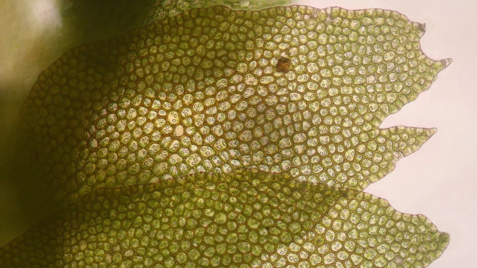
Telophase is the final stage of mitosis and of each meiotic division, when the separated chromosomes arrive at opposite poles of the cell, decondense back into chromatin, and new nuclear envelopes form around each set, creating two distinct nuclei. This stage directly reverses the events of prophase, which is why it is often described as "prophase in reverse." During telophase, the cell prepares to finish dividing its cytoplasm, a separate process called cytokinesis. The cell is now exiting mitosis and transitioning back into interphase, the normal state of a cell between divisions [1].
Key Takeaways
- Telophase reverses the events of prophase, rebuilding nuclei around the separated chromosomes.
- The mitotic spindle disassembles, and the chromosomes decondense into chromatin.
- New nuclear envelopes and nucleoli form, restoring the two nuclei of the daughter cells.
- Telophase is distinct from cytokinesis, which is the division of the cytoplasm that often overlaps with it.
- In meiosis, telophase I and telophase II differ in chromosome number and whether sister chromatids are separated.
What Happens in Telophase: Reversing Prophase
To understand telophase, it helps to recall prophase, the first stage of mitosis. In prophase, the cell condenses its chromatin into visible chromosomes, breaks down the nuclear envelope, and begins to build the mitotic spindle. Telophase is essentially the opposite. It is the cell's way of cleaning up after chromosome segregation and restoring the normal structure of the nucleus [1]. The key events of telophase are:
- Chromosomes arrive at the poles: The two sets of chromosomes, one for each daughter cell, have been pulled to opposite ends of the cell by the spindle fibers.
- Chromosomes decondense: The tightly packed chromosomes begin to unwind and spread out, returning to the diffuse form of chromatin. This makes them less visible under a light microscope.
- Nuclear envelope reforms: Membranes derived from the endoplasmic reticulum coat the surface of each chromosome set, fusing to create a new nuclear envelope around each group [3][4].
- Nucleoli reappear: The nucleolus, the site of ribosome production, reforms within each new nucleus.
- Spindle disassembles: The microtubules of the mitotic spindle break down, and their building blocks are recycled for use in the new cells.
The Molecular Machinery of Mitotic Exit
The physical changes you see in telophase are driven by molecular changes inside the cell. This transition from mitosis back to interphase is called mitotic exit. During mitosis, a group of enzymes called cyclin-dependent kinases (CDKs) are highly active. They add phosphate groups to many proteins, which drives events like chromosome condensation and nuclear envelope breakdown. For the cell to exit mitosis, this CDK activity must fall [2].
As CDK activity drops, opposing enzymes called protein phosphatases become relatively more active. These phosphatases remove the phosphate groups that the CDKs added. This removal reverses the mitotic state, allowing chromosomes to decondense and the nuclear envelope to reform [2]. The key phosphatases in animal cells are called PP1 and PP2A [2]. This coordinated process of kinase inactivation and phosphatase activation ensures that the cell does not try to rebuild the nucleus while the chromosomes are still being moved. To understand how CDKs are regulated throughout the cell cycle, you can review our guide on cyclin-dependent kinases.
Nuclear Envelope Reformation: A Detailed Look
The reformation of the nuclear envelope is a complex process that is critical for the cell to function properly. In cells that undergo open mitosis, the nuclear envelope is completely dismantled during prophase. During telophase, it must be rebuilt from scratch around each set of chromosomes. This process is tightly coordinated with the chromosomes themselves [3].
The process begins when proteins that are destined for the nuclear envelope bind to the surface of the condensed chromosomes. These proteins then recruit membrane from the endoplasmic reticulum, which is the cell's extensive membrane system. This membrane spreads out to coat the chromosomes [3][4]. As the membrane pieces fuse, they form a new nuclear envelope. The final step is a sealing process called annular fusion, which closes any remaining holes in the envelope. This step is carried out by a protein complex called ESCRT-III, which is the same machinery used to pinch off the connection between two cells during cytokinesis [4]. This shows how the processes of nuclear division and cytoplasmic division are linked at a molecular level.
Telophase vs. Cytokinesis: Two Separate Processes
Telophase and cytokinesis are often mentioned together, but they are distinct processes. Telophase is the final stage of karyokinesis, which is the division of the nucleus. Cytokinesis is the division of the cytoplasm, which includes the organelles and other cellular contents. In most animal and plant cells, cytokinesis begins during late anaphase or telophase, but it is a separate step in the cell cycle [1].
| Feature | Telophase | Cytokinesis |
|---|---|---|
| What is divided | The nucleus | The cytoplasm |
| Main event | Nuclear envelope reforms, chromosomes decondense | Cell membrane pinches in (animals) or cell plate forms (plants) |
| Result | Two distinct nuclei within one cell | Two completely separate daughter cells |
| Timing | Final stage of mitosis | Overlaps with telophase, but finishes after it |
It is possible for mitosis to happen without cytokinesis. In such cases, the cell will have multiple nuclei and is called a multinucleate cell [1]. However, in normal cell division, cytokinesis cannot happen without a preceding nuclear division. The cell must first ensure that the genetic material is correctly divided before it splits the cytoplasm.
Telophase 1 and Telophase 2 in Meiosis
Telophase also occurs during meiosis, the process that produces gametes (sex cells). Meiosis has two rounds of division, so it has two telophase stages: telophase I and telophase II. While they share the basic features of any telophase, they differ in what they accomplish.
- Telophase I follows the separation of homologous chromosomes. Homologous chromosomes are pairs of chromosomes, one from each parent, that carry the same genes. In anaphase I, these pairs are pulled apart. So, at telophase I, each new nucleus contains one chromosome from each homologous pair. The chromosomes may still consist of two sister chromatids. The cell then divides, and the resulting cells are haploid, meaning they have half the number of chromosomes as the original cell.
- Telophase II follows the separation of sister chromatids. Sister chromatids are the two identical copies of a single chromosome that are joined at the centromere. In anaphase II, these sister chromatids are finally pulled apart. At telophase II, each new nucleus contains a single set of chromosomes, each consisting of just one chromatid. This completes the reduction in chromosome number, producing four haploid daughter cells.
In both cases, the nuclear envelope reforms and the chromosomes decondense, just as in mitosis. For a deeper comparison of the two processes, see our guide on mitosis vs meiosis. You can also review the full sequence of events in our stages of meiosis guide.
Telophase in the Bigger Picture
Telophase is the final step in a carefully orchestrated process that ensures genetic stability. By rebuilding the nucleus around each set of chromosomes, telophase ensures that each daughter cell receives a complete and correct copy of the genetic information. This is essential for the survival of the cell and the organism. The process is so fundamental that it is conserved across many species, from yeast to humans. Understanding telophase is therefore a key part of understanding cell biology. You can review the entire process of mitosis in our phases of mitosis guide, or start with the basics in our Cell Biology 101 guide. The events of telophase are also relevant to understanding the nucleus function in a cell.
Frequently Asked Questions
What happens in the telophase of mitosis?
In the telophase of mitosis, the two sets of chromosomes have reached opposite poles of the cell. The chromosomes begin to decondense, unwinding from their tightly packed mitotic form back into the diffuse chromatin of interphase. New nuclear envelopes form around each set of chromosomes, and the nucleoli reappear. The mitotic spindle, which was responsible for moving the chromosomes, disassembles. This process restores two distinct nuclei within the cell, preparing it for cytokinesis [1].
What are four things that happen during telophase?
Four key events occur during telophase. First, the chromosomes arrive at the poles and begin to decondense into chromatin. Second, a new nuclear envelope reforms around each set of chromosomes, using membrane from the endoplasmic reticulum [3]. Third, the nucleoli, which are the sites of ribosome production, reappear within the new nuclei. Fourth, the mitotic spindle disassembles, breaking down the microtubules that were used for chromosome movement.
Why is telophase important?
Telophase is important because it completes the process of nuclear division. It ensures that each daughter cell receives a fully formed, functional nucleus with the correct genetic material. By rebuilding the nuclear envelope, telophase re-establishes the compartmentalization between the nucleus and the cytoplasm, which is essential for normal cellular functions like gene expression and RNA processing. Without telophase, the cell would have two sets of chromosomes in a single, shared cytoplasm, which would disrupt normal cell function.
How does cytokinesis relate to telophase?
Cytokinesis is the division of the cytoplasm, while telophase is the division of the nucleus. They are separate processes but are closely coordinated. Cytokinesis often begins during late anaphase or telophase, after the chromosomes have started to move to the poles. While telophase rebuilds the nuclei, cytokinesis pinches the cell in two to create two separate daughter cells. In normal cells, cytokinesis depends on the successful completion of nuclear division, but mitosis can occur without cytokinesis, resulting in multinucleate cells [1].
What is the difference between telophase 1 and telophase 2?
Telophase I and telophase II are the final stages of the two divisions in meiosis. Telophase I follows the separation of homologous chromosomes, so each new nucleus contains one chromosome from each pair, but the chromosomes still have two sister chromatids. Telophase II follows the separation of sister chromatids, so each new nucleus contains chromosomes with only a single chromatid. The result is that telophase I produces two haploid cells, while telophase II produces four haploid cells.
References
- Clark MA, Douglas M, Choi J (2018). 10.2 The Cell Cycle. Biology 2e, OpenStax.
- Wurzenberger C, Gerlich DW (2011). Phosphatases: providing safe passage through mitotic exit. Nature Reviews Molecular Cell Biology 12:469-482.
- LaJoie D, Ullman KS (2017). Coordinated events of nuclear assembly. Current Opinion in Cell Biology 46:39-45.
- Olmos Y, Hodgson L, Mantell J et al. (2015). ESCRT-III controls nuclear envelope reformation. Nature 522:236-239.