Sister Chromatid vs Chromosome: Key Differences
By Dr. Zubair Khalid, DVM, MS, PhD ·
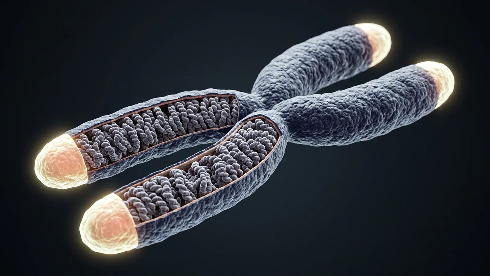
A chromosome is the complete DNA-protein structure that carries part of a cell's genetic information, while a chromatid is one of the two identical DNA-protein copies that a chromosome carries after it has been replicated. Sister chromatids are the two copies produced by replication of a single chromosome, held together at the centromere by a ring-shaped protein complex called cohesin until the cell divides.
This distinction matters because most confusion in genetics comes from mixing up a structure with a state. A chromosome does not stop being a chromosome when it copies itself. It simply changes state, from a chromosome with one chromatid to a chromosome with two sister chromatids. Students who miss that point misread karyotypes, misjudge DNA content on exam questions, and misunderstand why meiosis produces four haploid cells while mitosis produces two diploid ones.
The Core Definitions


The National Human Genome Research Institute defines a chromatid as one of the two identical halves of a replicated chromosome, with the two halves joined at a region called the centromere [1]. That definition contains the whole concept in one sentence.
A chromosome is the entire thread-like structure of DNA wrapped around histone proteins, plus the associated non-histone proteins that package it. Humans have 46 chromosomes in a typical somatic cell, arranged as 23 pairs. Each chromosome carries a specific set of genes, and the physical arrangement of those genes along the chromosome is what geneticists call a linkage map.
A chromatid is one copy of that chromosome after DNA replication. Before replication, the chromosome exists as a single chromatid. After replication, it exists as two chromatids, each containing one complete double helix of the original DNA sequence.
Sister chromatids are the two chromatids of one replicated chromosome. They are identical in sequence because they were produced by semiconservative replication of the same template. They are not identical to the chromatids of the homologous chromosome that came from the other parent, which may carry different alleles at many loci.
The word "sister" is doing real work here. It signals shared origin from one chromosome, not shared origin from one parent. Homologous chromosomes are "homologs" or "partners," and they come from two parents. Sister chromatids come from one chromosome and one replication event.
Why This Distinction Matters
Cell division is the process that distributes genetic material to daughter cells, and every error in that process traces back to how chromatids and chromosomes are handled. Mitosis separates sister chromatids so that each daughter cell receives one complete copy of every chromosome. Meiosis separates homologous chromosomes in the first division and sister chromatids in the second, cutting the chromosome number in half and shuffling alleles between homologs.
Cohesin sits at the center of this process. Cohesin is a ring-shaped protein complex that topologically entraps DNA. During DNA replication, cohesin rings that were already loaded onto the template DNA become co-entrapped around both newly synthesized sister chromatids, which is how cohesion is established [2]. A reconstitution study using purified budding yeast proteins showed that cohesin rings remain DNA bound through complete DNA synthesis, and that some rings encircle both replication products while others end up embracing only one, pointing to a two-step capture mechanism [2].
That molecular detail has clinical weight. Cohesin variants are linked to Cornelia de Lange syndrome, Roberts syndrome, premature ovarian insufficiency, non-obstructive azoospermia, aneuploidy, and several cancers [3]. When cohesion fails, chromosomes mis-segregate, and mis-segregation is the cellular event behind miscarriage, congenital aneuploidy, and much of reproductive aging [4].
The Comparison Table
| Feature | Chromosome | Chromatid | Sister Chromatid |
|---|---|---|---|
| Definition | Complete DNA-protein structure carrying genetic information | One of the two identical copies of a replicated chromosome | Either one of the two chromatids joined at the centromere of a single replicated chromosome |
| DNA molecules | One before S phase, two after S phase | One | One each, two total per replicated chromosome |
| Number in a human G1 somatic cell | 46 | 46 | 0 (no sisters exist yet) |
| Number in a human G2 somatic cell | 46 | 92 | 46 pairs |
| Joined by cohesin | Not applicable before replication | No, once separated | Yes, along arms and at pericentromeric regions |
| Stage where it is the dominant term | Throughout the cell cycle | After sister separation until the next S phase | S phase through metaphase of mitosis or meiosis II |
| Separates from its partner | Not applicable | Yes, at anaphase of mitosis or anaphase II of meiosis | Yes, at anaphase of mitosis or anaphase II of meiosis |
| Homolog relationship | Pairs with its homolog | Not applicable | Not a homolog, an identical copy |
| Centromere | One per chromosome | One per chromatid | One shared centromere per pair |
The table captures the single most important idea. Chromosome and chromatid are not two different molecules. They are two descriptions of the same DNA-protein thread at different points in the cell cycle.
How a Chromosome Becomes Two Sister Chromatids
Step 1: G1 Phase, One Chromosome, One Chromatid
In G1, before DNA replication, each chromosome is a single DNA double helix packaged with histones. At this stage, "chromosome" and "chromatid" describe the same object, and most biologists simply say chromosome. Cohesin rings are already loaded onto the DNA at this point, positioned along the chromosome arms and enriched near the centromere.
Step 2: S Phase, Replication Creates Sisters
During S phase, the DNA replication machinery copies each chromosome. The replisome, the molecular machine that synthesizes DNA, travels along the template. As it passes, cohesin rings that were loaded on the template can become co-entrapped around both daughter duplexes, establishing sister chromatid cohesion [2]. Additional cohesin is recruited to chromatin during replication as a second route to cohesion [2].
The result is a replicated chromosome with two sister chromatids, each carrying one full copy of the original sequence. The two sisters remain physically linked.
Step 3: Cohesin Locks the Sisters Together
Cohesin is built around an SMC1-SMC3-RAD21 ring, and the ring topology is what holds the sisters [5]. Several accessory proteins regulate the ring. Pds5 is a cohesin subunit required for complex stability on chromatin, and it controls cohesin ATPase activity through the Eco1-Smc3 acetylation pathway [6]. Sororin acts as a direct structural lock on the DNA exit gate of cohesin, engaging the RAD21-SMC3 interface to keep the ring closed [5]. Acetylation of the Smc3 subunit by Eco1 is a separate regulatory layer, and cohesin's ATPase activity and its acetylation state control cohesion and loop architecture through distinct mechanisms [7].
Two pools of cohesin exist in replicated cells. One pool performs loop extrusion, folding DNA into loops that organize the genome. The other pool performs cohesion, physically tethering sister chromatids [8]. These pools share core subunits but associate with different regulatory subunits and interact with chromosomes in fundamentally different ways [8].
Step 4: Mitosis, Condensation and Alignment
As the cell enters mitosis, chromosomes condense. The two sister chromatids become visible under a microscope as parallel rods joined at a primary constriction, the centromere. Kinetochores assemble at the centromere of each sister and attach to spindle microtubules. Bipolar attachment, meaning each sister connects to the opposite spindle pole, is monitored by the spindle assembly checkpoint.
Pericentric cohesion is what makes bipolar attachment possible. Cohesin at the pericentromeric region holds the sisters together so that the two kinetochores can face opposite poles, and this arrangement prevents aneuploidy [9].
Step 5: Anaphase, Sisters Separate
At anaphase, the protease separase cleaves the RAD21 subunit of cohesin, the ring opens, and the sisters are pulled to opposite poles. Each sister is now an independent chromosome in a daughter cell. The chromosome count per cell stays at 46, but the chromatid count per cell drops from 92 to 46.
How Sister Chromatids Behave in Meiosis
Meiosis is the specialized division that produces gametes. It has two rounds of chromosome segregation after a single round of DNA replication.
Meiosis I: Homologs Separate, Sisters Stay Together
In meiosis I, homologous chromosomes pair, recombine, and then separate. Sister chromatids remain attached. This is the reductional division, and it is what halves the chromosome number.
Meiosis uses specialized cohesin complexes. Instead of the canonical SMC1α, SMC3, RAD21, and STAG1/2 combination used in mitosis, germ cells deploy SMC1β, REC8, RAD21L, and STAG3 [10]. REC8 provides the primary replication-coupled cohesion that maintains chromosome axis integrity, while RAD21L supports homolog pairing and recombination [4]. These meiosis-specific cohesins form the chromosome axis and organize chromatin loops, providing the framework for homolog recognition, synapsis, and crossover control [10].
The Rec8 cohesin complex is required both for pairing and recombination of homologous chromosomes and for sister chromatid cohesion [11]. A fission yeast study identified a rec8 mutant that lost the ability to assemble axis-loop chromatin structure without losing sister chromatid cohesion, and it showed reduced meiotic recombination, which separates the two functions [11]. A companion mis4 mutant reproduced the same phenotype, and the interaction surface between Mis4 and Rec8 turned out to be the critical interface [11].
At centromeres, meiotic cohesin complexes cooperate with shugoshin and PP2A to protect cohesion during meiosis I, which is what allows the reductional division to occur while sisters stay linked [4].
Meiosis II: Sisters Separate
In meiosis II, the cell divides again, and this time sister chromatids separate. The result is four haploid cells, each with 23 chromosomes, each consisting of a single chromatid.
The stepwise release of cohesion across the two divisions is a defining feature of meiosis. Premature separation of sister chromatids during meiotic prophase causes aneuploid gametes and inviable zygotes [12]. In C. elegans, loss of the deubiquitinase DUO-1 leads to impaired assembly of the synaptonemal complex, loss of meiotic chromosome axis integrity, ineffective homolog pairing, and premature sister chromatid separation [12].
What Cohesin Actually Does Beyond Holding Sisters Together
Cohesin has a second job in genome maintenance. It organizes the genome during interphase by extruding chromatin loops, and those loops have key roles in gene regulation [13]. Recent work shows that cohesin-mediated looping is closely linked to repair of DNA double-strand breaks [13].
Cohesin also guides homology search during DNA repair. A high-resolution method called sister-pore-C mapped intra- and intermolecular interactions in replicated chromosomes and showed that two functionally distinct pools of cohesin act at double-strand breaks [14]. Loop-extruding cohesin accumulates across megabase-scale domains surrounding the break to control local homology sampling, while cohesive cohesin concentrates at the break site to tether the broken DNA end to the sister chromatid [14]. This restricts the search space and helps preserve genomic integrity.
Arm cohesion biases DNA repair toward the sister chromatid rather than the homolog, which prevents loss of heterozygosity [9]. Cohesin also suppresses unequal sister chromatid exchange between repetitive sequences. In yeast, the rate of unequal sister chromatid exchange between repeats 4 kilobases apart was 15 times higher than between repeats 68 kilobases apart, and the distal rate increased 4 to 7 fold in mutants with reduced arm cohesion [9]. Higher densities of cohesion sites confine repair to local sequences [9].
How Scientists Observe Chromatids and Chromosomes
Karyotyping
A karyotype is a photograph of a cell's condensed chromosomes arranged in pairs by size and banding pattern. Karyotyping is done on cells arrested in metaphase, when sister chromatids are still paired. Each visible chromosome in a normal human metaphase spread therefore shows two chromatids joined at the centromere.
Sister Chromatid Cohesion Assays
Sister chromatid cohesion defects are diagnosed by examining chromosome shape after depletion of a target protein. A 2026 study trained object detection models to automate this classification, and a YOLOv8-based model reached 89.40 percent concordance with manual analysis and successfully distinguished wild-type cells from DDX11-knockout cells [15]. This matters because manual shape classification is time-consuming and subject to individual interpretation [15].
Chromosome Conformation Capture
Methods that map physical contacts between DNA regions reveal how cohesin organizes replicated chromosomes. Polymer modeling combined with inter-chromatid contact maps in budding yeast showed that extruding and cohesive cohesins are sparsely distributed, producing mildly compacted and loosely aligned sister chromatids [16]. The same analysis suggested that cohesion is asymmetric, favoring tethering between non-homologous cohesin-enriched regions [16].
Fluorescence Microscopy
Labeled probes against specific DNA sequences, combined with immunofluorescence against cohesin subunits, let researchers watch cohesion in living or fixed cells. Time-lapse imaging of meiosis has been used to track the stepwise loss of cohesion across meiosis I and meiosis II.
Clinical and Comparative Relevance
Cohesin variants cause a spectrum of human disorders. These include Cornelia de Lange syndrome, Roberts syndrome, cancer, neuropsychiatric disease, premature ovarian insufficiency, reproductive aging, aneuploidy, and non-obstructive azoospermia [3]. Identifying cohesin mutations is a priority for precision diagnostics [3].
A striking feature of mammalian oocytes is the extraordinary stability of meiotic cohesin. It must persist from fetal stages until adulthood without efficient turnover, which underpins female reproductive longevity [4]. Progressive loss of cohesin integrity with age contributes to aneuploidy, infertility, and congenital disorders [10]. This is one of the clearest examples of how a molecular detail about sister chromatid cohesion translates directly into clinical reproductive medicine.
The same principles apply across eukaryotes with variation in detail. Rice meiosis operates under a homologous-recombination-dominant double-strand break repair regime that is independent of KU-mediated non-homologous end joining, in contrast to mitosis where KU is essential [17]. That finding shows that the balance between repair pathways differs between mitotic and meiotic divisions, and it reinforces why sister chromatid cohesion is regulated differently in the two contexts.
Common Mistakes and Limitations
Treating chromatid and chromosome as different molecules. They are the same DNA-protein thread described at different cell cycle stages. A chromosome with one chromatid is still a chromosome. A chromosome with two sister chromatids is also still one chromosome.
Saying a replicated chromosome has two chromosomes. It does not. It has one chromosome and two chromatids. The chromosome count per cell does not change when DNA replicates. The chromatid count does.
Confusing sister chromatids with homologous chromosomes. Sister chromatids are identical copies from one parent chromosome. Homologs are similar but not identical chromosomes from two parents. Sister chromatids separate in mitosis and meiosis II. Homologs separate in meiosis I.
Assuming cohesion is uniform along the chromosome. Cohesion is enriched at pericentromeric regions and distributed along arms at varying density, and the density affects how far a broken DNA end can search for a repair template [9]. Cohesion is also asymmetric, favoring tethering between non-homologous cohesin-enriched regions in budding yeast [16].
Ignoring the distinction between loop extrusion and cohesion. These are two separable activities carried out by two distinct cohesin pools [8]. A cohesin mutant can lose cohesion while retaining positioned loops, and vice versa [7]. Treating cohesin as a single-function complex leads to wrong predictions.
Overgeneralizing from model organisms. Much of the mechanistic work on cohesion establishment comes from budding yeast [2], and much of the work on meiotic cohesin comes from yeast, worms, and mice [11][12][10]. The core principles are conserved, but subunit composition and regulation differ between organisms and between mitosis and meiosis.
Forgetting that individual cases vary. Any clinical question about a cohesin variant, a reproductive diagnosis, or a suspected chromosomal abnormality needs evaluation by a qualified clinician. This article explains the biology, not the diagnosis.
Quick Review
- A chromosome is the whole DNA-protein structure. A chromatid is one of the two identical copies present after replication.
- Sister chromatids are the two copies of one replicated chromosome, joined at the centromere by cohesin.
- A chromosome with one chromatid becomes a chromosome with two sister chromatids after S phase. The chromosome count does not change. The chromatid count doubles.
- Cohesin is a ring-shaped complex that topologically entraps both sister chromatids during replication [2]. Sororin locks the DNA exit gate [5], and Pds5 controls cohesin ATPase activity through the Eco1-Smc3 acetylation pathway [6].
- Sister chromatids separate in mitosis and in meiosis II. Homologous chromosomes pair and separate in meiosis I.
- Meiosis uses specialized cohesin subunits including REC8, RAD21L, SMC1β, and STAG3 [10][4].
- "Chromatid versus chromosome" describes a state, not a different molecule.
Frequently Asked Questions
What is the difference between a chromosome and a chromatid?
A chromosome is the full DNA-protein structure that carries genetic information. A chromatid is one of the two identical copies of that structure after DNA replication. The chromosome count stays the same after replication, while the chromatid count doubles.
How many chromatids are in a human chromosome after replication?
Two. Each replicated human chromosome consists of two sister chromatids joined at a single centromere, so a human somatic cell in G2 has 46 chromosomes and 92 chromatids.
What holds sister chromatids together?
Cohesin, a ring-shaped protein complex that topologically entraps both sister chromatids. Sororin locks the ring's DNA exit gate, and Pds5 regulates the complex through the Eco1-Smc3 acetylation pathway [6][5].
Do sister chromatids separate in mitosis or meiosis?
Both. Sister chromatids separate at anaphase of mitosis and at anaphase II of meiosis. In meiosis I, homologous chromosomes separate while sister chromatids stay together.
Are sister chromatids identical?
Yes, in sequence. They are produced by semiconservative replication of one template chromosome, so they carry the same alleles. Homologous chromosomes are not identical because they come from two different parents.
Is a chromatid a chromosome?
A chromatid is one copy of a chromosome, so the same physical thread can be called a chromatid or a chromosome depending on context. The two terms describe states of the same structure, not two different molecules.
Related Articles
- Epigenetics vs Epigenesis: Key Differences Explained
- Point Mutation vs Missense: Key Differences Explained
- Epigenetics vs Genetics: Key Differences and Interactions
- Operon Inducer vs Activator: Key Differences Explained
- Epigenetics vs Mutation: Key Differences and Mechanisms
- Small RNA vs microRNA: Key Differences Explained
- Animal vs Plant Cell: Key Differences Compared
- In Vitro vs In Vivo: Key Differences Explained
Sources
- Chromatid
- Biochemical reconstitution of sister chromatid cohesion establishment during DNA replication.
- Cohesin variants associated with human reproductive and developmental disorders.
- Meiosis-Specific Cohesin in Mammalian Germ Cells.
- Sororin locks the DNA-exit gate of cohesin to preserve sister-chromatid cohesion.
- Pds5 regulates sister chromatid cohesion by controlling cohesin ATPase activity through the Eco1-Smc3 acetylation pathway.
- Cohesin acetylation and ATPase activity control cohesion and loop architecture through distinct mechanisms.
- Organization of replicated chromosomes by DNA loops and sister chromatid cohesion.
- Cohesin promotes genomic stability by suppressing unequal sister chromatid exchange.
- Chromosome Architecture Defined by the Meiosis-Specific Cohesin in Mammalian Germ Cells.
- Interaction Between Rec8 and Mis4 Is Required for Axis-Loop Chromatin Formation and Homologous Chromosome Recombination During Meiosis.
- Active maintenance of meiosis-specific chromosome structures in Caenorhabditis elegans by the deubiquitinase DUO-1.
- Folding a broken genome: the versatile roles of cohesin in genome maintenance.
- Cohesin guides homology search during DNA repair using loops and sister chromatid linkages.
- Detection and classification of chromosomes with sister chromatid cohesion defects using object detection models.
- Modeling the spatial organization of replicated chromosomes in yeast reveals a loose asymmetric cohesion between sister chromatids.
- The telomere-binding protein KU is required for DSB repair in rice mitosis but not in meiosis.