Metaphase: Chromosome Alignment and the Spindle Checkpoint
By Dr. Zubair Khalid, DVM, MS, PhD ·
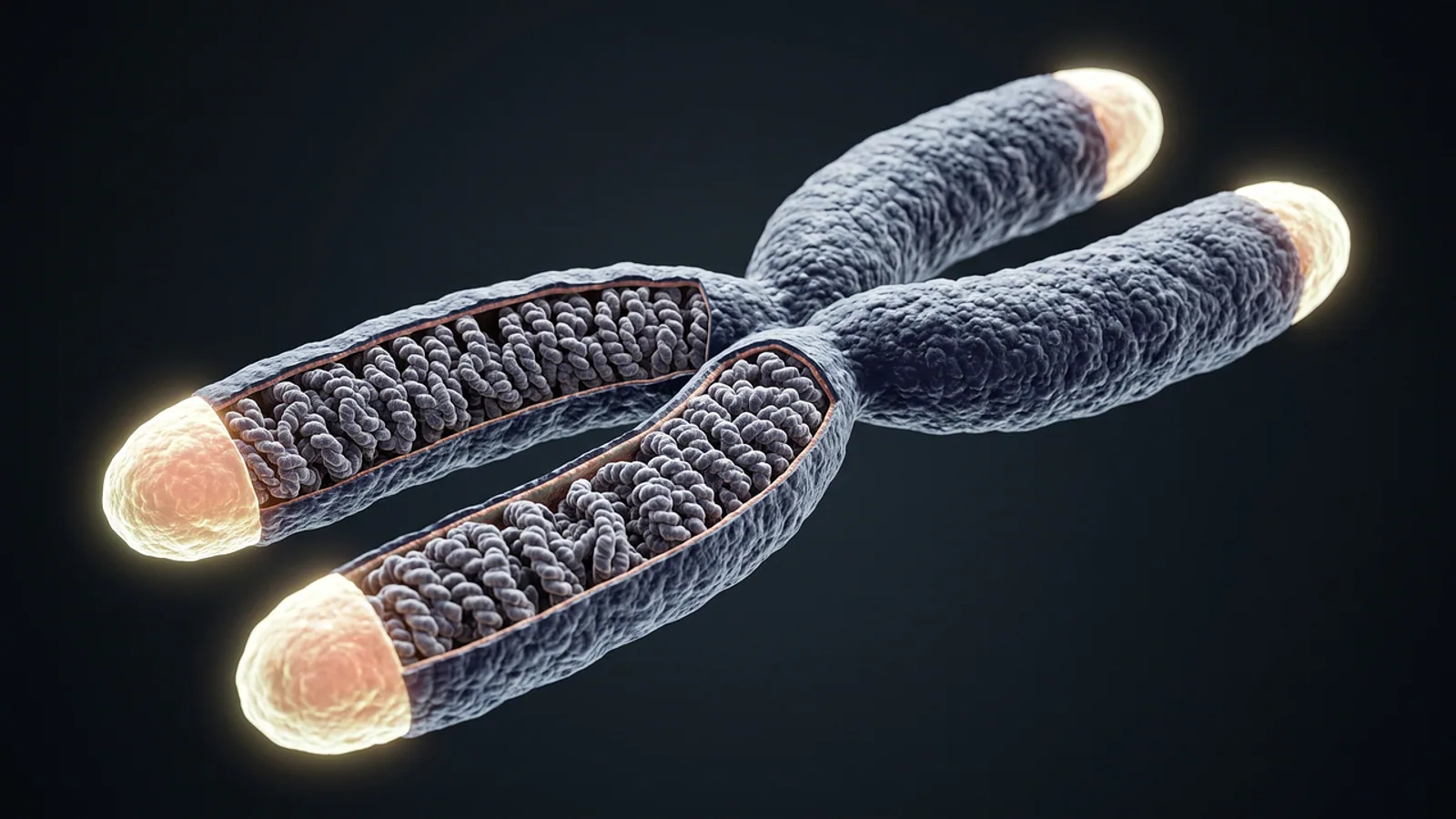
Metaphase is the stage of cell division when duplicated chromosomes line up along the middle of the cell, called the metaphase plate, before they are pulled apart. This alignment ensures that each new daughter cell receives one complete copy of the genetic material. During metaphase, the cell’s spindle fibers attach to the chromosomes and hold them in place under tension. The cell verifies that every chromosome is correctly attached before it proceeds to anaphase. This careful positioning is a defining event in both mitosis and meiosis.
Key Takeaways
- Metaphase follows prometaphase in mitosis, where chromosomes align at the cell’s equator.
- The metaphase plate is an imaginary plane at the cell’s center, not a physical structure.
- Kinetochores are protein complexes on the centromere that connect chromosomes to spindle microtubules.
- The spindle assembly checkpoint delays anaphase until every kinetochore is properly attached to spindle fibers.
- Metaphase I of meiosis aligns homologous pairs, while metaphase II aligns individual chromosomes like mitosis.
What Is the Metaphase Plate?
The metaphase plate is the imaginary plane located equidistant from the two spindle poles. It is not a real cellular structure, but rather a geometric reference point. During metaphase, chromosomes are moved by spindle microtubules until their centromeres rest on this plane. The arms of the chromosomes may extend to either side, but the centromeres are aligned.
The position of the metaphase plate is determined by the balance of forces from the two poles. Microtubules from opposite poles pull on each chromosome. When the pulling forces are equal, the chromosome settles at the center. This balanced tension is critical for accurate chromosome segregation. If the forces are unequal, the chromosome would be pulled toward one pole prematurely. The cell monitors this tension as part of its quality control.
Kinetochores and Spindle Microtubules
Each sister chromatid has a kinetochore, a protein structure that forms at the centromere. The kinetochore serves as the attachment point for spindle microtubules [1]. Microtubules from opposite poles attach to the kinetochores of sister chromatids. One sister chromatid is connected to the top pole, and the other is connected to the bottom pole. This arrangement is called bi-orientation.
| Feature | Kinetochore | Centromere |
|---|---|---|
| Location | On the surface of the centromere | Region of DNA on the chromosome |
| Composition | Proteins | DNA and associated proteins |
| Function | Binds spindle microtubules | Holds sister chromatids together |
| Role in metaphase | Attachment point for pulling forces | Site where kinetochores assemble |
The attachment of microtubules to kinetochores is dynamic. Microtubules grow and shrink, and they can detach and reattach. This dynamic behavior allows the cell to correct errors. If a microtubule attaches to the wrong pole, the tension will be abnormal. The cell can detect this and release the incorrect attachment. Proper attachment creates tension that stabilizes the connection. The kinetochore is the central player in this process, linking the chromosome to the spindle machinery [2].
The Spindle Assembly Checkpoint
The spindle assembly checkpoint (SAC) is a surveillance mechanism that ensures chromosome segregation is accurate [2]. The SAC prevents the cell from entering anaphase until all chromosomes are correctly attached to the spindle. It monitors the state of kinetochore attachment and tension. If any kinetochore is unattached or improperly attached, the checkpoint sends a "wait" signal.
The SAC works through a series of protein signals. Unattached kinetochores generate an inhibitory signal that blocks the activity of the anaphase-promoting complex. This complex is responsible for triggering the separation of sister chromatids. Once every kinetochore is properly attached, the inhibitory signal stops. The anaphase-promoting complex becomes active, and the cell proceeds to anaphase [3].
This checkpoint is essential for preventing aneuploidy, which is an abnormal number of chromosomes. Errors in chromosome segregation can lead to conditions like cancer or developmental disorders. The SAC is a safeguard that gives the cell time to fix mistakes. It is a key reason why metaphase can last longer than other stages if problems are detected. The cell prioritizes accuracy over speed.
Metaphase in Mitosis vs. Meiosis
Metaphase occurs in both mitosis and meiosis, but the arrangement of chromosomes differs. In mitosis, individual chromosomes line up at the metaphase plate. Each chromosome consists of two sister chromatids. The sister chromatids are pulled apart in anaphase. This process produces two genetically identical daughter cells.
In meiosis, there are two rounds of division, so metaphase occurs twice. Metaphase I and metaphase II have distinct arrangements.
| Stage | What lines up | What separates in anaphase | Result |
|---|---|---|---|
| Mitotic metaphase | Individual chromosomes | Sister chromatids | Two identical cells |
| Meiosis metaphase I | Homologous chromosome pairs | Homologous chromosomes | Two cells with half the chromosome number |
| Meiosis metaphase II | Individual chromosomes | Sister chromatids | Four genetically distinct cells |
In metaphase I of meiosis, homologous chromosomes pair up. Each pair, called a tetrad, lines up at the metaphase plate. The orientation of each pair is random. This is the basis of independent assortment. In anaphase I, the homologous chromosomes are separated, not the sister chromatids. This reduces the chromosome number by half.
Metaphase II is more similar to mitotic metaphase. Individual chromosomes, each with two sister chromatids, line up at the plate. In anaphase II, the sister chromatids are finally separated. The key difference is that the cells entering metaphase II are haploid. For a detailed comparison, see our Mitosis vs Meiosis guide.
Why Are Karyotypes Prepared from Metaphase Cells?
A karyotype is a photograph of an individual’s chromosomes arranged in pairs. Karyotypes are prepared from cells in metaphase because chromosomes are most condensed at this stage. During interphase, chromosomes are spread out as chromatin and are not visible as distinct structures. As the cell enters mitosis, the chromosomes condense. By metaphase, they are at their maximum condensation level.
This condensation makes individual chromosomes visible under a light microscope. The characteristic banding patterns on each chromosome can be seen. These patterns allow cytogeneticists to identify each chromosome pair. They can also detect structural abnormalities, such as deletions or duplications. The alignment at the metaphase plate is not relevant for the karyotype, but the condensation state is.
To prepare a karyotype, cells are stimulated to divide and then stopped at metaphase. A chemical called colchicine is often used to arrest the cells in metaphase. The cells are then stained and photographed. The chromosomes are cut out of the photograph and arranged in pairs. This technique is used to diagnose genetic disorders. You can learn more about reading these images in our guide on What Is a Karyotype.
The Role of Metaphase in the Cell Cycle
Metaphase is part of the mitotic phase, which follows interphase [1]. During interphase, the cell grows, replicates its DNA, and prepares for division. The mitotic phase includes mitosis and cytokinesis. Mitosis itself is divided into prophase, prometaphase, metaphase, anaphase, and telophase. Metaphase is the stage where the cell confirms that everything is ready for separation.
The transition from metaphase to anaphase is a major commitment point. Once the cell passes this checkpoint, it cannot reverse the process. The sister chromatids will be separated, and the cell will divide. This is why the spindle assembly checkpoint is so important. It is the last chance to correct errors before the irreversible step of segregation. The cell uses the time in metaphase to ensure that every chromosome is properly positioned. This process is part of the larger regulation of the cell cycle, which you can explore in our Cell Biology 101 guide.
Frequently Asked Questions
What happens in metaphase?
In metaphase, chromosomes are moved by spindle microtubules until their centromeres align at the metaphase plate. Each chromosome’s sister chromatids are attached to microtubules from opposite poles. The spindle assembly checkpoint verifies that all attachments are correct. Once the checkpoint is satisfied, the cell is cleared to enter anaphase. The entire process ensures that each daughter cell will receive an identical set of chromosomes.
What is a kinetochore?
A kinetochore is a protein complex that assembles on the centromere of a chromosome. It is the physical site where spindle microtubules attach to the chromosome. Each sister chromatid has its own kinetochore. The kinetochore also plays a role in signaling to the spindle assembly checkpoint. It can detect whether it is properly attached to microtubules and send signals to delay anaphase if it is not.
What is the metaphase plate?
The metaphase plate is an imaginary plane located at the equator of the cell, equidistant from the two spindle poles. It is not a physical structure. Chromosomes align their centromeres on this plane during metaphase. The position of the plate is determined by the balance of pulling forces from the spindle microtubules. This alignment ensures that the sister chromatids are positioned to be pulled apart evenly in anaphase.
What is the difference between metaphase 1 and metaphase 2?
Metaphase I occurs during the first division of meiosis. Homologous chromosomes, each made of two sister chromatids, pair up and align at the metaphase plate. In anaphase I, the homologous chromosomes separate. Metaphase II occurs during the second division. Individual chromosomes, not pairs, align at the plate. In anaphase II, sister chromatids separate. Metaphase II is similar to mitotic metaphase, but the cells are haploid.
Why are chromosomes easiest to see in metaphase?
Chromosomes are easiest to see in metaphase because they are at their highest level of condensation. During interphase, the DNA is loosely packed as chromatin. As mitosis begins, the chromosomes condense. By metaphase, they are short, thick, and visible under a light microscope. This condensation, combined with the alignment at the metaphase plate, makes them ideal for counting and analysis. This is why karyotypes are prepared from metaphase cells.
References
- Clark MA, Douglas M, Choi J (2018). 10.2 The Cell Cycle. Biology 2e, OpenStax.
- Musacchio A, Salmon ED (2007). The spindle-assembly checkpoint in space and time. Nature Reviews Molecular Cell Biology 8:379-393.
- Musacchio A (2015). The Molecular Biology of Spindle Assembly Checkpoint Signaling Dynamics. Current Biology 25:R1002-R1018.