# Pronation vs Supination: Definitions and Examples
Pronation is rotation of a limb segment about its own longitudinal axis so that the palmar or plantar surface turns downward or medially. Supination is rotation about the same longitudinal axis so that the palmar or plantar surface turns upward or laterally.
Those two sentences are the whole core of the topic, and almost every error students make comes from forgetting the second half of each definition. Pronation and supination are rotations. They are not flexion, not extension, not abduction, and not adduction. A limb can be fully flexed and still be pronated, and a limb can be fully extended and still be supinated. The rotation happens around an axis that runs lengthwise through the bone or bone pair, which is why the surface facing the ground or the midline is the landmark that defines the movement.
This matters in veterinary practice because pronation and supination are used constantly in imaging, orthopedic examination, surgical planning, and rehabilitation. Elbow congruity in dogs changes measurably when the antebrachium is rotated into pronation or supination, and computed tomography measurements of radioulnar incongruity shift by double-digit percentages at rotation angles that are easy to produce by hand [1]. A radiograph or CT scan taken in the wrong rotational position can make a normal joint look abnormal or hide a real problem. Understanding the definitions prevents that class of error.
## The Defining Feature: Rotation About a Longitudinal Axis
### What "longitudinal axis" means
A longitudinal axis is an imaginary line running from the proximal end of a bone or bone segment to its distal end, roughly parallel to the shaft. Rotation about that axis is called axial rotation or simply rotation. When a limb segment rotates around this line, the segment does not swing forward, backward, inward, or outward. It twists in place.
The practical test is simple. If the distal end of the segment moves through space in an arc, that is flexion, extension, abduction, or adduction. If the distal end stays roughly where it is and the segment turns like a key in a lock, that is axial rotation. Pronation and supination are the two named directions of axial rotation in the antebrachium, the carpus, the tarsus, and the pes.
### Why the palmar and plantar surfaces are the reference
In quadrupeds, the standard anatomical position has the palmar surface of the forepaw and the plantar surface of the hindpaw facing the ground. That gives anatomists a stable reference surface. Rotation that brings this surface toward the ground, or toward the midline in a limb held away from the body, is pronation. Rotation that brings it away from the ground, or away from the midline, is supination.
This surface-based definition is more useful than a bone-based definition in comparative anatomy because the bones differ so much between species. A goat has a fused radius and ulna with a single metacarpal bone, while a dog has two separate antebrachial bones and five metacarpals [2]. The bones are not comparable, but the palmar surface is.
## Pronation in Detail
Pronation is the rotation that turns the palmar or plantar surface down or medially. In the classic human demonstration, the arm hangs at the side with the elbow flexed to 90 degrees and the palm facing forward. Rotating the palm to face the floor is pronation. The radius crosses over the ulna in the process, which is why the movement is sometimes described as "turning the thumb inward."
Two features define pronation in any species:
1. The reference surface (palmar or plantar) rotates toward the ground or toward the body's midline.
2. The rotation occurs around the longitudinal axis of the segment, not around a transverse or craniocaudal axis.
In the dog, the pronator teres and pronator quadratus muscles produce this rotation, and the movement is limited by the anatomy of the radioulnar joints. In the cat, the same muscles act on a more mobile antebrachium, which is why cats can rotate the paw further than dogs can.
## Supination in Detail
Supination is the rotation that turns the palmar or plantar surface up or laterally. In the same human demonstration, rotating the palm from facing the floor back to facing the ceiling is supination. The radius and ulna uncross and return to parallel.
The supinator muscle and the biceps brachii produce supination in species that have meaningful forearm rotation. The biceps is a powerful supinator when the elbow is flexed, which is why supination strength is greatest in that position in humans [3]. The same principle applies in animals with mobile antebrachia, though the magnitude is smaller.
Supination is also the position that exposes the dorsal surface of the paw and the cranial surface of the antebrachium. In imaging, this matters because the dorsal and palmar surfaces of the carpus have different soft tissue and bone profiles.
## The Comparison Table
| Joint or segment | Axis of rotation | Pronation direction | Supination direction | Species notes |
|--|--|--|--|--|
| Radioulnar (proximal and distal) | Longitudinal axis of the radius and ulna | Radius crosses over ulna, palmar surface turns down or medially | Radius and ulna uncross, palmar surface turns up or laterally | Full range in primates. Limited in dogs. Moderate in cats [4][5] |
| Antebrachium as a whole | Longitudinal axis through the forearm | Paw rotates so palmar surface faces down or medially | Paw rotates so palmar surface faces up or laterally | Rotation depends on radioulnar joint mobility [1] |
| Carpus | Longitudinal axis through the carpus | Carpal bones rotate so palmar surface turns down or medially | Carpal bones rotate so palmar surface turns up or laterally | Often confused with carpal flexion and abduction |
| Tarsus | Longitudinal axis through the tarsus | Tarsal bones rotate so plantar surface turns down or medially | Tarsal bones rotate so plantar surface turns up or laterally | Rotation is small in most domestic species |
| Pes (whole foot) | Longitudinal axis through the pes | Plantar surface turns down or medially | Plantar surface turns up or laterally | Rotation is coupled with tarsal and digital movement |
## The Common Confusion: Carpal and Tarsal Rotation Terms
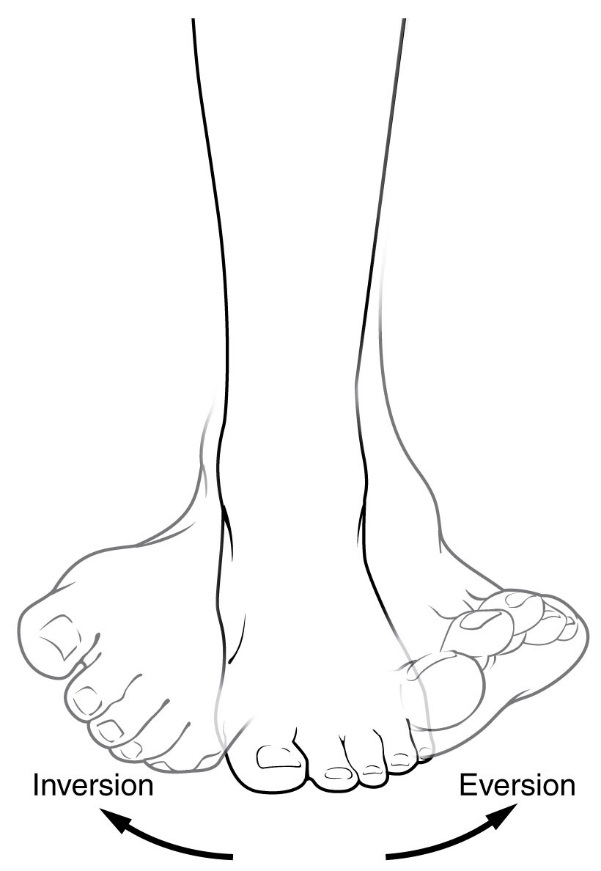
In the foot, pronation and supination are often described as eversion and inversion, a common source of confusion. Image: Connexions, CC BY 3.0, via Wikimedia Commons.
The terms pronation and supination are used in two different ways in veterinary anatomy, and this is the single biggest source of student confusion.
### Use 1: Radioulnar pronation and supination
This is the classic definition. The radius rotates around the ulna (or the ulna rotates around the radius, depending on the species and the fixation point). The movement happens at the proximal and distal radioulnar joints. In humans and other primates, the range is large, typically around 150 to 180 degrees of total rotation. In dogs, the range is much smaller because the radius and ulna are less mobile relative to each other. In cats, the range is intermediate, and the anatomy of the distal radioulnar ligament allows more rotation than in dogs [5].
### Use 2: Carpal and tarsal pronation and supination
In this usage, the terms describe rotation of the carpus or tarsus around its own longitudinal axis, often as part of a composite movement that includes flexion and abduction. A carpus that is pronated has its palmar surface turned medially. A carpus that is supinated has its palmar surface turned laterally. This is not the same as radioulnar rotation, even though the same words are used.
The confusion arises because in a standing animal, radioulnar rotation and carpal rotation are coupled. When the antebrachium pronates, the carpus follows, and the paw turns with it. A student watching a dog turn its paw inward may attribute the whole movement to the carpus when most of it is happening at the radioulnar joints, or vice versa.
### How to keep them separate
The fix is to name the joint. Write "radioulnar pronation" or "carpal pronation" rather than just "pronation." When reading a clinical report, check which joint the author means. When writing your own notes, always specify the segment.
A second fix is to remember that radioulnar rotation moves the radius relative to the ulna, while carpal rotation moves the carpus relative to the antebrachium. These are different mechanical events even when they happen together.
## The Classic Example: Radioulnar Pronation and Supination in Primates
The primate forearm is the textbook example because the radius and ulna are separate, mobile, and connected by a well-developed interosseous membrane. The proximal radioulnar joint is a pivot joint in which the radial head rotates within the radial notch of the ulna. The distal radioulnar joint is a second pivot joint in which the ulnar head rotates within the sigmoid notch of the radius.
In this arrangement, pronation and supination are pure axial rotations. The hand can be turned palm down or palm up without changing the angle of the elbow. The total range in a healthy adult human is roughly 150 to 180 degrees, and the movement is used in almost every manual task.
The clinical importance of this arrangement is well documented. In patients with lateral elbow pain, pronation and supination torques are significantly reduced in the affected limb compared with the sound limb, and the deficit is largest at extreme rotation positions [3]. In patients with Parkinson's disease, the speed, acceleration, and frequency of forearm pronation and supination correlate with gait and posture scores, and the correlation is strongest when the movement is performed with the non-dominant hand [6]. These findings exist because the primate radioulnar joint is a precise, measurable, and clinically informative structure.
## Pronation and Supination in Dogs and Cats
### Dogs: limited antebrachial rotation
In dogs, the radius and ulna are separate bones, but they are less mobile relative to each other than in primates. The interosseous space is narrow and extends the length of the bones, and the radioulnar joints are shallower [2]. The result is that dogs have a limited range of radioulnar pronation and supination. The movement is present, but it is measured in tens of degrees rather than in the 150-degree range seen in humans.
This limitation has direct clinical consequences. When a dog's elbow is imaged with computed tomography, the rotational position of the antebrachium changes the measured radioulnar incongruity. In a cadaveric study of 20 thoracic limbs from medium-sized dogs, radioulnar incongruity measured at the apex of the medial coronoid process deviated by 18.14% at 15 degrees of pronation or supination and by 31.31% at 35 degrees [1]. At the base of the coronoid process, 15 degrees of pronation produced no significant difference, but 15 degrees of supination did [1]. The practical message is that limb position must be controlled during CT evaluation of the elbow, and the rotational angle must be recorded.
A separate study of 101 elbow joints with arthroscopically confirmed medial coronoid disease and 20 sound control joints found that dogs with medial coronoid disease had decreased lateral, laterocentral, and central radioulnar ratios, indicating radioulnar joint malformation in the transverse plane [7]. The medial sections showed no difference, which means the malformation is lateral and central rather than medial. This is a structural finding, not a rotational one, but it shows how closely radioulnar geometry and elbow disease are linked in dogs.
### Cats: intermediate rotation
Cats have more antebrachial rotation than dogs. The distal radioulnar ligament in cats is triangular and extends between the dorsal surface of the distal radius and ulna, with a less extensive interosseous component than in dogs and cheetahs [5]. This anatomical difference is thought to account for the increased rotation of the feline antebrachium.
The range is measurable. In a cadaveric study of 58 feline thoracic limbs mounted with the elbow and carpus flexed at 90 degrees, the mean angle of rotation without any surgical alteration was 129.5 degrees, with a standard deviation of 15.9 degrees [4]. When a radioulnar synostosis was simulated with a 2 mm cortical screw placed through the radius into the ulna, the mean angle dropped to 37.5 degrees, with a standard deviation of 14.5 degrees, and the reduction was statistically significant [4]. The position of the screw (proximal, middle, or distal radial diaphysis) did not change the result [4]. This study shows two things: cats have substantial antebrachial rotation, and any radioulnar synostosis, regardless of location, reduces it by roughly 70 percent.
### Why the difference matters
The difference between dogs and cats is not just a comparative anatomy curiosity. It affects how each species compensates for forelimb injury, how much rotational stress the carpus and elbow absorb during normal activity, and how much rotational correction is possible in surgical planning. A dog with a radioulnar problem has less rotational reserve than a cat with the same problem.
## How Pronation and Supination Are Tested and Observed
### Physical examination
In a conscious animal, pronation and supination are assessed by holding the limb proximal to the joint being tested and gently rotating the distal segment. The examiner watches the orientation of the palmar or plantar surface. The endpoint is reached when the surface has turned as far as it will go without resistance or signs of pain. The range is recorded in degrees from the neutral position.
In dogs, the examination is usually performed with the elbow flexed to about 90 degrees, which relaxes the collateral ligaments and allows the radioulnar joints to rotate more freely. In cats, the same position is used, and the greater range is immediately apparent.
### Imaging
Computed tomography is the standard method for measuring radioulnar incongruity, and the rotational position of the limb must be standardized. A custom fixation device is used to hold the limb at a known angle, and scans are repeated at several pronation and supination angles [1]. Without this standardization, measurements are not comparable between scans or between patients.
The effect of rotation on imaging is not trivial. In the canine elbow study, the deviation at 35 degrees of rotation was more than 30 percent at the apex of the medial coronoid process [1]. A clinician who does not control rotation could misdiagnose incongruity or miss it entirely.
### Biomechanical testing
In cadaveric studies, pronation and supination are measured with the limb mounted in a jig that fixes the elbow and carpus at known angles. A controlled rotational force is applied to the metacarpus, and the resulting angle is recorded. This is how the feline rotation range of 129.5 degrees was established [4]. The same method is used to test the effect of surgical implants, synostoses, and ligament repairs.
### Gait and posture assessment
In humans, pronation and supination of the forearm are used as a proxy for gait and posture stability. A study of 24 patients with idiopathic Parkinson's disease found that the frequency of forearm pronation and supination contributed to about one third of the best-performing regression models for predicting gait and posture scores, and that speed and acceleration also contributed significantly [6]. The correlation was strongest when the movement was performed with the non-dominant hand [6]. This is a human application, but it illustrates the principle that forearm rotation is a sensitive indicator of motor control.
## Comparative and Clinical Relevance
### Elbow congruity and disease
The relationship between radioulnar rotation and elbow congruity is one of the most clinically important applications of these definitions. In dogs, the radioulnar joint is a load-bearing structure, and small changes in rotational position change the contact geometry between the radius and ulna. The CT study of 20 cadaveric limbs showed that incongruity measurements at the medial coronoid apex vary by up to 31.31 percent depending on rotation [1]. The study of 101 elbows with medial coronoid disease showed that affected joints have a different radioulnar ratio than sound joints, with the difference concentrated in the lateral and central regions [7]. Together, these findings suggest that rotational position and radioulnar geometry are both relevant to elbow disease, and that both must be assessed.
### Nerve alignment
The deep branch of the radial nerve passes through the supinator muscle at the superior arcade. A high-resolution ultrasound study of 110 nerves from 55 asymptomatic participants found that the angle of the nerve at this arcade changes significantly between maximal pronation and maximal supination, with a mean difference of about 7 degrees [8]. This is a human study, but the anatomical principle applies to any species with a supinator muscle and a radial nerve passing through it. The nerve is not fixed in place. It moves with forearm rotation, and that movement can affect nerve function and imaging.
### Ligament tension
The anterior bundle of the medial collateral ligament of the elbow changes length with forearm rotation. In a study of six healthy volunteers, the medial and middle parts of the ligament decreased in length from pronation to supination, both with and without axial load [9]. The lateral ulnar collateral ligament remained nearly static [9]. This means that pronation increases tension on the medial collateral ligament, which is relevant to elbow stability and to the interpretation of stress radiographs.
### Surgical planning
Radioulnar synostosis is a surgical procedure that fuses the radius to the ulna to eliminate painful or dysfunctional rotation. It is used in humans for conditions such as congenital radioulnar synostosis and brachial plexus birth injury. In a series of 21 children with brachial plexus birth injury and complete pronation palsy, radioulnar synostosis using a vascularized radial periosteal flap fixed the forearm in 10 to 20 degrees of pronation, and the procedure was followed by radiographic union and parental satisfaction [10]. A separate series of 20 children used a vascularized ulnar periosteal flap and fixed the forearm in slight pronation [11]. These procedures work precisely because pronation and supination are axial rotations at a defined joint. Fusing the joint eliminates the rotation.
### Prosthetic replacement
When the distal radioulnar joint is destroyed by disease or trauma, prosthetic arthroplasty can restore rotation. A systematic review of 43 case series found that both semi-constrained bipolar prostheses and unconstrained ulnar head replacements produced statistically significant and clinically meaningful improvements in function, including range of pronation and supination [12]. The weighted mean long-term survival was 89 percent for the bipolar prosthesis and 87 percent for ulnar head replacement [12]. These outcomes are measured in degrees of rotation, which is only possible because pronation and supination are defined as measurable axial movements.
## Clinical Relevance, Limitations and Common Mistakes
The clinical relevance of pronation and supination is broad. They are measured in orthopedic examinations, controlled in imaging, altered in surgery, and used as indicators of neurological function. The limitations are equally important. Range of motion varies with species, breed, age, and individual anatomy. A single measurement in a single position does not capture the full picture. Imaging findings depend on limb position, and a scan taken at one rotational angle may not be comparable to a scan taken at another. Surgical outcomes depend on the underlying condition and on the skill of the surgeon.
The most common mistakes are definitional. Students confuse pronation with flexion, supination with abduction, and radioulnar rotation with carpal rotation. They forget that the palmar or plantar surface is the reference and that the axis is longitudinal. They assume that dogs have the same forearm rotation as humans, when the actual range is much smaller. They assume that cats have the same range as dogs, when cats have more. They read a clinical report that says "pronation" without checking which joint is meant.
A second class of mistakes is procedural. Clinicians who do not control rotational position during CT imaging may get measurements that are not comparable between patients or between visits. Clinicians who test pronation and supination without stabilizing the proximal limb may get false endpoints. Clinicians who interpret nerve or ligament findings without considering forearm rotation may miss the effect of position on the structure being examined.
Individual cases require veterinary assessment. This article is educational and is not a substitute for veterinary diagnosis or treatment.
## Quick Review
1. Pronation turns the palmar or plantar surface down or medially. Supination turns it up or laterally.
2. Both are rotations about a longitudinal axis, not flexion, extension, abduction, or adduction.
3. The classic example is radioulnar pronation and supination in primates, where the radius and ulna are separate and mobile.
4. Dogs have limited antebrachial rotation because the radius and ulna are less mobile relative to each other.
5. Cats have more rotation than dogs, with a mean of 129.5 degrees in a cadaveric study, and any radioulnar synostosis reduces it to about 37.5 degrees [4].
6. Carpal and tarsal pronation and supination are different from radioulnar rotation, even though the same words are used. Always name the joint.
7. Rotational position changes imaging measurements. In dogs, radioulnar incongruity at the medial coronoid apex deviates by up to 31.31 percent at 35 degrees of rotation [1].
## Frequently Asked Questions
### What is the difference between pronation and supination?
Pronation turns the palmar or plantar surface down or medially, and supination turns it up or laterally. Both are rotations about a longitudinal axis.
### Is pronation the same as flexion?
No. Flexion decreases the angle between two bones at a joint, while pronation rotates a segment around its own long axis. A limb can be flexed and pronated at the same time.
### Do dogs have pronation and supination?
Yes, but the range is limited. The radius and ulna are separate bones, but they are less mobile relative to each other than in primates [2].
### Do cats have more forearm rotation than dogs?
Yes. Cats have a less extensive interosseous component of the distal radioulnar ligament, which allows more rotation [5]. A cadaveric study measured a mean of 129.5 degrees of rotation in feline forelimbs [4].
### Why does limb position matter in CT scans of the elbow?
Because radioulnar incongruity changes with rotation. In dogs, measurements at the medial coronoid apex deviate by up to 31.31 percent at 35 degrees of pronation or supination [1].
### What is the difference between radioulnar and carpal pronation?
Radioulnar pronation rotates the radius relative to the ulna. Carpal pronation rotates the carpus relative to the antebrachium. They are different joints and different mechanical events.
## Related Articles
- [What Is Research Misconduct? Definitions, Examples, and How to Recognize It](/blog/research-skills/what-is-research-misconduct-definitions-examples-and-how-to-recognize-it)
- [cell biology definitions](/blog/careers/cell-biology-definitions)
- [Operational Definitions in Quantitative Research](/blog/research-skills/operational-definitions-in-quantitative-research-how-to-define-your-variables-for-measurable-outcome)
- [Designing Case Definitions for Veterinary Surveillance](/knowledge/veterinary-medicine/veterinary-epidemiology/designing-case-definitions-veterinary-surveillance)
- [What Are Research Ethics? Core Principles and Definitions](/blog/guides/what-are-research-ethics-core-principles-and-definitions)
- [Poultry Diseases: Definitions, Classifications, and Economic Impact](/knowledge/bacteria/avian-bacteria/poultry-diseases-meaning)
- [Dehydration Synthesis: Definition and Examples](/knowledge/molecular-biology/dehydration-synthesis-definition-and-examples)
- [Mean Square Displacement: Formula and Examples](/blog/research-skills/mean-square-displacement-formula-and-examples)
## Sources
1. [Computed Tomography Assessment of Healthy Elbow Joint Congruity in Dogs Being Affected by Pronation and Supination Angulation: A Cadaveric Study.](https://pubmed.ncbi.nlm.nih.gov/40218315/)
2. [COMPARATIVE MACRO ANATOMY OF FORELIMB BONES OF BLACK BENGAL GOAT AND INDIGENOUS DOG: AN OVER VIEW](https://doi.org/10.54536/ajaset.v3i1.16)
3. [Impact of Lateral Elbow Pain on Pronation and Supination Torques and Influence of Forearm Rotation Angle.](https://pubmed.ncbi.nlm.nih.gov/40829037/)
4. [Effects of simulated radioulnar synostosis on supination and pronation in cats. A cadaveric study.](https://pubmed.ncbi.nlm.nih.gov/38366794/)
5. [Anatomy of the distal radioulnar ligament in cats.](https://pubmed.ncbi.nlm.nih.gov/36779780/)
6. [Hand Pronation-Supination Movement as a Proxy for Remotely Monitoring Gait and Posture Stability in Parkinson's Disease.](https://pubmed.ncbi.nlm.nih.gov/35270972/)
7. [Assessment of the Conformation of the Radioulnar Joint Comparing Dogs with and without Medial Coronoid Disease.](https://pubmed.ncbi.nlm.nih.gov/40889765/)
8. [Deep branch of the radial nerve: effect of pronation/supination on longitudinal nerve alignment.](https://pubmed.ncbi.nlm.nih.gov/37010538/)
9. [In vivo changes in length of elbow collateral ligaments during pronation and supination on an outstretched arm.](https://pubmed.ncbi.nlm.nih.gov/32170363/)
10. [Radioulnar Synostosis Using a Vascularized Pedicled Radial Periosteal Flap for Supination Deformity in Residual Brachial Plexus Birth Injury.](https://pubmed.ncbi.nlm.nih.gov/42568170/)
11. [Forearm Synostosis Using A Vascularized Pedicled Ulnar Periosteal Graft for Supination Deformity in Brachial Plexus Birth Injury.](https://pubmed.ncbi.nlm.nih.gov/40622056/)
12. [Efficacy and safety of prosthetic arthroplasty of the distal radioulnar joint: a systematic review.](https://pubmed.ncbi.nlm.nih.gov/41766119/)