# Cuboid Bone: Anatomy, Function, and Comparative Notes
The cuboid bone is a short, stout tarsal bone of the distal row that sits on the lateral side of the foot, articulating behind with the calcaneus, medially with the navicular (central tarsal), and in front with the lateral metatarsals. It acts as the bony keystone of the lateral longitudinal arch and carries a groove on its plantar surface that redirects the tendon of the fibularis (peroneus) longus muscle across the sole.
Understanding the cuboid matters because the lateral column of the foot transmits a large share of the load during standing and propulsion, and because the fibularis longus tendon wraps directly around this bone. In species as different as the dog, horse, and human, the same basic plan is present but the size, fusion, and joint mobility of the cuboid vary widely. For students, the cuboid is a reliable test of whether you can read a tarsal bone by its surfaces rather than by memorizing a picture.
## Where the Cuboid Sits in the Tarsus
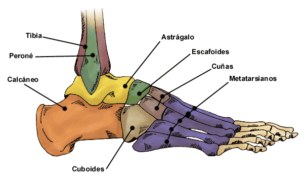
Surface view of the left foot, with the cuboid located on the lateral midfoot between the calcaneus and the fourth and fifth metatarsals. Image: Ryxi10, CC BY-SA 3.0, via Wikimedia Commons.
### The tarsus as three functional columns
The tarsus (the collective bones of the ankle and proximal foot) is organized into a proximal row, an intermediate or central region, and a distal row. The calcaneus and talus form the proximal row. The distal row contains the cuboid and the bones that articulate with the metatarsals. Functionally, anatomists divide the foot into a medial column, a central column, and a lateral column. The cuboid is the defining bone of the lateral column.
### Position and neighbors
The cuboid lies lateral and slightly plantar to the talus. Its posterior surface faces the calcaneus, its medial surface faces the navicular or central tarsal bone, and its anterior surface faces the fourth and fifth metatarsals. Because it sits at the junction of the ankle and the midfoot, the cuboid is loaded from behind by the calcaneus and delivers that load forward into the lateral metatarsals. This position explains why the bone is thick and block-like rather than slender.
### Why the name fits
The term cuboid means "cube-like," and the bone earns the name because it presents roughly six surfaces in a compact block. Not every surface is a true square, but the six-surface convention is the standard way to describe it in comparative anatomy and is the framework used in this article.
## The Six Surfaces of the Cuboid
Describing a short bone by its surfaces is the fastest route to identifying it in a skeleton or on imaging. The cuboid has six named surfaces.
1. **Dorsal surface.** Faces upward toward the skin of the instep. It is roughened for ligament attachment and is the surface most easily seen on a dorsoplantar radiograph.
2. **Plantar surface.** Faces the sole. This surface carries the oblique groove for the fibularis longus tendon and, in many species, a bony prominence called the peroneal tubercle.
3. **Lateral surface.** Faces the outer edge of the foot. It is often narrow and provides attachment for ligaments and, in some species, for part of the plantar fascia.
4. **Medial surface.** Faces the navicular or central tarsal bone and bears a flat articular facet for the cuboideonavicular joint.
5. **Proximal (posterior) surface.** Faces the calcaneus and bears the concave or saddle-shaped facet of the calcaneocuboid joint.
6. **Distal (anterior) surface.** Faces the metatarsals and is divided into facets for the fourth and fifth metatarsals, and in some species for the third.
### The groove for the fibularis longus tendon
The most functionally important feature of the cuboid is the groove on its plantar surface. The fibularis longus muscle arises on the lateral side of the crus, its tendon passes behind the lateral malleolus, and then turns sharply beneath the foot. The cuboid groove acts as a pulley: it holds the tendon against the bone and changes the direction of pull from vertical to horizontal. This redirection lets the muscle support the transverse arch of the foot and, in species with a mobile lateral column, helps control the midfoot during push-off.
### The peroneal tubercle
The peroneal tubercle is a small bony ridge or projection on the lateral or plantar aspect of the cuboid, near the groove. It increases the mechanical advantage of the tendon by deepening the pulley and provides an attachment point for the plantar ligaments and the short plantar calcaneocuboid ligament. In humans the tubercle is a well-known landmark on the lateral wall of the foot. In domestic species it is present but varies in prominence.
## Articulations of the Cuboid
The cuboid participates in three named joint complexes. Each has a different role in load transfer and mobility.
### Calcaneocuboid joint
The calcaneocuboid joint is the articulation between the proximal surface of the cuboid and the distal articular surface of the calcaneus. It is part of the transverse tarsal (midtarsal) joint complex. In species with a mobile midfoot, this joint allows a small amount of gliding and rotation that lets the foot adapt to uneven ground. In species with a rigid, cursorial foot, the joint is stabilized by strong plantar and dorsal ligaments and permits little motion.
Subchondral bone density studies show that the pattern of loading across the calcaneocuboid joint differs with locomotor mode. In primates that use a mobile midfoot, peak compressive loads concentrate dorsally at the calcaneocuboid interface during the midtarsal break of support, while human calcanei show a different, less localized density pattern consistent with a rigid, weight-bearing foot [1]. This is a useful reminder that joint surfaces are shaped by habitual loading, not just by phylogeny.
### Cuboideonavicular joint
The cuboideonavicular joint connects the medial surface of the cuboid to the navicular or central tarsal bone. It is a small plane joint reinforced by dorsal, plantar, and interosseous ligaments. In the dog and cat, the corresponding articulation is between the cuboid and the central tarsal bone. In the horse, the cuboid articulates with the central and third tarsal bones. The joint contributes to the transverse arch and allows limited gliding during loading.
### Cuboideometatarsal articulations
The distal surface of the cuboid articulates with the lateral metatarsals. In the dog, the cuboid articulates with the fourth and fifth metatarsals. In the horse, it articulates with the fourth metatarsal, and in some individuals with the third. In humans, the cuboid articulates with the fourth and fifth metatarsals. These joints are plane joints supported by dorsal, plantar, and interosseous ligaments, and they allow only slight movement under normal conditions.
### Summary table of articulations and relations
| Surface | Articulates with | Joint name | Notes |
|--|--|--|--|
| Proximal | Calcaneus | Calcaneocuboid | Part of transverse tarsal complex |
| Medial | Navicular or central tarsal | Cuboideonavicular | Reinforced by interosseous ligaments |
| Distal | Lateral metatarsals | Cuboideometatarsal | Plane joints, minimal motion |
| Plantar | Fibularis longus tendon | Not a joint | Groove acts as a pulley |
| Lateral | Soft tissues | Not a joint | Peroneal tubercle and ligament attachments |
| Dorsal | Soft tissues | Not a joint | Ligament attachment, palpable landmark |
## Function of the Cuboid
### Keystone of the lateral longitudinal arch
The lateral longitudinal arch runs from the calcaneus, through the cuboid, to the lateral metatarsals. The cuboid is the keystone of this arch: its wedge shape and its position between the calcaneus behind and the metatarsals in front mean that compressive load is transferred through it rather than around it. When the arch is loaded, the cuboid is compressed between the calcaneus and the metatarsals, and the plantar ligaments resist the tendency of the arch to flatten.
### Load transfer and shock absorption
During standing, the cuboid transmits body weight from the calcaneus to the lateral metatarsals. During locomotion, it also absorbs and redistributes impact. In cursorial species, the lateral column is stiffened so that energy is stored and returned efficiently. In arboreal or climbing species, the same column is more mobile so the foot can conform to branches. Comparative work on caviomorph rodents shows exactly this split: arboreal species have tarsal-metatarsal features that permit movement at multiple levels of the complex, while cursorial species have features that stabilize joints, interlock the tarsal bones, and restrict motion to the parasagittal plane [2].
### Tendon redirection
The fibularis longus tendon relies on the cuboid groove to change direction. Without a bony pulley, the tendon would bowstring away from the foot and lose mechanical efficiency. The groove and the peroneal tubercle together keep the tendon in place and allow it to act as a dynamic support for the transverse arch and as a stabilizer of the lateral column.
## How the Cuboid Is Observed and Tested
### Gross anatomy and radiography
In the anatomy laboratory, the cuboid is identified by its six surfaces and by the groove on its plantar aspect. On standard dorsoplantar and lateral radiographs, the cuboid is superimposed on the other distal tarsal bones, so it is best evaluated on oblique views. The calcaneocuboid joint space is visible on a lateral projection, and the cuboideometatarsal joints are seen on dorsoplantar views.
### Cross-sectional imaging
Computed tomography (CT) and magnetic resonance imaging (MRI) provide detailed views of the cuboid and its joints. In a study of the normal tarsal joint of a Bengal tiger, CT with bone and soft tissue window settings allowed differentiation of cortical, subchondral, and trabecular bone, while MRI with T1-weighted and STIR sequences showed the ligaments, muscles, and tendons around the tarsus [3]. The same principles apply in dogs, cats, and horses: CT is best for bone detail and MRI is best for the soft tissue structures that cross the cuboid.
### Subchondral bone density mapping
Computed tomography osteoabsorptiometry is a technique that maps the density of subchondral bone as a proxy for long-term joint loading. It has been used to compare the talus across Canidae species and to study the calcaneocuboid joint across primates [4][1]. These methods are research tools rather than routine clinical tests, but they show how the cuboid and its neighbors adapt to the loads placed on them.
### Ossification and development
The tarsal bones ossify after birth in most mammals, and the timing differs between species and between sexes. A review of foot ossification found that tarsal bones generally show significant differences in ossification age between sexes, with later and slower ossification in males [5]. In foals, incomplete ossification of the cuboidal bones (the small tarsal bones, including the cuboid) is a recognized radiographic finding and is linked to immature or premature birth [6]. This is why radiographs of the tarsus are often taken in neonatal foals before they are allowed unrestricted exercise.
## Comparative Notes Across Species
### Dog
In the dog, the cuboid is a small distal tarsal bone. It articulates proximally with the calcaneus, medially with the central tarsal bone, and distally with the fourth and fifth metatarsals. In some breeds and individuals, the cuboid is partially or completely fused with the fourth tarsal bone, which can make it difficult to identify as a separate structure on radiographs. The dog's tarsus is adapted for digitigrade locomotion, and the lateral column is relatively rigid compared with the mobile midfoot of primates.
### Cat and other felids
The domestic cat has a tarsal arrangement similar to the dog. In the Bengal tiger, CT and MRI studies have documented the normal appearance of the tarsal joint, including the bones and the soft tissue structures that cross the cuboid [3]. The cuboid in felids is small and block-like, and its groove accommodates the fibularis longus tendon in the same way as in the dog.
### Horse
In the horse, the cuboid is a distinct small tarsal bone in the distal row. It articulates with the calcaneus, the central and third tarsal bones, and the fourth metatarsal. The horse has a highly specialized, digitigrade (technically unguligrade) limb, and the tarsus acts as a major shock-absorbing and propulsive joint. The cuboid contributes to the lateral stability of the hock. In foals, incomplete ossification of the cuboidal bones is a clinically important finding because it predisposes to collapse of the tarsus under load [6].
### Human
In humans, the cuboid is a keystone of the lateral column of the foot. It articulates with the calcaneus behind, the navicular and lateral cuneiform medially, and the fourth and fifth metatarsals in front. The human cuboid is larger and more robust than that of most domestic mammals, reflecting the demands of bipedal weight-bearing. The peroneal tubercle is a consistent landmark on the lateral aspect of the bone, and the groove for the fibularis longus tendon is well developed. The human foot is rigid compared with that of nonhuman primates, and this rigidity is attributed to the propulsive and stability requirements of bipedalism [1].
### Other mammals
Comparative studies across mammals show that the cuboid is a conserved element but varies in shape and mobility. In caviomorph rodents, the tarsal-metatarsal complex ranges from a mobile pattern in arboreal species to a stabilized, interlocking pattern in cursorial species [2]. In the southern giant pouched rat, seven tarsal bones are visible radiographically, and a tarsal sesamoid bone is present in all individuals [7]. In the greater cane rat, the tarsal sesamoid bone is also consistently visualized [8]. These findings illustrate that the cuboid is part of a larger tarsal complex whose organization reflects locomotor behavior.
### Species comparison table
| Feature | Dog | Horse | Human |
|--|--|--|--|
| Size relative to tarsus | Small | Small | Large |
| Fusion with other tarsals | Sometimes fused with fourth tarsal | Distinct | Distinct |
| Articulates with | Calcaneus, central tarsal, metatarsals IV and V | Calcaneus, central and third tarsal, metatarsal IV | Calcaneus, navicular, lateral cuneiform, metatarsals IV and V |
| Fibularis longus groove | Present | Present | Present, well developed |
| Peroneal tubercle | Variable | Variable | Consistent landmark |
| Lateral column mobility | Low | Low | Low (rigid for bipedalism) |
## Clinical Relevance, Limitations and Common Mistakes
The cuboid is not a common site of primary disease in small animals, but it is involved in several important conditions. In foals, incomplete ossification of the cuboidal bones can lead to collapse of the tarsus, and radiographic assessment is used to guide exercise restriction [6]. In dogs, the cuboid can be affected by fractures, luxations, and degenerative joint disease of the tarsus, although these are less common than injuries to the talus and calcaneus. In humans, the calcaneocuboid joint is a well-known site of arthritis and injury, and subchondral bone density patterns there have been studied as a marker of loading history [1].
Students frequently make three mistakes with the cuboid. First, they confuse it with the navicular or central tarsal bone. The cuboid is lateral, the navicular is medial. Second, they assume the cuboid is always a separate bone. In some dogs it is fused with the fourth tarsal, and in some species the distal tarsal bones are partially fused. Third, they overlook the plantar groove and the fibularis longus tendon, which is the single most functionally important feature of the bone.
This article is educational and is not a substitute for veterinary diagnosis or treatment. Individual animals vary, and any lameness or suspected tarsal injury should be evaluated by a veterinarian.
## Quick Review
1. The cuboid is a distal-row tarsal bone on the lateral side of the foot.
2. It has six surfaces: dorsal, plantar, lateral, medial, proximal, and distal.
3. The plantar surface carries the groove for the fibularis longus tendon and often a peroneal tubercle.
4. It articulates with the calcaneus, the navicular or central tarsal, and the lateral metatarsals.
5. It is the keystone of the lateral longitudinal arch and transfers load from the calcaneus to the metatarsals.
6. In dogs it is small and may fuse with the fourth tarsal, in horses it is a distinct small tarsal, and in humans it is a robust keystone of the lateral column.
7. In foals, incomplete ossification of the cuboidal bones is a clinically important radiographic finding.
## Frequently Asked Questions
### What is the cuboid bone?
The cuboid bone is a short, block-like tarsal bone on the lateral side of the foot that articulates with the calcaneus behind and the lateral metatarsals in front.
### What does the cuboid bone do?
It transfers load through the lateral column of the foot, acts as the keystone of the lateral longitudinal arch, and provides a pulley groove for the fibularis longus tendon.
### Is the cuboid bone the same in dogs and humans?
No. In dogs the cuboid is small and may be fused with the fourth tarsal bone, while in humans it is a large, robust bone that is a keystone of the lateral column.
### What is the groove on the cuboid for?
The groove on the plantar surface holds the tendon of the fibularis longus muscle and redirects its pull across the sole of the foot.
### What is the peroneal tubercle?
The peroneal tubercle is a small bony prominence on the lateral or plantar aspect of the cuboid that deepens the tendon groove and anchors plantar ligaments.
### Why is cuboidal bone ossification important in foals?
Incomplete ossification of the cuboidal bones in foals can lead to collapse of the tarsus under load, so radiographic assessment is used to guide exercise restriction.
## Related Articles
- [Comparative Anatomy of the Mammalian Kidney](/knowledge/veterinary-medicine/veterinary-anatomy-physiology/comparative-anatomy-mammalian-kidney)
- [Comparative Anatomy of the Mammalian Heart](/knowledge/veterinary-medicine/veterinary-anatomy-physiology/comparative-anatomy-mammalian-heart)
- [Equine Hindlimb Anatomy: Bones, Joints, and Ligaments](/knowledge/veterinary-medicine/veterinary-anatomy-physiology/equine-hindlimb-anatomy-bones-joints-ligaments)
- [Do Frogs Have Bones? Skeletal Anatomy and Pet Care](/knowledge/veterinary-medicine/amphibian-care/do-frogs-have-bones-skeletal-anatomy-and-pet-care)
- [Equine Larynx Anatomy and Function in Respiration](/knowledge/veterinary-medicine/veterinary-anatomy-physiology/equine-larynx-anatomy-function-respiration)
- [Canine Lymphatic System: Anatomy and Function](/knowledge/veterinary-medicine/veterinary-anatomy-physiology/canine-lymphatic-system-anatomy-function)
## Sources
1. [Apparent density of the primate calcaneo-cuboid joint and its association with locomotor mode, foot posture, and the "midtarsal break".](https://pubmed.ncbi.nlm.nih.gov/19918985/)
2. [The tarsal-metatarsal complex of caviomorph rodents: Anatomy and functional-adaptive analysis.](https://pubmed.ncbi.nlm.nih.gov/28345764/)
3. [Computed tomography and magnetic resonance imaging study of a normal tarsal joint in a Bengal tiger (Panthera tigris).](https://pubmed.ncbi.nlm.nih.gov/31035987/)
4. [Spatial Subchondral Bone Density Reflecting Joint Loading of the Talus in Different Canidae.](https://pubmed.ncbi.nlm.nih.gov/30965371/)
5. [Resources for innovative learning of anatomy and foot ossification: Graphic design and virtual reality.](https://pubmed.ncbi.nlm.nih.gov/39340794/)
6. [Image:Cuboidal bone hypoplasia, radiograph, foal-Merck Veterinary Manual](https://www.merckvetmanual.com/multimedia/image/cuboidal-bone-hypoplasia-radiograph-foal)
7. [Radiological Anatomy of the Pelvis and Hind Limb of the Southern Giant Pouched Rat (Cricetomys ansorgei).](https://pubmed.ncbi.nlm.nih.gov/42261804/)
8. [Radiological Anatomy of the Pelvis and Pelvic Limb of the Greater Cane Rat (Thryonomys swinderianus).](https://pubmed.ncbi.nlm.nih.gov/38420289/)