Rabbit Haemorrhagic Disease Virus 2 (RHDV2)
By Dr. Zubair Khalid, DVM, MS, PhD ·
Key Takeaways
- RHDV2 is a highly virulent calicivirus causing acute necrotising hepatitis in European rabbits and hares, distinguished from RHDV1 by its broader host range and ability to infect younger animals (as young as 11 days).
- Transmission occurs via faecal-oral, oral-nasal, and conjunctival routes, with significant indirect spread through contaminated fomites, feed, bedding, and insect vectors, and the virus is environmentally stable for months.
- Molecular diagnostics, primarily RT-PCR targeting the VP60 gene, are the gold standard for RHDV2 detection, but results in vaccinated rabbits require careful interpretation due to potential RNA detection for up to 21 days post-vaccination.
- Clinical signs range from peracute death (12-72 hours post-infection) to subacute disease with jaundice, lethargy, and respiratory distress, with gross pathology characterised by hepatomegaly, splenomegaly, and hepatic necrosis.
- Vaccination with inactivated bivalent (RHDV1 + RHDV2) or monovalent RHDV2 vaccines is the primary preventive measure, though breakthrough infections can occur, often with milder clinical signs.
- Serological assays (ELISA) detect anti-RHDV2 antibodies but cannot differentiate between infection and vaccination and are not useful for diagnosing active infections.
Introduction
Rabbit Haemorrhagic Disease Virus 2 (RHDV2) is a highly virulent calicivirus that causes acute, often fatal, necrotising hepatitis in European rabbits (Oryctolagus cuniculus) and several hare species [<a href="#ref-1">1</a>]. First identified in France in 2010, RHDV2 rapidly spread across Europe and other continents, becoming a major threat to domestic and wild lagomorph populations [<a href="#ref-1">1</a>]. Unlike the classical RHDV (RHDV1), RHDV2 affects younger rabbits and exhibits a broader host range [<a href="#ref-1">1</a>, <a href="#ref-2">2</a>]. This article provides a detailed, evidence-based review of RHDV2, with emphasis on molecular diagnostics, pathobiology, and control strategies, drawing exclusively on published peer-reviewed data [1, 2, 3] and standard veterinary texts.
Virology and Genomic Organisation
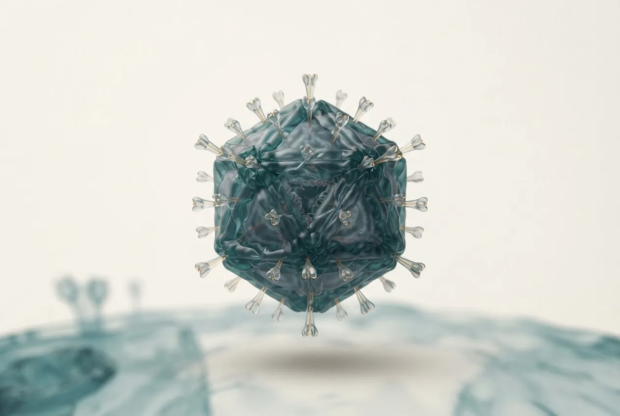
RHDV2 is a non-enveloped, positive-sense single-stranded RNA virus belonging to the family Caliciviridae, genus Lagovirus [<a href="#ref-2">2</a>]. The genome is approximately 7.4 kb and encodes two open reading frames (ORFs). ORF1 produces a polyprotein that is cleaved into non-structural proteins and the major capsid protein VP60, while ORF2 encodes a minor structural protein VP10 [<a href="#ref-3">3</a>]. The VP60 capsid protein is the primary immunogen and the target of both host neutralising antibodies and vaccine-induced immunity [<a href="#ref-3">3</a>]. RHDV2 is antigenically distinct from RHDV1, meaning that vaccines based on RHDV1 VP60 may not provide full cross-protection against RHDV2 [<a href="#ref-2">2</a>, <a href="#ref-3">3</a>]. The virus is highly resistant to environmental inactivation, surviving for months in organic matter and on fomites [<a href="#ref-1">1</a>].
Host Range and Transmission
RHDV2 infects both domestic and wild European rabbits, as well as several hare species (e.g., Lepus europaeus, Lepus timidus) and cottontail rabbits [<a href="#ref-1">1</a>]. Unlike RHDV1, which primarily affects rabbits older than 8 weeks, RHDV2 causes disease in rabbits as young as 11 days, likely due to differences in viral receptor tropism [<a href="#ref-1">1</a>]. Transmission occurs via the faecal-oral, oral-nasal, and conjunctival routes [<a href="#ref-1">1</a>]. Infected rabbits shed virus in faeces, urine, and respiratory secretions before the onset of clinical signs and for several days post-mortem [<a href="#ref-1">1</a>]. Indirect transmission via contaminated feed, bedding, cages, insects (e.g., flies), and human clothing is a significant route of spread [<a href="#ref-1">1</a>]. The incubation period ranges from 1 to 3 days [<a href="#ref-1">1</a>].
Clinical Signs and Pathology
Clinical presentation in RHDV2 ranges from peracute death (often with no premonitory signs) to subacute disease with jaundice, lethargy, anorexia, pyrexia, and respiratory signs (dyspnoea, nasal haemorrhage) [<a href="#ref-1">1</a>]. In peracute cases, rabbits may die within 12 to 72 hours post-infection with minimal clinical signs [<a href="#ref-1">1</a>]. Subacute or chronic infection can last 1 to 2 weeks and is characterised by hepatic necrosis, icterus, and weight loss [<a href="#ref-1">1</a>]. Gross pathological findings include hepatomegaly, splenomegaly, and multifocal hepatic necrosis with diffuse congestion. Histopathology reveals acute necrotising hepatitis with haemorrhage and intravascular fibrin thrombi in multiple organs, consistent with disseminated intravascular coagulation [<a href="#ref-1">1</a>]. Compared to RHDV1, RHDV2 may produce a longer disease course and a higher proportion of subclinical carriers [<a href="#ref-2">2</a>].
Molecular Diagnostics
Accurate and timely diagnosis of RHDV2 is essential for disease control and differentiation from RHDV1 and other causes of acute hepatitis in rabbits. Molecular methods, particularly reverse-transcription polymerase chain reaction (RT-PCR), are the gold standard [<a href="#ref-2">2</a>].
RT-PCR and Real-Time RT-PCR
Conventional RT-PCR targeting the VP60 gene can detect RHDV2 RNA in liver, spleen, blood, and faecal samples [<a href="#ref-2">2</a>]. Real-time RT-PCR (RT-qPCR) using fluorescent probes (e.g., TaqMan) provides higher sensitivity, quantification, and the ability to discriminate RHDV2 from RHDV1 through specific primer-probe sets targeting sequence differences in the capsid gene [<a href="#ref-2">2</a>]. A critical challenge arises in vaccinated rabbits. Commercially available RT-PCR tests may cross-react with vaccine-derived RNA, leading to false-positive results in recently vaccinated animals [<a href="#ref-2">2</a>]. Carvalho et al. demonstrated that detection of RHDV2 RNA in vaccinated rabbits is possible for at least 14 to 21 days post-vaccination, and this window depends on the vaccine type and the target amplicon [<a href="#ref-2">2</a>]. This diagnostic limitation is especially relevant when evaluating outbreaks in vaccinated populations [<a href="#ref-2">2</a>].
Interpretation of PCR Results
The decision tree for interpreting RHDV2 RT-PCR results in the context of vaccination status is illustrated in the Mermaid diagram below.
flowchart TD
 A["Rabbit with clinical signs or died suddenly"] --> B{"Collect liver/spleen or blood"}
 B --> C["Perform RT-qPCR for RHDV2"]
 C --> D{"Positive?"}
 D -->|"Yes"| E{"Vaccination status known?"}
 D -->|"No"| F["RHDV2 unlikely. Consider other pathogens"]
 E -->|"Vaccinated within 21 days"| G["Possible vaccine RNA interference"]
 G --> H{"Confirm with sequencing or differential RHDV1 PCR"}
 E -->|"Not vaccinated or >21 days post-vaccination"| I["Confirmed RHDV2 infection"]
 H --> I
 H --> J["Consider alternative diagnoses if sequencing negative"]
This algorithm emphasises the need to correlate PCR results with vaccination history and, when necessary, confirm with sequencing to avoid diagnostic confusion [<a href="#ref-2">2</a>].
Serological Assays
Serology using enzyme-linked immunosorbent assays (ELISAs) based on recombinant VP60 protein can detect anti-RHDV2 antibodies in serum [<a href="#ref-3">3</a>]. Yang et al. demonstrated that recombinant VP60 expressed in baculovirus induces strong humoral immunity and is used in serological monitoring [<a href="#ref-3">3</a>]. However, serology cannot distinguish between infection and vaccination and is not useful for diagnosing active infections [<a href="#ref-3">3</a>].
Vaccination Strategies
Vaccination remains the cornerstone of prevention. Inactivated vaccines containing RHDV2 antigen, either alone or in combination with RHDV1, are widely used [<a href="#ref-1">1</a>, <a href="#ref-3">3</a>]. The VP60 capsid protein is the primary immunogen; recombinant VP60 vaccines have shown protective efficacy in experimental challenges [<a href="#ref-3">3</a>]. In the UK, an inactivated bivalent vaccine (RHDV1 + RHDV2) is licensed for rabbits from 5 weeks of age [<a href="#ref-1">1</a>]. Vaccination is generally recommended annually, but booster intervals may be shorter in high-risk areas [<a href="#ref-1">1</a>]. Harcourt-Brown et al. reported that vaccinated rabbits in an RHDV2 epidemic had a significantly lower mortality rate than unvaccinated rabbits, although breakthrough infections occurred, likely due to incomplete immunity or waning vaccine-induced protection [<a href="#ref-1">1</a>]. Vaccinated rabbits that become infected often exhibit milder clinical signs, and their reduced viral shedding may contribute to herd immunity [<a href="#ref-1">1</a>].
Challenges in Molecular Diagnosis of Vaccinated Rabbits
As alluded to above, one of the most significant diagnostic challenges is the interference of live-attenuated or inactivated vaccine material in RT-PCR assays used for RHDV2 detection [<a href="#ref-2">2</a>]. Inactivated vaccines may contain residual viral nucleic acid that is detectable by PCR for a variable period after administration [<a href="#ref-2">2</a>]. Carvalho et al. systematically evaluated this issue by testing rabbits after vaccination with different commercial vaccines [<a href="#ref-2">2</a>]. They found that RT-PCR targeting the VP60 gene could amplify vaccine-derived RNA for up to 21 days post-vaccination, depending on the vaccine dose and route [<a href="#ref-2">2</a>]. To mitigate this, diagnostic laboratories must request vaccination history and, if possible, test samples collected more than three weeks post-vaccination [<a href="#ref-2">2</a>]. Alternatively, using RT-qPCR assays that amplify a region of the genome that differs between vaccine strains and field strains (e.g., a hypervariable region of VP60) can improve specificity [<a href="#ref-2">2</a>]. Sequencing of the PCR product remains the definitive method to distinguish field virus from vaccine RNA [<a href="#ref-2">2</a>].
Epidemiological Considerations
RHDV2 has become the dominant RHDV strain in many regions, including the UK, where it has largely replaced RHDV1 [<a href="#ref-1">1</a>]. The virus spreads efficiently in both pet and wild rabbit populations, with outbreaks peaking in the warmer months when insect vector activity is high [<a href="#ref-1">1</a>]. Unlike RHDV1, RHDV2 can infect young rabbits, which has increased the population of susceptible animals and complicated control [<a href="#ref-1">1</a>]. The ability of RHDV2 to survive for months in the environment and to be transmitted via fomites contributes to its persistence [<a href="#ref-1">1</a>]. Biosecurity measures, including quarantine of new rabbits, disinfection with calcium hypochlorite (10%) or 10% sodium hydroxide, and insect control, are critical for outbreak containment [<a href="#ref-1">1</a>].
Frequently Asked Questions
What is the difference between RHDV1 and RHDV2?
RHDV2 is a distinct viral variant that infects younger rabbits and has a broader host range including hares, whereas RHDV1 primarily affects rabbits older than 8 weeks and is largely restricted to O. cuniculus [<a href="#ref-1">1</a>, <a href="#ref-2">2</a>]. Antigenic differences mean that RHDV1-based vaccines do not fully protect against RHDV2 [<a href="#ref-1">1</a>].
How long can a vaccinated rabbit test positive by PCR?
Vaccine-derived RNA can be detected by RT-PCR for up to 21 days post-vaccination [<a href="#ref-2">2</a>]. Therefore, a positive PCR result within this window must be interpreted with caution, and confirmatory sequencing is recommended [<a href="#ref-2">2</a>].
What samples are best for RHDV2 molecular diagnosis?
Liver and spleen tissues from deceased rabbits have the highest viral loads [<a href="#ref-2">2</a>]. Whole blood or serum from live rabbits is also suitable. Faecal samples can be used but may yield lower sensitivity [<a href="#ref-2">2</a>].
Can recombinant VP60 protein be used as a vaccine?
Yes, recombinant VP60 expressed in baculovirus or other systems induces protective immunity and has been used in experimental vaccines [<a href="#ref-3">3</a>]. Some commercial inactivated vaccines also use native VP60 from cell culture [<a href="#ref-1">1</a>].
Is there cross-protection between RHDV1 and RHDV2 vaccines?
Bivalent vaccines containing both RHDV1 and RHDV2 antigens provide the broadest protection [<a href="#ref-1">1</a>]. Monovalent RHDV1 vaccines do not reliably protect against RHDV2 challenge [<a href="#ref-1">1</a>].
How is RHDV2 transmitted in a rabbitry?
Transmission occurs via direct contact with infected rabbits, contaminated fomites (feed, bedding, cages), and mechanical vectors such as flies [<a href="#ref-1">1</a>]. The virus is stable in the environment for months [<a href="#ref-1">1</a>].
What are the key pathological findings in RHDV2?
Post-mortem examination reveals an enlarged, mottled liver, splenomegaly, and pulmonary congestion [<a href="#ref-1">1</a>]. Histology shows severe hepatic necrosis, haemorrhage, and fibrin thrombi [<a href="#ref-1">1</a>].
How can a practitioner differentiate vaccine RNA from field virus in PCR?
Vaccination history is essential. Samples collected more than 21 days post-vaccination are unlikely to contain vaccine RNA [<a href="#ref-2">2</a>]. If doubt persists, sequencing of the VP60 amplicon is required, as vaccine and field strains differ in defined regions of the capsid gene [<a href="#ref-2">2</a>].
Tables
Table 1. Key Diagnostic Features of RHDV2
| Feature | Description |
|---|---|
| Incubation period | 1-3 days [<a href="#ref-1">1</a>] |
| Clinical signs | Peracute death, jaundice, lethargy, anorexia, dyspnoea, nasal haemorrhage [<a href="#ref-1">1</a>] |
| Target organs | Liver, spleen, lung [<a href="#ref-1">1</a>] |
| Sample types for PCR | Liver, spleen, whole blood, faeces [<a href="#ref-2">2</a>] |
| PCR detection window post-vaccination | Up to 21 days [<a href="#ref-2">2</a>] |
| Confirmation method | VP60 sequencing [<a href="#ref-2">2</a>] |
Table 2. Comparison of RHDV1 and RHDV2
| Parameter | RHDV1 | RHDV2 |
|---|---|---|
| Susceptible age | >8 weeks | All ages including young [<a href="#ref-1">1</a>] |
| Host range | O. cuniculus | Rabbits and hares [<a href="#ref-1">1</a>] |
| Vaccine cross-protection | RHDV1 vaccine protects | Requires RHDV2 or bivalent vaccine [<a href="#ref-1">1</a>] |
| PCR cross-reactivity | Limited overlap | Specific primers needed [<a href="#ref-2">2</a>] |