Bluetongue Virus Serotype 8
By Dr. Zubair Khalid, DVM, MS, PhD ·
Key Takeaways
- Bluetongue virus serotype 8 (BTV-8) is an Orbivirus transmitted by Culicoides midges, causing bluetongue disease in ruminants, notably exhibiting severe clinical signs in sheep and cattle, unlike many other serotypes.
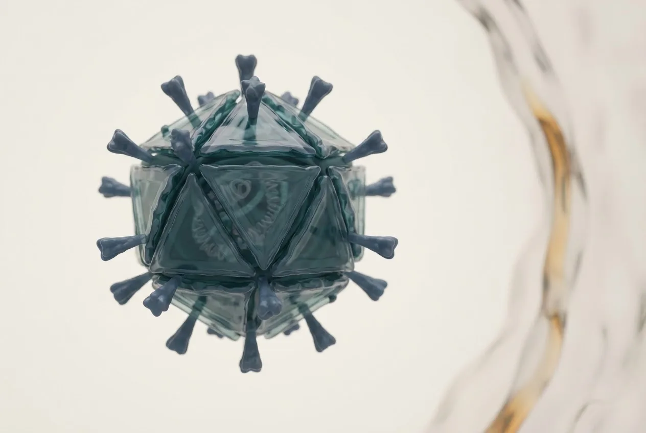
- BTV-8 virions are non-enveloped, icosahedral particles with a dsRNA genome organized into ten segments, with outer capsid proteins VP2 and VP5 mediating cell attachment and entry, and VP2 determining serotype specificity.
- Diagnosis relies on RT-qPCR for rapid viral RNA detection, serotype-specific RT-qPCR targeting Seg-2, virus isolation for confirmation, and serological tests like competitive ELISA for antibody detection.
- Pathogenesis involves viremia and infection of vascular endothelial cells, leading to increased vascular permeability, hemorrhage, and edema, with characteristic clinical signs including fever, oral lesions, and coronitis.
- Control strategies encompass vector control measures, movement restrictions of animals from infected areas, and vaccination using inactivated BTV-8 vaccines to induce neutralizing antibodies.
- The European BTV-8 epizootic of 2006 was characterized by severe disease in cattle and significant teratogenic effects, including congenital malformations, attributed to the specific strain's virulence and the naive immune status of European livestock.
Introduction
Bluetongue virus serotype 8 (BTV-8) is a member of the species Bluetongue virus within the genus Orbivirus, family Sedoreoviridae [<a href="#ref-1">1</a>]. Bluetongue virus (BTV) is the causative agent of bluetongue disease, a non-contagious, arthropod-borne viral infection of domestic and wild ruminants [<a href="#ref-1">1</a>, <a href="#ref-2">2</a>]. BTV-8 gained particular prominence following its emergence in northern Europe in 2006, where it caused a major epizootic with severe clinical disease in both sheep and cattle, a departure from the typical subclinical infection observed in cattle for most other serotypes [<a href="#ref-2">2</a>, <a href="#ref-3">3</a>]. This article provides a detailed reference on BTV-8, covering its virological properties, pathogenesis, diagnostic approaches, and control strategies, with emphasis on the molecular and biophysical mechanisms underlying host-virus interactions.
Taxonomy and Classification
BTV-8 is one of at least 29 recognized serotypes of Bluetongue virus [<a href="#ref-1">1</a>]. Serotype classification is based on the antigenic properties of the outer capsid protein VP2, which is the primary target of neutralizing antibodies [<a href="#ref-1">1</a>, <a href="#ref-4">4</a>]. The virus is classified within the genus Orbivirus, which also includes other arthropod-borne viruses of veterinary importance such as African horse sickness virus and epizootic hemorrhagic disease virus [<a href="#ref-1">1</a>]. BTV-8 belongs to the species Bluetongue virus and is further grouped into topotypes based on genome segment 2 (Seg-2) nucleotide sequences, which correlate with geographic origin [<a href="#ref-4">4</a>]. The European BTV-8 strain that emerged in 2006 is classified within the western topotype and is closely related to strains from sub-Saharan Africa [<a href="#ref-2">2</a>, <a href="#ref-4">4</a>].
Virion Structure and Genome Organization
The BTV-8 virion is a non-enveloped, icosahedral particle approximately 80 nm in diameter [<a href="#ref-1">1</a>]. The capsid is composed of three concentric protein layers: an outer capsid layer, a core layer, and a subcore layer [<a href="#ref-1">1</a>, <a href="#ref-5">5</a>]. The outer capsid consists of two major proteins, VP2 and VP5, which are responsible for cell attachment and membrane penetration, respectively [<a href="#ref-1">1</a>, <a href="#ref-5">5</a>]. VP2 determines serotype specificity and is the primary target of neutralizing antibodies [<a href="#ref-4">4</a>]. The core layer is composed of VP7, which forms the major structural scaffold, while the subcore layer is made of VP3 [<a href="#ref-1">1</a>, <a href="#ref-5">5</a>]. Inside the core, the genome is organized as ten linear double-stranded RNA (dsRNA) segments, designated Seg-1 through Seg-10 [<a href="#ref-1">1</a>, <a href="#ref-5">5</a>]. Each segment encodes one or more viral proteins. The coding assignments are as follows:
| Genome Segment | Encoded Protein(s) | Function |
|---|---|---|
| Seg-1 | VP1 | RNA-dependent RNA polymerase (RdRp) |
| Seg-2 | VP2 | Outer capsid, serotype determinant, cell attachment |
| Seg-3 | VP3 | Subcore scaffold protein |
| Seg-4 | VP4 | Capping enzyme (guanylyltransferase, methyltransferase) |
| Seg-5 | VP5 | Outer capsid, membrane penetration |
| Seg-6 | VP6 | Helicase, RNA unwinding |
| Seg-7 | VP7 | Core surface protein |
| Seg-8 | NS1 | Non-structural, tubule formation, viral factory assembly |
| Seg-9 | NS2 | Non-structural, viral inclusion body matrix protein |
| Seg-10 | NS3/NS3a | Non-structural, virus release via budding or lysis |
The dsRNA genome is fully encapsidated, and the viral RdRp (VP1) synthesizes mRNA transcripts within the core particle [<a href="#ref-1">1</a>, <a href="#ref-5">5</a>]. The capping enzyme VP4 adds a 5' cap structure to viral mRNAs, enabling efficient translation by host ribosomes [<a href="#ref-5">5</a>].
Replication Cycle
BTV-8 replication occurs entirely in the cytoplasm of infected cells [<a href="#ref-1">1</a>]. The replication cycle can be divided into the following stages:
- Attachment and Entry: VP2 binds to host cell receptors, which are thought to include sialic acid residues and an unidentified protein receptor on the surface of endothelial cells and mononuclear phagocytes [<a href="#ref-1">1</a>, <a href="#ref-5">5</a>]. Following attachment, the virus is internalized via clathrin-mediated endocytosis [<a href="#ref-5">5</a>]. The low pH within endosomes triggers a conformational change in VP5, which mediates fusion of the viral outer capsid with the endosomal membrane, releasing the transcriptionally active core into the cytoplasm [<a href="#ref-1">1</a>, <a href="#ref-5">5</a>].
- Transcription and Translation: Within the core, VP1 transcribes each dsRNA segment into positive-sense mRNA transcripts, which are capped by VP4 and extruded through pores in the core [<a href="#ref-1">1</a>, <a href="#ref-5">5</a>]. These mRNAs are translated by host ribosomes to produce viral proteins. Non-structural proteins NS1 and NS2 accumulate in the cytoplasm and form viral inclusion bodies (VIBs), which serve as sites for genome replication and core assembly [<a href="#ref-1">1</a>, <a href="#ref-5">5</a>].
- Genome Replication: Newly synthesized VP1, VP3, VP4, VP6, and VP7 assemble into subcore and core structures. The dsRNA genome is replicated within these nascent cores using the negative-sense strand as a template for positive-sense strand synthesis, followed by complementary strand synthesis [<a href="#ref-1">1</a>, <a href="#ref-5">5</a>].
- Maturation and Release: Outer capsid proteins VP2 and VP5 are added to the core to form mature virions. Virus release occurs primarily through cell lysis, but NS3/NS3a facilitates non-lytic budding from the plasma membrane, particularly in insect cells [<a href="#ref-1">1</a>, <a href="#ref-5">5</a>]. This non-lytic release is critical for efficient transmission by Culicoides vectors.
Pathogenesis and Clinical Disease
BTV-8 is pathogenic primarily in sheep, but the European strain that emerged in 2006 caused significant clinical disease in cattle as well [<a href="#ref-2">2</a>, <a href="#ref-3">3</a>]. The virus replicates initially in regional lymph nodes following the bite of an infected Culicoides midge [<a href="#ref-1">1</a>]. Subsequent viremia leads to infection of vascular endothelial cells, particularly in small blood vessels of the skin, oral mucosa, and coronary band [<a href="#ref-1">1</a>, <a href="#ref-2">2</a>]. Endothelial damage results in increased vascular permeability, hemorrhage, and edema [<a href="#ref-1">1</a>, <a href="#ref-2">2</a>].
In sheep, clinical signs include fever, depression, salivation, nasal discharge, facial edema, hyperemia and ulceration of the oral mucosa, coronitis, and lameness [<a href="#ref-1">1</a>, <a href="#ref-2">2</a>]. Severe cases may involve cyanosis of the tongue (hence "bluetongue"), although this is not pathognomonic [<a href="#ref-1">1</a>]. Mortality in susceptible sheep flocks can exceed 50% [<a href="#ref-2">2</a>].
In cattle, BTV-8 infection is often subclinical for most serotypes, but the European BTV-8 strain produced overt disease characterized by fever, nasal discharge, conjunctivitis, salivation, oral erosions, teat lesions, and coronitis [<a href="#ref-2">2</a>, <a href="#ref-3">3</a>]. Reproductive consequences include abortion, stillbirth, and congenital malformations (e.g., hydranencephaly, cerebellar hypoplasia) in calves and lambs [<a href="#ref-2">2</a>, <a href="#ref-3">3</a>]. The pathogenesis of these teratogenic effects involves infection of the fetal central nervous system during mid-gestation [<a href="#ref-1">1</a>, <a href="#ref-2">2</a>].
Transmission and Epidemiology
BTV-8 is transmitted exclusively by biting midges of the genus Culicoides (Diptera: Ceratopogonidae) [<a href="#ref-1">1</a>, <a href="#ref-2">2</a>]. The principal vector in northern Europe is Culicoides obsoletus complex, while Culicoides imicola is the primary vector in southern Europe and Africa [<a href="#ref-2">2</a>]. The virus replicates in the midge vector, and after an extrinsic incubation period of 7-14 days (temperature-dependent), the midge becomes infectious [<a href="#ref-1">1</a>]. Transmission is mechanical and biological; no transovarial transmission occurs in the vector [<a href="#ref-1">1</a>].
The emergence of BTV-8 in northern Europe in 2006 was unprecedented, as the virus had previously been confined to tropical and subtropical regions [<a href="#ref-2">2</a>]. The incursion was attributed to a combination of factors: climatic changes allowing northward expansion of Culicoides vectors, introduction of infected ruminants, and possible long-distance windborne dispersal of infected midges [<a href="#ref-2">2</a>, <a href="#ref-3">3</a>]. The epizootic spread rapidly across the Netherlands, Belgium, Germany, France, Luxembourg, and the United Kingdom [<a href="#ref-2">2</a>, <a href="#ref-3">3</a>]. Subsequent overwintering of the virus was observed, likely due to persistent infection in adult midges or transplacental transmission in cattle [<a href="#ref-2">2</a>].
BTV-8 is not directly contagious between animals; transmission requires vector activity [<a href="#ref-1">1</a>]. The disease is seasonal, with peak incidence in late summer and autumn when Culicoides populations are highest [<a href="#ref-2">2</a>].
Diagnostic Approaches
Diagnosis of BTV-8 infection relies on a combination of clinical observation, molecular detection, serology, and virus isolation [<a href="#ref-1">1</a>, <a href="#ref-6">6</a>]. The following table summarizes the principal diagnostic methods:
| Method | Target | Application | Advantages | Limitations |
|---|---|---|---|---|
| RT-qPCR | Viral RNA (Seg-1 or Seg-10) | Acute infection, surveillance | High sensitivity, serotype-specific assays available | Requires specialized equipment, cannot distinguish live vs. inactivated virus |
| Conventional RT-PCR | Viral RNA (Seg-2) | Serotype identification | Amplicon sequencing for topotyping | Lower throughput than qPCR |
| Virus isolation | Infectious virus | Reference confirmation, strain characterization | Gold standard for live virus | Requires cell culture (e.g., BHK-21, Vero), biosafety level 2 facilities, time-consuming (3-7 days) |
| ELISA (competitive) | Anti-VP7 antibodies | Serological surveillance | Detects group-specific antibodies, independent of serotype | Cannot differentiate serotypes, may cross-react with other orbiviruses |
| Serum neutralization test | Neutralizing antibodies | Serotype-specific serology | Gold standard for serotype identification | Labor-intensive, requires live virus, 5-7 days |
| Agar gel immunodiffusion | Group-specific antibodies | Screening | Simple, inexpensive | Lower sensitivity, subjective interpretation |
Molecular diagnostics, particularly real-time reverse transcription polymerase chain reaction (RT-qPCR), are the primary tools for rapid detection of BTV-8 RNA in blood, tissue, or semen samples [<a href="#ref-1">1</a>, <a href="#ref-6">6</a>]. Serotype-specific RT-qPCR assays targeting Seg-2 allow direct identification of BTV-8 [<a href="#ref-6">6</a>]. Virus isolation in cell culture followed by serotype confirmation via neutralization or sequencing remains the reference standard for definitive serotyping [<a href="#ref-1">1</a>].
Serological methods detect antibodies against BTV group antigens (e.g., VP7) using competitive ELISA, which is suitable for surveillance and trade testing [<a href="#ref-1">1</a>, <a href="#ref-6">6</a>]. The serum neutralization test (SNT) provides serotype-specific results but is more laborious [<a href="#ref-1">1</a>].
Diagnostic Workflow for Suspected BTV-8 Infection
The following Mermaid diagram illustrates a typical diagnostic decision tree for a suspect case of bluetongue in a ruminant:
flowchart TD
 A["Clinical suspicion: fever, oral lesions, coronitis, edema"] --> B{"Collect whole blood in EDTA"}
 B --> C["RT-qPCR for BTV group (Seg-1 or Seg-10)"]
 C --> D{"Positive?"}
 D -->|"Yes"| E["Serotype-specific RT-qPCR (Seg-2) for BTV-8"]
 D -->|"No"| F["Consider other differentials: FMD, EHD, BVD, MCF"]
 E --> G{"BTV-8 positive?"}
 G -->|"Yes"| H["Report to veterinary authority; confirm by virus isolation if needed"]
 G -->|"No"| I["Test for other BTV serotypes or other orbiviruses"]
 H --> J["Serology (cELISA) for retrospective surveillance"]
 J --> K["Serum neutralization for serotype confirmation"]
Control and Prevention
Control of BTV-8 relies on vector control, movement restrictions, and vaccination [<a href="#ref-1">1</a>, <a href="#ref-2">2</a>]. Because the virus is vector-borne, reducing exposure to Culicoides midges is critical. Insecticide treatment of animals, housing animals during peak vector activity (dusk to dawn), and use of insecticide-impregnated nets can reduce biting rates [<a href="#ref-2">2</a>]. Movement restrictions on ruminants from infected zones to free zones are enforced to prevent geographic spread [<a href="#ref-2">2</a>].
Vaccination is the most effective long-term control measure. Inactivated vaccines against BTV-8 were developed and deployed during the European epizootic [<a href="#ref-2">2</a>, <a href="#ref-3">3</a>]. These vaccines contain purified, inactivated whole virus adjuvanted with saponin or oil emulsions [<a href="#ref-1">1</a>]. They induce neutralizing antibodies against VP2 and provide protection against clinical disease and viremia [<a href="#ref-1">1</a>, <a href="#ref-2">2</a>]. Modified live virus (MLV) vaccines exist for other serotypes but are not recommended for BTV-8 due to safety concerns, including reversion to virulence and reassortment with field strains [<a href="#ref-1">1</a>].
Surveillance programs combining sentinel animal serology, vector trapping, and RT-qPCR testing of bulk milk or blood samples are used to monitor virus circulation and demonstrate freedom from infection [<a href="#ref-2">2</a>, <a href="#ref-6">6</a>].
Frequently Asked Questions
What is the origin of the BTV-8 strain that emerged in Europe in 2006?
The European BTV-8 strain is believed to have originated from sub-Saharan Africa, possibly from West Africa, based on phylogenetic analysis of Seg-2 sequences [<a href="#ref-2">2</a>, <a href="#ref-4">4</a>]. The exact mechanism of introduction remains unclear, but likely involved the movement of infected ruminants or windborne dispersal of infected Culicoides midges across the Mediterranean [<a href="#ref-2">2</a>].
Why did BTV-8 cause severe disease in cattle while other serotypes typically do not?
The European BTV-8 strain exhibited enhanced virulence for cattle compared to most other BTV serotypes [<a href="#ref-2">2</a>, <a href="#ref-3">3</a>]. The molecular basis for this increased pathogenicity is not fully understood but may involve specific amino acid residues in VP2 and VP5 that affect receptor binding and endothelial cell tropism [<a href="#ref-2">2</a>, <a href="#ref-5">5</a>]. Additionally, the naive immune status of European cattle populations contributed to the severity of clinical signs [<a href="#ref-2">2</a>].
Can BTV-8 be transmitted vertically?
Yes, transplacental transmission of BTV-8 has been documented in both sheep and cattle [<a href="#ref-2">2</a>, <a href="#ref-3">3</a>]. This route of transmission can lead to abortion, stillbirth, and congenital anomalies such as hydranencephaly and cerebellar hypoplasia [<a href="#ref-2">2</a>]. Vertical transmission is unusual for most BTV serotypes and was a notable feature of the European BTV-8 epizootic [<a href="#ref-2">2</a>].
How is BTV-8 differentiated from other bluetongue virus serotypes?
Serotype differentiation is achieved through serotype-specific RT-qPCR targeting Seg-2 (encoding VP2) or by serum neutralization testing using serotype-specific antisera [<a href="#ref-1">1</a>, <a href="#ref-6">6</a>]. Sequencing of Seg-2 provides definitive serotype identification and allows topotyping [<a href="#ref-4">4</a>].
What is the role of NS3/NS3a in BTV-8 transmission?
NS3/NS3a is a non-structural protein that facilitates virus release from infected cells without causing immediate cell lysis [<a href="#ref-1">1</a>, <a href="#ref-5">5</a>]. This non-lytic budding is particularly important for efficient replication in insect cells, allowing sustained virus production without destroying the vector midge [<a href="#ref-1">1</a>]. NS3/NS3a interacts with host cellular proteins involved in exocytosis [<a href="#ref-5">5</a>].
Are there any wildlife reservoirs for BTV-8?
Wild ruminants, including deer, elk, and bison, can be infected with BTV-8, but they typically develop subclinical infections [<a href="#ref-1">1</a>]. These species may serve as sentinels or incidental hosts but are not considered major reservoirs for transmission to livestock [<a href="#ref-1">1</a>]. The role of wildlife in the epidemiology of BTV-8 in Europe is considered minimal [<a href="#ref-2">2</a>].
What are the differential diagnoses for bluetongue disease in sheep and cattle?
Differential diagnoses include foot-and-mouth disease (FMD), vesicular stomatitis, epizootic hemorrhagic disease (EHD), bovine viral diarrhea (BVD), malignant catarrhal fever (MCF), contagious ecthyma (orf), and photosensitization [<a href="#ref-1">1</a>, <a href="#ref-2">2</a>]. Laboratory confirmation via RT-qPCR is essential to distinguish BTV-8 from these conditions.
Related Clinical & Scientific Guides
References
[1] MacLachlan, N.J., and Dubovi, E.J. (eds.). *Fenner's Veterinary Virology*. 5th ed. Academic Press. [2] World Organisation for Animal Health (OIE). *Manual of Diagnostic Tests and Vaccines for Terrestrial Animals*. Chapter 3.1.3: Bluetongue (infection with bluetongue virus). [3] European [Food Safety](/knowledge/bacteria/livestock-bacteria/cooking-chicken-bacteria-prevention) Authority (EFSA). Scientific opinion on bluetongue serotype 8. *EFSA Journal*. [4] Maan, S., Maan, N.S., Samuel, A.R., et al. Analysis and phylogenetic comparisons of full-length VP2 sequences of bluetongue virus serotypes. *Virus Research*. [5] Roy, P. Bluetongue virus: structure, replication, and pathogenesis. In: *Bluetongue*. Academic Press. [6] Hoffmann, B., Beer, M., Reid, S.M., et al. A review of RT-PCR technologies used in veterinary virology and disease control: sensitive and specific diagnosis of five livestock diseases notifiable to the World Organisation for Animal Health. *Veterinary Microbiology*.Disclaimer: This article is for educational and informational purposes only. It is not intended to substitute for professional veterinary advice, diagnosis, treatment, or regulatory guidance. Always consult a licensed veterinarian or qualified specialist regarding animal health, disease diagnosis, and therapeutic decisions.