Viral Nervous Necrosis Virus (Betanodavirus)
By Dr. Zubair Khalid, DVM, MS, PhD ·
Key Takeaways
- Viral Nervous Necrosis Virus (VNNV), a Betanodavirus, causes significant neurological disease (VNN/VER) and high mortality, particularly in larval and juvenile marine and freshwater fish, with RGNNV being a highly virulent genotype.
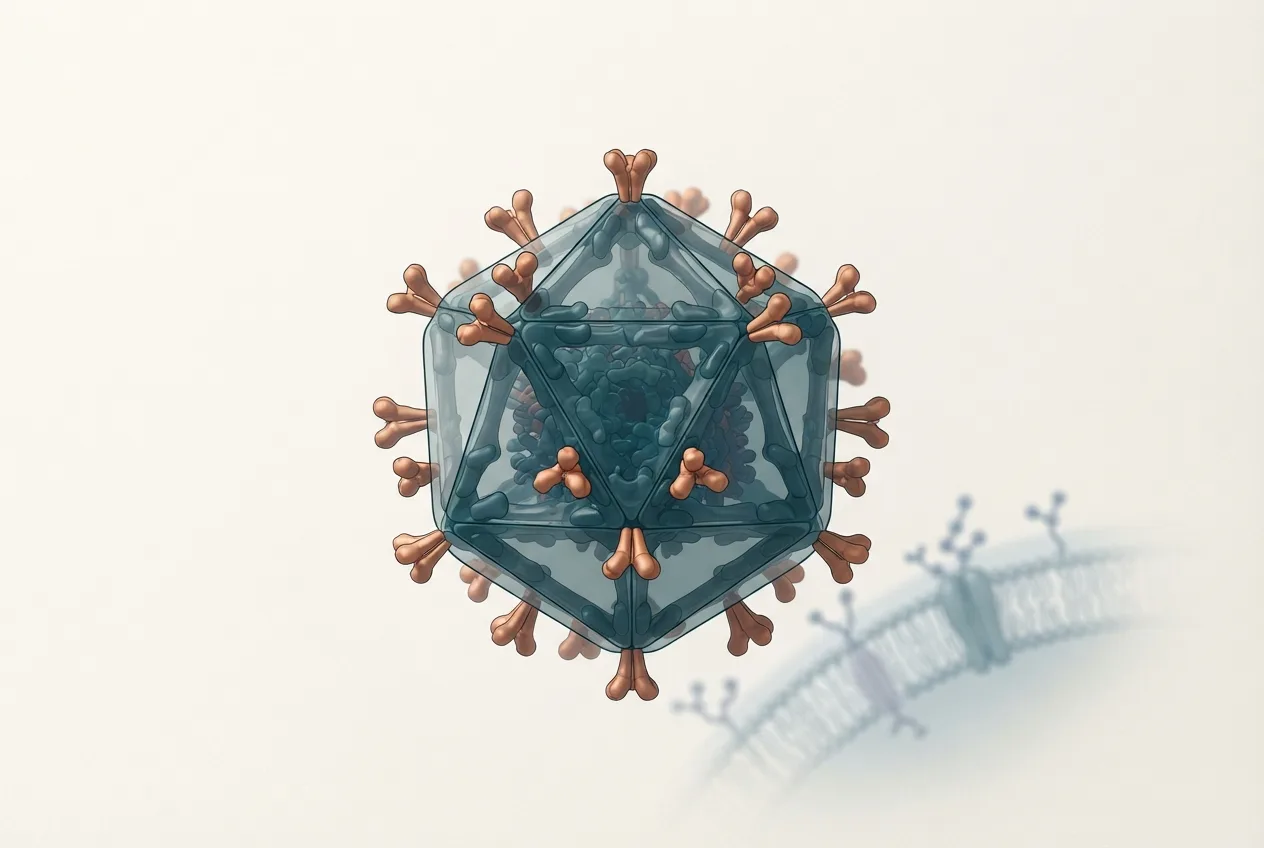
- The virus possesses a non-enveloped, icosahedral structure with a bipartite, single-stranded, positive-sense RNA genome (RNA1 encoding RdRp, RNA2 encoding capsid protein) and a subgenomic RNA3 encoding a B2 protein that suppresses RNA interference.
- Diagnosis relies on sensitive molecular methods like RT-qPCR targeting the RNA2 segment, alongside histopathology revealing characteristic CNS vacuolation and necrosis, and immunohistochemistry for viral antigen localization.
- Control strategies encompass stringent biosecurity, stress reduction through optimized management practices (e.g., lower stocking densities), and the development of effective vaccines, including inactivated, recombinant protein, and virus-like particle (VLP) platforms.
- Transmission is primarily horizontal via waterborne exposure (gills, skin) and can be vertical from infected broodstock, with clinical signs including abnormal swimming, exophthalmia, and anorexia.
Introduction
Viral Nervous Necrosis Virus (VNNV), classified within the genus Betanodavirus of the family Nodaviridae, is a significant pathogen affecting many marine and freshwater fish species worldwide [<a href="#ref-1">1</a>]. The disease, commonly referred to as viral nervous necrosis (VNN) or viral encephalopathy and retinopathy (VER), is characterized by severe neurological signs and high mortality rates, particularly in larval and juvenile fish [<a href="#ref-2">2</a>, <a href="#ref-3">3</a>]. The economic impact on aquaculture, especially for species such as groupers, sea bass, and flatfish, is substantial [<a href="#ref-4">4</a>]. This article provides a detailed reference on the virology, pathogenesis, diagnostic methodologies, and control strategies for VNNV, with a focus on the red-spotted grouper nervous necrosis virus (RGNNV) genotype, which is one of the most prevalent and virulent [<a href="#ref-2">2</a>, <a href="#ref-5">5</a>].
Virology and Taxonomy
Betanodaviruses are small, non-enveloped, icosahedral viruses with a diameter of approximately 25 to 30 nm [<a href="#ref-5">5</a>]. The viral genome consists of two single-stranded, positive-sense RNA segments: RNA1 (approximately 3.1 kb) and RNA2 (approximately 1.4 kb) [<a href="#ref-6">6</a>]. RNA1 encodes the RNA-dependent RNA polymerase (RdRp), also known as protein A, which is essential for viral replication [<a href="#ref-7">7</a>]. RNA2 encodes the capsid protein (CP), the primary structural component that determines antigenicity and host specificity [<a href="#ref-5">5</a>, <a href="#ref-8">8</a>]. A subgenomic RNA3, transcribed from RNA1, encodes a non-structural protein (B2) that functions as a suppressor of RNA interference, a key host antiviral defense mechanism [<a href="#ref-9">9</a>].
Four major genotypes have been identified based on the nucleotide sequence of the RNA2 segment: striped jack nervous necrosis virus (SJNNV), tiger puffer nervous necrosis virus (TPNNV), barfin flounder nervous necrosis virus (BFNNV), and red-spotted grouper nervous necrosis virus (RGNNV) [<a href="#ref-2">2</a>]. The RGNNV genotype is particularly notable for its broad host range and high virulence in warm-water fish species [<a href="#ref-2">2</a>, <a href="#ref-4">4</a>]. The capsid protein is the primary target for neutralizing antibodies and is therefore a central focus for vaccine development [<a href="#ref-5">5</a>, <a href="#ref-8">8</a>].
Host Range and Clinical Disease
Betanodaviruses infect over 120 species of fish, encompassing both marine and freshwater environments [<a href="#ref-1">1</a>]. The most severe outbreaks occur in larval and juvenile fish, with mortality rates often exceeding 90% [<a href="#ref-3">3</a>, <a href="#ref-10">10</a>]. Clinical signs are primarily neurological and include abnormal swimming behavior such as spiraling, corkscrew swimming, and whirling [<a href="#ref-3">3</a>]. Affected fish may also exhibit exophthalmia (pop-eye), skin darkening, and anorexia [<a href="#ref-11">11</a>]. Histopathological examination reveals characteristic vacuolation and necrosis in the brain, retina, and spinal cord tissues [<a href="#ref-3">3</a>, <a href="#ref-11">11</a>].
The susceptibility to VNNV is influenced by multiple factors, including host species, age, and environmental conditions. A study on shi drum juveniles demonstrated that increased rearing densities significantly enhanced susceptibility to betanodavirus infection, a process mediated by the neuroactive ligand-receptor interaction pathway [<a href="#ref-1">1</a>]. This finding highlights the importance of stress management in aquaculture settings to mitigate disease risk.
Pathogenesis and Immune Response
Following entry, typically via the mucosal surfaces of the gills or skin, the virus spreads to the central nervous system (CNS) via the bloodstream or peripheral nerves [<a href="#ref-7">7</a>]. Replication occurs primarily in neurons and glial cells of the brain, retina, and spinal cord, leading to the characteristic cytopathic effects of vacuolation and necrosis [<a href="#ref-3">3</a>, <a href="#ref-11">11</a>]. The host immune response to VNNV involves both humoral and cellular components. In giant groupers, vaccination with inactivated virus elicited significant humoral and cytokine responses, including the upregulation of interferon-related genes and the production of specific antibodies [<a href="#ref-7">7</a>]. Similarly, studies on orange-spotted grouper demonstrated that virus-like particles (VLPs) produced in Escherichia coli could induce protective immune responses [<a href="#ref-10">10</a>].
The role of antimicrobial peptides (AMPs) in antiviral defense has also been investigated. Certain AMPs, such as epinecidin-1, have demonstrated antiviral activity against fish nodavirus, suggesting a potential therapeutic avenue [<a href="#ref-12">12</a>]. Transcriptome analysis of medaka following treatment with epinecidin-1 and TH1-5 peptides during NNV infection revealed modulation of immune-related gene expression, further supporting the potential of AMPs as antiviral agents [<a href="#ref-9">9</a>].
Diagnostic Methods
Accurate and timely diagnosis of VNNV is critical for disease management and control. A range of diagnostic techniques are available, each with specific advantages and limitations.
Molecular Diagnostics
Reverse transcription polymerase chain reaction (RT-PCR) and real-time quantitative RT-PCR (RT-qPCR) are the most widely used molecular methods for VNNV detection due to their high sensitivity and specificity [<a href="#ref-6">6</a>]. These assays target conserved regions of the RNA2 segment, allowing for detection across multiple genotypes. A novel quantitative immunomagnetic reduction (IMR) assay has been developed for the detection of NNV, offering a rapid and sensitive alternative to conventional PCR [<a href="#ref-13">13</a>, <a href="#ref-14">14</a>]. The IMR assay uses antibody-coated magnetic nanoparticles to detect viral antigens in solution, with the reduction in magnetic signal being proportional to the virus concentration [<a href="#ref-13">13</a>, <a href="#ref-14">14</a>].
Isothermal amplification methods, such as recombinase polymerase amplification (RPA), have also been developed for VNNV detection [<a href="#ref-6">6</a>]. RPA offers the advantage of rapid amplification at a constant temperature, making it suitable for field-based diagnostics without the need for thermal cycling equipment [<a href="#ref-6">6</a>].
Serological and Immunological Assays
Enzyme-linked immunosorbent assays (ELISAs) and immunochromatographic tests are used for the detection of viral antigens or specific antibodies in fish serum or tissue homogenates [<a href="#ref-3">3</a>, <a href="#ref-13">13</a>]. These methods are valuable for large-scale surveillance and for assessing vaccine efficacy. The IMR assay, as mentioned above, represents a novel immunological approach that combines high sensitivity with rapid turnaround times [<a href="#ref-13">13</a>, <a href="#ref-14">14</a>].
Histopathology and Immunohistochemistry
Histopathological examination of brain and eye tissues remains a cornerstone for confirming VNNV infection [<a href="#ref-3">3</a>]. The characteristic vacuolation and necrosis in the CNS are pathognomonic for the disease. Immunohistochemistry (IHC) using specific antibodies against the viral capsid protein allows for the precise localization of viral antigens within affected tissues, providing both diagnostic and research utility [<a href="#ref-3">3</a>].
The following table summarizes the key diagnostic methods for VNNV.
| Diagnostic Method | Target | Sensitivity | Application |
|---|---|---|---|
| RT-PCR / RT-qPCR | Viral RNA (RNA2) | High | Routine detection, quantification |
| Recombinase Polymerase Amplification (RPA) | Viral RNA (RNA2) | High | Field-based, rapid detection |
| Immunomagnetic Reduction (IMR) Assay | Viral capsid protein | High | Rapid antigen detection |
| ELISA | Viral antigen or antibody | Moderate to High | Surveillance, vaccine studies |
| Histopathology / IHC | Tissue lesions / viral antigen | Moderate | Confirmatory diagnosis, research |
Control and Prevention
Control of VNNV in aquaculture relies on a combination of biosecurity measures, management practices, and vaccination.
Biosecurity and Management
Strict biosecurity protocols, including disinfection of equipment, quarantine of new stock, and screening of broodstock, are essential to prevent the introduction and spread of VNNV [<a href="#ref-2">2</a>]. Reducing stress factors, such as high stocking densities, poor water quality, and handling, can decrease susceptibility to infection [<a href="#ref-1">1</a>]. The administration of poly(I:C), a synthetic double-stranded RNA analog that induces interferon production, has been shown to reduce viral titers in the brain tissue of infected fish, suggesting a potential prophylactic strategy [<a href="#ref-11">11</a>].
Vaccine Development
Significant efforts have been directed towards developing effective vaccines against VNNV. Several vaccine platforms have been explored, including inactivated whole-virus vaccines, recombinant protein vaccines, virus-like particles (VLPs), and DNA vaccines [<a href="#ref-2">2</a>, <a href="#ref-5">5</a>, <a href="#ref-8">8</a>, <a href="#ref-10">10</a>, <a href="#ref-15">15</a>].
Inactivated vaccines have shown promise in laboratory and field trials. Bath immunization of grouper larvae with inactivated betanodavirus conferred significant protection against subsequent challenge [<a href="#ref-15">15</a>]. A study on the protective effects of a killed vaccine in stellate sturgeon demonstrated reduced histopathological lesions in the brain and eye tissues, as confirmed by IHC [<a href="#ref-3">3</a>]. The optimization of inactivation procedures is critical for vaccine development, as improper inactivation can lead to reduced immunogenicity or safety concerns [<a href="#ref-2">2</a>].
Recombinant protein vaccines, including those produced using cell-free protein synthesis systems, have been developed for sevenband grouper [<a href="#ref-8">8</a>]. These vaccines offer advantages in terms of safety and scalability. VLPs, which are non-infectious structures that mimic the native virus, have been produced in E. coli and shown to induce protective immune responses in orange-spotted grouper [<a href="#ref-10">10</a>]. The stability of VLPs in the aqueous state and the potential for lyophilization to produce stable vaccine formulations have also been investigated [<a href="#ref-5">5</a>].
Novel approaches, such as the use of aptamers for targeted delivery of small interfering RNAs (siRNAs), represent a cutting-edge strategy for antiviral therapy [<a href="#ref-4">4</a>]. Aptamers specifically directed to RGNNV-infected cells can mediate the delivery of siRNAs, leading to the inhibition of viral replication [<a href="#ref-4">4</a>].
The following Mermaid diagram illustrates a decision tree for VNNV control in an aquaculture setting.
graph TD
 A["VNNV Outbreak Suspected"] --> B{"Clinical Signs & History"}
 B -->|"Neurological signs, high mortality"| C["Collect Samples"]
 C --> D{"Diagnostic Confirmation"}
 D -->|"RT-PCR / RT-qPCR Positive"| E["Confirm VNNV"]
 D -->|"Histopathology / IHC Positive"| E
 E --> F{"Control Strategy"}
 F --> G["Biosecurity Measures"]
 F --> H["Management Practices"]
 F --> I["Vaccination"]
 G --> J["Disinfection, Quarantine, Screening"]
 H --> K["Reduce Stocking Density, Improve Water Quality"]
 I --> L["Inactivated / Recombinant / VLP Vaccine"]
 J & K & L --> M["Monitor & Evaluate"]
 M -->|"Outbreak Controlled"| N["Maintain Surveillance"]
 M -->|"Persistent Infection"| O["Review & Adjust Strategy"]
Frequently Asked Questions
What is the primary host range of Viral Nervous Necrosis Virus?
Viral Nervous Necrosis Virus infects over 120 species of marine and freshwater fish, with the most severe disease observed in larval and juvenile stages of species such as groupers, sea bass, and flatfish [<a href="#ref-1">1</a>, <a href="#ref-3">3</a>].
How is VNNV transmitted in aquaculture settings?
Transmission occurs horizontally through waterborne exposure via the gills or skin, and vertically from infected broodstock to offspring [<a href="#ref-7">7</a>].
What are the key clinical signs of VNNV infection?
Clinical signs include abnormal swimming behavior (spiraling, whirling), exophthalmia, skin darkening, and anorexia, with high mortality rates in young fish [<a href="#ref-3">3</a>, <a href="#ref-11">11</a>].
Which diagnostic method is considered the gold standard for VNNV detection?
Real-time quantitative RT-PCR (RT-qPCR) is widely regarded as the gold standard due to its high sensitivity and specificity for detecting viral RNA [<a href="#ref-6">6</a>].
Are there effective vaccines available for VNNV?
Yes, several vaccine platforms have been developed, including inactivated whole-virus vaccines, recombinant protein vaccines, and virus-like particles, with varying degrees of efficacy demonstrated in laboratory and field trials [<a href="#ref-2">2</a>, <a href="#ref-5">5</a>, <a href="#ref-8">8</a>, <a href="#ref-10">10</a>, <a href="#ref-15">15</a>].
Can stress factors influence the severity of VNNV outbreaks?
Yes, stress factors such as high rearing densities have been shown to increase susceptibility to VNNV infection, mediated by neuroactive ligand-receptor interaction pathways [<a href="#ref-1">1</a>].
What is the role of the capsid protein in VNNV biology?
The capsid protein, encoded by RNA2, is the primary structural component of the virus, determines antigenicity and host specificity, and is the main target for neutralizing antibodies and vaccine development [<a href="#ref-5">5</a>, <a href="#ref-8">8</a>].
Is there any antiviral therapy available for VNNV?
While no licensed antiviral drugs are available, experimental therapies such as antimicrobial peptides and aptamer-mediated siRNA delivery have shown antiviral activity in research settings [<a href="#ref-4">4</a>, <a href="#ref-12">12</a>].