Tilapia Parvovirus
By Dr. Zubair Khalid, DVM, MS, PhD ·
Key Takeaways
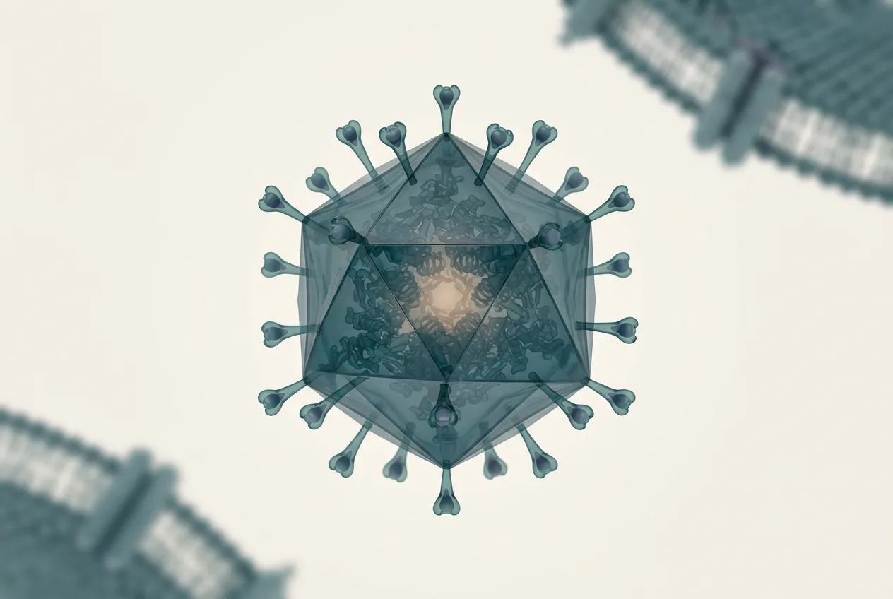
- Tilapia parvovirus (TiPV) is an emerging pathogen in farmed tilapia, characterized by a small, non-enveloped icosahedral capsid and a single-stranded linear DNA genome, sharing characteristics with other parvoviruses.
- The pathognomonic histopathological lesion for TiPV infection is the presence of Cowdry type A inclusion bodies (CAIB) within the nuclei of pancreatic acinar cells, offering a specific diagnostic target.
- While CAIB are exclusively found in the pancreas, TiPV DNA can be detected by in situ hybridization in multiple tissues including the liver, kidney, spleen, gills, and ovarian oocyte membranes, indicating broad tissue tropism.
- TiPV infections frequently co-occur with Streptococcus agalactiae, with multifocal granulomas secondary to bacterial infection being a common concurrent histopathological finding that requires differentiation from primary viral lesions.
- Definitive diagnosis relies on a combination of histopathology (CAIB in pancreas) and molecular confirmation via in situ hybridization (ISH) for viral nucleic acid detection or PCR/sequencing for genomic amplification.
Introduction
Tilapia parvovirus (TiPV) is an emerging viral pathogen associated with significant morbidity and mortality in farmed tilapia, a globally important aquaculture species [<a href="#ref-1">1</a>]. As a member of the family Parvoviridae, TiPV shares fundamental biophysical and genomic characteristics with other parvoviruses, including a small, non-enveloped icosahedral capsid and a single-stranded linear DNA genome. The virus was first identified in association with disease outbreaks in tilapia aquaculture systems, where it has been implicated as a primary or contributing etiological agent [<a href="#ref-1">1</a>]. Understanding the pathobiology of TiPV is critical for developing effective diagnostic strategies and control measures. This article provides a detailed review of TiPV, with a focus on its histopathological features, tissue tropism, diagnostic methodologies, and the implications of co-infection with bacterial pathogens such as Streptococcus agalactiae.
Virological and Genomic Characteristics
Parvoviruses are characterized by their small size (approximately 18-26 nm in diameter) and a genome of roughly 4-6 kilobases [<a href="#ref-1">1</a>]. The TiPV genome, based on partial sequencing, demonstrates high nucleotide identity (greater than 99%) to previously reported isolates, suggesting a high degree of genetic stability among circulating strains [<a href="#ref-1">1</a>]. The genome encodes non-structural (NS) proteins involved in replication and structural (VP) proteins that form the viral capsid. The replication strategy of parvoviruses is dependent on host cell DNA polymerase machinery, typically requiring cells in the S-phase of the cell cycle for efficient replication. This dependence on actively dividing cells influences the tissue tropism and pathogenesis observed in infected fish.
Clinical Presentation and Pathogenesis
TiPV infection in Nile tilapia (Oreochromis niloticus) is associated with non-specific clinical signs, including lethargy, anorexia, and increased mortality rates [<a href="#ref-1">1</a>]. Gross pathological findings are often not pathognomonic, necessitating histopathological and molecular examination for definitive diagnosis. The pathogenesis of TiPV involves viral entry into susceptible cells, nuclear translocation of the viral genome, and subsequent replication leading to cellular damage and necrosis. The virus has been detected in multiple tissue types, indicating a broad cellular tropism in vivo [<a href="#ref-1">1</a>].
Histopathological Diagnosis: The Pancreas as a Prime Target
A landmark finding in the histopathological diagnosis of TiPV infection is the presence of Cowdry type A inclusion bodies (CAIB) in pancreatic acinar cells [<a href="#ref-1">1</a>]. These intranuclear inclusions are a well-established histopathological hallmark for parvoviral infections in other species, including shrimp and terrestrial mammals. In the context of TiPV, CAIB are observed exclusively in the exocrine pancreas, both within the hepatopancreas and in pancreatic tissue distributed along the intestine [<a href="#ref-1">1</a>]. This specific localization is a critical diagnostic feature.
The following table summarizes the key histopathological features of TiPV infection in Nile tilapia.
| Feature | Description | Diagnostic Significance |
|---|---|---|
| , - | , - | , - |
| Cowdry Type A Inclusion Bodies (CAIB) | Intranuclear, eosinophilic inclusions that displace chromatin to the nuclear periphery. | Pathognomonic for TiPV when observed in pancreatic acinar cells. |
| Affected Tissue | Exocrine pancreas (hepatopancreas and intestinal pancreatic tissue). | Highly specific; not observed in other parenchymal organs. |
| Cellular Tropism | Pancreatic acinar cells. | Indicates a specific cellular target for viral replication and inclusion formation. |
| Co-infection Lesions | Multifocal granulomas (secondary to S. agalactiae). | Indicates common co-infection scenario; granulomas are not directly caused by TiPV. |
The presence of CAIB in the pancreas provides a practical and accessible target for histopathological diagnosis, particularly in regions where advanced molecular diagnostics may not be readily available [<a href="#ref-1">1</a>]. This pattern is considered critical for determining the presence of TiPV infection in new geographic areas [<a href="#ref-1">1</a>].
Tissue Tropism and Viral Distribution
In situ hybridization (ISH) using a TiPV-specific DNA probe has demonstrated the intranuclear presence of TiPV DNA in many tissues [<a href="#ref-1">1</a>]. These include the liver, pancreas, kidney, spleen, gills, and the membrane of oocytes in the ovary [<a href="#ref-1">1</a>]. This broad distribution confirms that TiPV can replicate in multiple tissue types, despite the exclusive manifestation of CAIB in the pancreas [<a href="#ref-1">1</a>]. The detection of viral nucleic acid in ovarian tissues raises important questions regarding potential vertical transmission routes, a phenomenon observed in other parvoviruses such as Porcine Parvovirus.
The following Mermaid diagram illustrates the diagnostic workflow for TiPV infection, emphasizing the role of histopathology and molecular confirmation.
flowchart TD
 A["Clinical Suspicion: Morbidity/Mortality in Tilapia"] --> B{"Histopathology of Pancreas"}
 B -->|"Cowdry Type A Inclusion Bodies Present"| C["Presumptive TiPV Diagnosis"]
 B -->|"No Inclusion Bodies"| D["Consider Other Etiologies"]
 C --> E{"Confirmatory Testing"}
 E -->|"In Situ Hybridization (ISH)"| F["TiPV DNA Detection in Multiple Tissues"]
 E -->|"PCR/Sequencing"| G["Partial Genome Amplification & Identity Confirmation"]
 F --> H["Definitive TiPV Diagnosis"]
 G --> H
 D --> I["Test for TiPV by PCR/ISH"]
 I -->|"Positive"| H
 I -->|"Negative"| J["Investigate Bacterial Co-infections or Other Viruses"]
 J --> K["Consider Tilapia Lake Virus or Streptococcus agalactiae"]
Co-infection Dynamics with Streptococcus agalactiae
A significant observation in TiPV-affected populations is the high prevalence of co-infection with Streptococcus agalactiae [<a href="#ref-1">1</a>]. This bacterium is a well-known pathogen in tilapia aquaculture, causing streptococcosis, a disease characterized by meningoencephalitis and multifocal granulomatous inflammation. In TiPV-infected fish, multifocal granulomas secondary to S. agalactiae infection are commonly observed histopathologically [<a href="#ref-1">1</a>]. The interaction between TiPV and S. agalactiae is complex and may involve viral-induced immunosuppression, which predisposes fish to secondary bacterial infections. Alternatively, concurrent infections may simply reflect the high environmental burden of both pathogens in intensive aquaculture systems. The presence of granulomas does not directly indicate TiPV infection but is a common concurrent finding that must be differentiated from primary TiPV lesions [<a href="#ref-1">1</a>]. For further details on this bacterial pathogen, refer to the article on Streptococcus agalactiae in Farmed Tilapia: Clinical Outbreaks and Molecular Detection.
Diagnostic Approaches
The diagnosis of TiPV infection relies on a combination of histopathological examination and molecular techniques.
Histopathology
As detailed above, the identification of CAIB in pancreatic acinar cells is a highly specific histopathological marker for TiPV infection [<a href="#ref-1">1</a>]. This method is cost-effective and can be performed in basic histology laboratories, making it suitable for field-level surveillance.
In Situ Hybridization (ISH)
ISH using a TiPV-specific probe provides direct visualization of viral nucleic acid within tissue sections [<a href="#ref-1">1</a>]. This technique confirms the presence of the virus in specific cell types and tissues, offering insights into viral tropism and pathogenesis. ISH is particularly useful for confirming TiPV infection in cases where inclusion bodies are ambiguous or absent.
Molecular Detection and Sequencing
Polymerase chain reaction (PCR) amplification of TiPV genomic fragments, followed by sequencing, provides definitive confirmation of the virus [<a href="#ref-1">1</a>]. Partial genome amplification has revealed high nucleotide identity among isolates, facilitating molecular epidemiological studies [<a href="#ref-1">1</a>]. These molecular methods are essential for differentiating TiPV from other viral pathogens of tilapia, such as Tilapia Lake Virus.
Differential Diagnoses
TiPV infection must be differentiated from other causes of morbidity and mortality in tilapia. Key differential diagnoses include:
- Tilapia Lake Virus (TiLV): A negative-sense RNA virus that causes syncytial hepatitis and high mortality. Histopathological lesions differ significantly from TiPV, with TiLV characterized by syncytial cell formation in the liver.
- Streptococcosis: Caused by S. agalactiae or S. iniae, this bacterial disease presents with exophthalmia, corneal opacity, and meningoencephalitis. Histopathology reveals granulomas, not intranuclear inclusion bodies.
- Other Viral Infections: Other aquatic viruses such as Infectious Hematopoietic Necrosis Virus and Viral Hemorrhagic Septicemia Virus primarily affect salmonids and are not typically associated with tilapia, but should be considered in polyculture systems.
Implications for Aquaculture Health Management
The emergence of TiPV poses a challenge to tilapia aquaculture. The virus's ability to cause disease, particularly in conjunction with bacterial pathogens, underscores the need for comprehensive health management strategies. Biosecurity measures, including the screening of broodstock and fingerlings, are critical to prevent the introduction and spread of TiPV. The identification of the pancreas as a prime target for histopathological diagnosis facilitates rapid and cost-effective surveillance, enabling early detection and implementation of control measures [<a href="#ref-1">1</a>]. Further research into TiPV pathogenesis, transmission dynamics, and potential vaccine development is warranted.
Frequently Asked Questions
What is the primary histopathological feature of Tilapia parvovirus infection?
The primary histopathological feature is the presence of Cowdry type A inclusion bodies (CAIB) in the nuclei of pancreatic acinar cells, which is considered a pathognomonic lesion for TiPV infection in Nile tilapia [<a href="#ref-1">1</a>].
In which tissues can TiPV DNA be detected by in situ hybridization?
TiPV DNA has been detected by in situ hybridization in the liver, pancreas, kidney, spleen, gills, and the membrane of oocytes in the ovary, indicating a broad tissue tropism [<a href="#ref-1">1</a>].
Is TiPV infection typically found alone or with other pathogens?
TiPV infection is frequently found as a co-infection with the bacterium Streptococcus agalactiae, and affected fish often present with multifocal granulomas secondary to this bacterial infection [<a href="#ref-1">1</a>].
Why is the pancreas considered a prime target for TiPV diagnosis?
The pancreas is considered a prime target because Cowdry type A inclusion bodies manifest exclusively in pancreatic acinar cells, providing a specific and accessible histopathological marker for diagnosis, especially in areas without advanced molecular testing capabilities [<a href="#ref-1">1</a>].
How genetically similar are different TiPV isolates?
Partial genome amplification of TiPV has revealed high nucleotide identity (greater than 99%) among previously reported isolates, suggesting a high degree of genetic conservation [<a href="#ref-1">1</a>].