Spring Viraemia of Carp Virus
By Dr. Zubair Khalid, DVM, MS, PhD ·
Key Takeaways
- Spring Viraemia of Carp Virus (SVCV), a rhabdovirus, causes a contagious and often fatal hemorrhagic disease in cyprinid fish, particularly common carp, and is notifiable to the WOAH due to significant economic impact.
- The virus possesses a single-stranded, negative-sense RNA genome encoding five proteins (N, P, M, G, L), with the glycoprotein (G) serving as the major antigenic determinant for attachment and entry, and phylogenetic analysis based on the G gene divides SVCV into four genogroups.
- Transmission is horizontal via waterborne routes, with virus shed in feces, urine, and exudates, and outbreaks typically occur at water temperatures between 10°C and 17°C, with clinical signs including lethargy, exophthalmia, ascites, and hemorrhages.
- Diagnosis relies on virus isolation in EPC cell lines, rapid RT-qPCR targeting the G gene, and viability RT-qPCR (vqPCR) to distinguish infectious from non-infectious particles, with environmental surveillance employing concentration techniques.
- While no specific antiviral drugs are approved, various compounds like coumarin derivatives, bavachin, and Schisandrin A show in vitro and in vivo anti-SVCV activity by interfering with viral replication or modulating host immune responses, and experimental vaccines (DNA, subunit) are under development.
Introduction and Aetiology
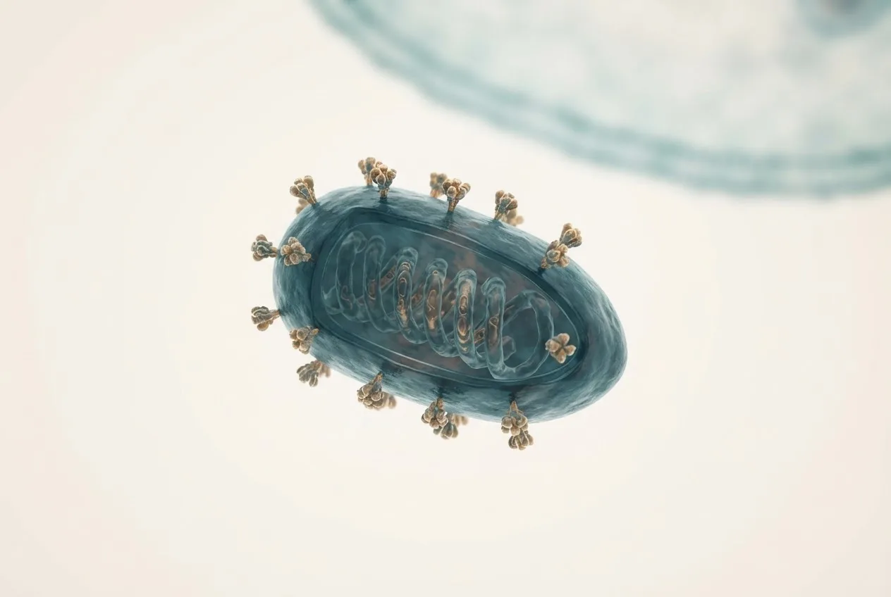
Spring Viraemia of Carp Virus (SVCV), historically designated Rhabdovirus carpio, is the causative agent of spring viraemia of carp (SVC), a contagious and often fatal hemorrhagic disease affecting cyprinid fishes, particularly common carp (Cyprinus carpio) [<a href="#ref-1">1</a>, <a href="#ref-2">2</a>]. The virus belongs to the genus Sprivivirus within the family Rhabdoviridae, order Mononegavirales [<a href="#ref-1">1</a>, <a href="#ref-3">3</a>]. SVCV is an enveloped, bullet-shaped virus characteristic of rhabdoviruses [<a href="#ref-2">2</a>]. The disease is notifiable to the World Organisation for Animal Health (WOAH) due to its significant economic impact on freshwater aquaculture globally [<a href="#ref-1">1</a>]. SVCV infection is distinct from other aquatic rhabdoviral diseases in its epidemiology and host range, which is largely restricted to cyprinid species.
Genomic Organization and Virion Structure
The SVCV genome is a single-stranded, negative-sense RNA molecule of approximately 11,000 nucleotides [<a href="#ref-2">2</a>, <a href="#ref-4">4</a>]. The genome is organized into five open reading frames encoding the nucleoprotein (N), phosphoprotein (P), matrix protein (M), glycoprotein (G), and viral RNA-dependent RNA polymerase (L) [<a href="#ref-2">2</a>, <a href="#ref-5">5</a>]. The genes are arranged in the canonical rhabdovirus order 3'-N-P-M-G-L-5' [<a href="#ref-2">2</a>]. The viral genome is encapsidated by the N protein to form a helical ribonucleoprotein (RNP) complex, which serves as the template for both transcription and replication [<a href="#ref-4">4</a>].
Cryo-electron microscopy (cryo-EM) studies have resolved the three-dimensional structure of the SVCV RNP complex at a resolution of 3.7 angstroms [<a href="#ref-4">4</a>]. The RNP assembly is stabilized by interactions between N and C loops, with the RNA genome wrapped in a groove between the N and C lobes, binding 9 nucleotides per protomer [<a href="#ref-4">4</a>]. The G protein, a trimeric class I transmembrane glycoprotein, is the major antigenic determinant and mediates viral attachment and entry into host cells [<a href="#ref-6">6</a>, <a href="#ref-7">7</a>]. Phylogenetic analysis based on the G gene partial nucleotide sequence divides SVCV into four principal genogroups: SVCV a, SVCV b, SVCV c, and SVCV d [<a href="#ref-3">3</a>, <a href="#ref-8">8</a>]. Genogroup Ia strains, including the highly virulent SVCV-CG01 strain isolated from zebrafish in China, have been characterized [<a href="#ref-8">8</a>]. Circulating strains in Serbia predominantly belong to genogroup SVCV d, with subgroups d1 and d2 and a proposed new subgroup d5 [<a href="#ref-3">3</a>].
Host Range and Transmission
The primary hosts for SVCV are cyprinid fish, with common carp being the most economically important species [<a href="#ref-1">1</a>, <a href="#ref-2">2</a>]. Other susceptible cyprinids include grass carp, silver carp, bighead carp, crucian carp, and ornamental koi. The host range is not strictly limited to cyprinids; experimental evidence demonstrates that SVCV can infect and be transmitted between common carp and Nile tilapia (Oreochromis niloticus) through cohabitation [<a href="#ref-9">9</a>]. Virus was isolated from gills, visceral organs, and intestine of both species from day 1 post-cohabitation up to day 35, and transmission from infected O. niloticus to healthy C. carpio was confirmed [<a href="#ref-9">9</a>].
Transmission occurs horizontally via waterborne exposure. The virus is shed in feces, urine, and exudates from infected fish [<a href="#ref-1">1</a>]. Entry routes include the gills, skin, and gastrointestinal tract [<a href="#ref-9">9</a>]. The disease typically manifests at water temperatures between 10°C and 17°C, with outbreaks occurring in the spring as water temperatures rise after winter [<a href="#ref-1">1</a>].
Pathogenesis and Host-Virus Interactions
Following entry, SVCV replicates in a variety of tissues, including kidney, spleen, liver, and gills [<a href="#ref-1">1</a>]. Viral replication induces profound cellular and systemic effects. Transcriptomic analysis of zebrafish cell lines infected with SVCV reveals significant modulation of genes involved in immune response, apoptosis, and cellular stress. The host's unfolded protein response (UPR) is a critical determinant of viral replication [<a href="#ref-10">10</a>]. SVCV exploits the UPR by targeting the immunoglobulin heavy chain-binding protein (BiP) and activating transcription factor 4 (ATF4) during early infection to enhance viral RNA synthesis and translation [<a href="#ref-10">10</a>]. At later stages, activation of the BiP, PKR-like ER kinase (PERK), and inositol-requiring enzyme 1 alpha (IRE1alpha) pathways supports viral progeny release and induces immune responses and apoptotic cell death [<a href="#ref-10">10</a>].
Reactive oxygen species (ROS) accumulate during SVCV infection. ROS activate the inflammatory response via the MAPK/AP-1 and PI3K signaling pathways, leading to upregulation of pro-inflammatory cytokines such as tumor necrosis factor alpha (TNF-alpha), cyclooxygenase-2 (COX-2), and interleukin-8 (IL-8) [<a href="#ref-11">11</a>]. The virus modulates p53 expression using two distinct mechanisms mediated by the N and P proteins. Early in infection, the N protein promotes p53 degradation via the ubiquitin-proteasome pathway, while late in infection, the P protein stabilizes p53 by enhancing K63-linked ubiquitination. Lysine residue 358 is the key site for K63-linked ubiquitination regulated by N and P proteins. SVCV also manipulates autophagy; viral replication requires autophagy induction, and inhibition of autophagy by compounds like arctigenin reduces viral replication.
Small non-coding RNAs play important regulatory roles in SVCV pathogenesis. Small RNA sequencing identified 111 differentially expressed microRNAs (DEmiRs) following SVCV infection in common carp [<a href="#ref-12">12</a>]. Specifically, miR-133b-3p_3 promotes SVCV replication by targeting p62, and miR-146b_1 facilitates viral proliferation through regulating TLR9 expression [<a href="#ref-12">12</a>]. Long non-coding RNAs (lncRNAs) are also differentially modulated following SVCV challenge, with distinct lncRNA expression profiles observed in wild-type versus rag1-heterozygous mutant zebrafish.
Clinical Signs and Pathology
The incubation period is typically 1 to 2 weeks depending on water temperature [<a href="#ref-1">1</a>]. Affected fish exhibit lethargy, darkening of the skin, exophthalmia, abdominal distension due to ascites, and hemorrhages on the skin, gills, and internal organs [<a href="#ref-1">1</a>]. Mortality rates can exceed 70% in epizootics. The disease is often referred to as acute hemorrhagic septicemia. Detailed pathological descriptions from the literature are consistent with a systemic viral infection affecting multiple organ systems.
Diagnostic Approaches
Diagnosis of SVCV relies on virus isolation in cell culture, molecular detection, and serological methods. The epithelioma papulosum cyprini (EPC) cell line is highly sensitive for virus isolation, with cytopathic effects (CPE) typically visible within 48 to 72 hours post-inoculation [<a href="#ref-8">8</a>].
Reverse transcription quantitative PCR (RT-qPCR) assays targeting the G gene or other conserved regions provide rapid and sensitive detection [<a href="#ref-13">13</a>]. A viability RT-qPCR (vqPCR) assay incorporating the dye PMAxx has been developed to differentiate infectious from non-infectious viral particles [<a href="#ref-13">13</a>]. This vqPCR assay achieved a 95% limit of detection (LoD95%) of 6.82 copies per reaction and showed no cross-reactivity with other tested aquatic pathogens [<a href="#ref-13">13</a>]. A rapid, portable dual-mode RAA-CRISPR/Cas12a system has also been described for one-pot detection of SVCV, enabling field-deployable testing [<a href="#ref-14">14</a>].
For environmental surveillance, virus concentration from water samples can be achieved through coagulation-flocculation-resuspension processes optimized with aluminum sulfate, and through immunomagnetic bead separation from large-volume samples [<a href="#ref-15">15</a>, <a href="#ref-16">16</a>].
Phylogenetic characterization of isolates is performed by sequencing the partial G gene and comparing to established genogroups [<a href="#ref-3">3</a>, <a href="#ref-8">8</a>].
Antiviral Strategies and Immunomodulation
No specific antiviral drugs are approved for use against SVCV in aquaculture [<a href="#ref-10">10</a>, <a href="#ref-17">17</a>]. Numerous compounds have demonstrated anti-SVCV activity in vitro and in vivo. Coumarin derivatives, including 7-(6-benzimidazole) coumarin (C10), inhibit SVCV replication in EPC cells with a maximum inhibitory rate greater than 97% by protecting mitochondria, reducing apoptosis, and enhancing interferon-related gene expression [<a href="#ref-18">18</a>]. The phenylpropanoid derivative 4-(4-methoxyphenyl)-3,4-dihydro-2H-chromeno[4,3-d]pyrimidine-2,5(1H)-dione (E2) blocks post-entry viral transport and activates interferon signaling, achieving over 90% inhibition of SVCV protein gene expression.
Bavachin, a major constituent of Psoralea corylifolia, inhibits SVCV replication by interfering with early replication events rather than adsorption, with half-maximal inhibitory concentrations (IC50) of 0.31 to 0.46 mg/L for viral protein expression. Arctigenin (ARG) exhibits high anti-SVCV activity (IC50 values of 0.29 to 0.35 mg/L) by blocking virus-induced autophagy. Schisandrin A (SA), a bioactive compound from Schisandra chinensis, inhibits SVCV replication by upregulating key antiviral genes including ifna1, ifnγ, isg15, and mx1, and by preserving mitochondrial integrity and reducing oxidative stress [<a href="#ref-17">17</a>].
Divalent cations also influence SVCV infection. Magnesium ions (Mg2+) enhance the interferon response by increasing IRF3 expression, thereby inhibiting SVCV replication in vitro and in vivo [<a href="#ref-19">19</a>]. Ferrous ions (Fe2+) inhibit SVCV proliferation via ATG14-dependent autophagy [<a href="#ref-20">20</a>]. Calcium ions (Ca2+) play an antiviral role by increasing p53 expression to protect against SVCV infection [<a href="#ref-21">21</a>].
Nanoparticle-based drug delivery systems improve antiviral efficacy. The metal-organic framework ZIF-8 loaded with 5-Fluorouracil (5-Fu@ZIF-8) achieved a maximum inhibition rate of 91.36% at 16 mg/L, with a reduced IC50 of 5.85 mg/L compared to 20.86 mg/L for free 5-Fu, and improved survival rates in infected fish [<a href="#ref-22">22</a>].
The fungicide azoxystrobin has been shown to increase SVCV infection in fish, an important consideration for integrated aquaculture practices [<a href="#ref-23">23</a>].
Vaccine Development
Vaccine development against SVCV has focused on DNA vaccines, subunit vaccines, and replicon-based systems. A DNA-layered salmonid alphavirus-based replicon vaccine (pSAV) encoding the SVCV glycoprotein induced 88% survival in common carp following challenge at 14 +/- 1 degrees Celsius, compared to approximately 50% in control groups [<a href="#ref-24">24</a>]. The pSAV vaccine induced innate immune genes at the injection site and upregulated IgM expression [<a href="#ref-24">24</a>].
Bacterial ghost-loaded DNA vaccines have been developed for immersion immunization. Common carp immunized with Escherichia coli DH5alpha ghosts loaded with a G protein-expressing plasmid showed increased serum antibody levels and relative percentage survival (RPS) of 59.57% after SVCV challenge [<a href="#ref-25">25</a>].
Subunit vaccines based on the G protein or its dominant epitope G3 (131 amino acids) have been enhanced through PEG modification. PEGylated G3 (PEG-G3) induced significantly stronger immune responses compared to unmodified G3, with an RPS of 53.6% versus 38.9% [<a href="#ref-7">7</a>]. Single-walled carbon nanotubes loaded with mannose-modified glycoprotein (SWCNTs-MG) as an immersion vaccine achieved an RPS of 77.9% under optimized immunization conditions (30 mg/L vaccine dose, 24 fish per liter density, 12-hour immunization time) [<a href="#ref-26">26</a>].
Immersion immunization with glycoprotein alone elicits robust early immune responses in the skin, with transcriptomic analysis revealing involvement of HSP70 and MAPK signaling pathways [<a href="#ref-6">6</a>]. The interaction between glycoprotein and HSP70 activates JNK, modulating mucosal and systemic immune responses [<a href="#ref-6">6</a>].
Control and Prevention
Biosecurity measures, including quarantine of introduced fish, disinfection of equipment and facilities, and temperature management during high-risk periods, are essential for preventing SVCV introduction and spread [<a href="#ref-1">1</a>]. Nutritional strategies such as dietary supplementation with solid-state fermentation products of yeast (SFPY) have been shown to enhance SVCV resistance by improving liver and intestinal health and modulating gut microbiota [<a href="#ref-27">27</a>].
Frequently Asked Questions
What is the primary host species for SVCV?
The primary host species for SVCV is the common carp (Cyprinus carpio), though the virus can infect many cyprinid and some non-cyprinid species such as Nile tilapia (Oreochromis niloticus) under experimental conditions [<a href="#ref-1">1</a>, <a href="#ref-9">9</a>].
How is SVCV transmitted between fish?
SVCV is transmitted horizontally through waterborne exposure to virus shed in feces, urine, and exudates from infected fish, with entry occurring via gills, skin, and the gastrointestinal tract [<a href="#ref-1">1</a>, <a href="#ref-9">9</a>].
What are the typical clinical signs of SVCV infection?
Typical clinical signs include lethargy, darkening of the skin, exophthalmia, abdominal distension due to ascites, and hemorrhages on the skin, gills, and internal organs, occurring primarily at water temperatures between 10 degrees and 17 degrees Celsius [<a href="#ref-1">1</a>].
How is SVCV diagnosed in a laboratory setting?
SVCV is diagnosed by virus isolation in EPC cell lines, RT-qPCR targeting the G gene, viability RT-qPCR (vqPCR) using PMAxx to differentiate infectious from non-infectious particles, and RAA-CRISPR/Cas12a systems for rapid detection [<a href="#ref-8">8</a>, <a href="#ref-13">13</a>, <a href="#ref-14">14</a>].
Are there any approved vaccines for SVCV?
No commercial vaccines are approved at present, but experimental DNA-layered alphavirus replicon vaccines, bacterial ghost-loaded DNA vaccines, PEGylated subunit vaccines, and carbon nanotube-based immersion vaccines have shown promising protection in laboratory trials [<a href="#ref-7">7</a>, <a href="#ref-24">24</a>, <a href="#ref-25">25</a>, <a href="#ref-26">26</a>].
What antiviral compounds are under investigation for SVCV?
Coumarin derivatives like C10, phenylpropanoid derivatives like E2, bavachin, arctigenin, and Schisandrin A have demonstrated significant anti-SVCV activity by targeting various stages of the viral life cycle or by modulating the host immune response [<a href="#ref-17">17</a>, <a href="#ref-18">18</a>].
Diagnostic Decision Workflow
flowchart TD
 A["Fish exhibiting clinical signs: lethargy, hemorrhages, ascites"] --> B{"Water temperature 10-17 C?"}
 B -->|"Yes"| C["Collect tissue samples: kidney, spleen, gills"]
 B -->|"No"| D["Consider other differential diagnoses"]
 C --> E["RT-qPCR or RAA-CRISPR/Cas12a screening"]
 E -->|"Positive"| F["Confirm by virus isolation in EPC cells"]
 E -->|"Negative"| G["Assess for other pathogens: Koi Herpesvirus, Carp Edema Virus, bacterial agents"]
 F --> H["Characterize isolate: G gene sequencing, genogroup assignment"]
 H --> I["Report to regulatory authorities per WOAH guidelines"]
 F --> J["Perform viability RT-qPCR if infectivity assessment needed"]