Vitreous Body: Anatomy, Function, and Clinical Notes
By Dr. Zubair Khalid, DVM, MS, PhD ·
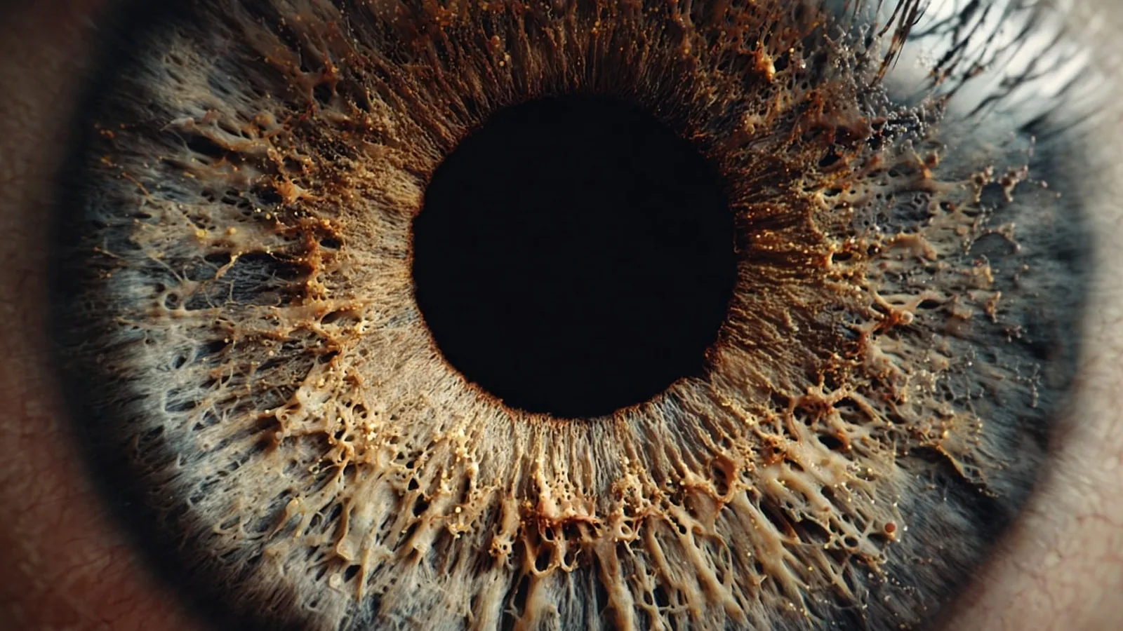
The vitreous body is a transparent, avascular gel that fills the space behind the lens and in front of the retina, occupying roughly four-fifths of the globe's volume in most domestic mammals. It is about 99% water, held in a scaffold of collagen fibrils and hyaluronic acid, and it transmits light while providing structural support to the retina and the lens.
This tissue matters because it is not simply packing material. It is a cell-regulated compartment with structural, mechanical, biological, and immunological functions that interact closely with the retina [1]. When the vitreous changes with age or disease, the consequences range from harmless floaters to blinding retinal detachment. For veterinary students, the vitreous is a favorite exam topic because it sits at the crossroads of ocular anatomy, optics, and clinical ophthalmology. For clinicians, it is the reason a dilated fundic examination is worth the extra two minutes.
This article is educational and is not a substitute for veterinary diagnosis or treatment.
Where the Vitreous Body Sits and What It Touches
Gross anatomy and boundaries
The vitreous body occupies the vitreous chamber, the large posterior compartment of the eye. Its boundaries are:
- Anteriorly: the posterior lens capsule and the posterior zonular fibers, with a small depression called the patellar fossa where the lens sits.
- Posteriorly: the internal limiting membrane of the retina.
- Peripherally: the pars plana and the ciliary body, with the anterior hyaloid membrane separating the vitreous from the posterior chamber.
The vitreous is not free-floating. It is anchored at several points, most importantly at the vitreous base, a band straddling the ora serrata where collagen fibrils insert firmly into the underlying retina and pars plana. This attachment is the anatomical reason why vitreous traction during aging or trauma can tear the retina at the ora serrata, producing a retinal dialysis or a horseshoe tear.
The vitreoretinal interface
The boundary between vitreous and retina is a specialized region called the vitreoretinal interface. It includes the cortical vitreous, the internal limiting membrane, and the intervening extracellular matrix. This interface is not a passive barrier. Hyalocytes, the resident immune cells of the vitreous, regulate the extracellular matrix and the immune response at this boundary, and when they become dysregulated they promote proliferative processes [1]. Structural phenomena such as vitreoschisis (splitting of the vitreous cortex) and retained vitreous cortex remnants act as scaffolds for fibrocellular proliferation on the retinal surface [1]. Optical coherence tomography now allows clinicians to visualize these changes in vivo, which is why OCT has become a standard tool in veterinary ophthalmology referral practice.
Why the anatomy matters clinically
Every vitreous disorder you will encounter in practice maps onto this anatomy:
- Posterior vitreous detachment (PVD) occurs when the cortical vitreous separates from the internal limiting membrane, except at the vitreous base.
- Vitreous hemorrhage accumulates in the spaces created by that separation.
- Vitreomacular traction arises when the separation is incomplete and residual adhesion pulls on the macula (or the area centralis in species that have one).
- Retinal tears occur when the vitreous base pulls on retina that is still firmly attached.
Composition: Water, Hyaluronic Acid, Collagen, and Cells
The vitreous is often described as "99% water," which is accurate but misleading. The remaining 1% is what gives the gel its optical clarity, its viscoelasticity, and its biological activity.
Water and the collagen-hyaluronan scaffold
The specific arrangement of collagen fibrils and water bound to hyaluronan minimizes light scattering while providing stability [2]. This is the central design principle of the vitreous. Collagen fibrils are spaced at a distance that keeps them optically transparent, and hyaluronic acid holds water in a hydrated network that fills the space between fibrils. If the fibril spacing changes, light scatters and the vitreous becomes hazy.
High-resolution structural work on porcine vitreous collagen has clarified this architecture in remarkable detail. Each 67-nanometer periodic unit of the native fibril contains type II, V/XI, and IX collagen triple helices together with opticin, at a stoichiometry of 8:4:4:4 [3]. Abundant galactose-glucose disaccharides modify hydroxylysine residues in conserved glycine-X-hydroxylysine motifs, and these glycans mediate fibril packing and structural stability [3]. This is the molecular basis for the transparency and mechanical resilience of the vitreous.
Composition table
| Component | Approximate share | Role | Notes |
|---|---|---|---|
| Water | ~99% of total mass | Solvent, optical medium | Bound to hyaluronan in the hydrated network [2] |
| Hyaluronic acid | Minor fraction of dry mass | Water binding, viscoelasticity, spacing of fibrils | Non-sulfated glycosaminoglycan, not covalently bound to collagen |
| Collagen fibrils | Minor fraction of dry mass | Structural scaffold, tensile strength | Heterotypic fibrils of type II, V/XI, and IX collagen with opticin [3] |
| Hyalocytes | Rare cells | Matrix regulation, immune surveillance, regression of embryonic vessels | Tissue-specific macrophage-like cells [2] |
| Other cells (rare) | Trace | Inflammatory or neoplastic infiltrates | Not normally resident |
The table is deliberately qualitative on percentages. The precise dry-mass fractions vary by species, age, and region of the vitreous, and no single figure covers dogs, cats, horses, and birds.
Hyalocytes: the forgotten cells
For most of the twentieth century, the vitreous was considered an inert transparent medium with no pathophysiological importance [1]. That view has changed. The vitreous harbors a tissue-specific immune cell population adapted to its particular needs, the hyalocytes [2]. These cells perform key tasks in the regression of embryonic vessels, tissue homeostasis, and immune surveillance, and they contribute significantly to the integrity and function of the vitreous [2]. Both alterations in vitreous composition or embryonic development and dysfunction of hyalocytes can form the basis of pathological processes [2].
Hyalocytes are not just bystanders. In disease states they can drive proliferative vitreoretinopathy, epiretinal membrane formation, and the fibrocellular proliferation that complicates retinal detachment surgery [1].
What else is in there
The vitreous also contains dissolved metabolites, electrolytes, proteins, and trace elements. Post-mortem multielemental analysis of human vitreous humor has detected elements at the ppm level (sodium, potassium, phosphorus, calcium, magnesium), the ppb level (aluminum, rubidium, zinc, iron, strontium, copper), and the ppt level (cerium, lanthanum, neodymium, terbium) [4]. Metabolic fingerprinting studies confirm that vitreous composition reflects physiological and pathophysiological processes occurring in the retina [5]. This is why vitreous humor is such a useful matrix in both forensic and biomarker research.
Vitreous Body vs Aqueous Humor: The Comparison Students Get Wrong
This is the single most common point of confusion in ocular anatomy, and it appears on nearly every veterinary licensing examination.
Aqueous humor is a clear, watery fluid produced by the ciliary body (specifically the ciliary epithelium). It flows from the posterior chamber, through the pupil, into the anterior chamber, and drains primarily through the iridocorneal angle via the trabecular meshwork and aqueous veins. It is continuously produced and continuously drained, turning over many times per hour. Its job is to nourish the avascular cornea and lens and to maintain intraocular pressure.
Vitreous humor is a gel, not a fluid, and it is produced during embryonic development. It is not continuously renewed. It fills the space behind the lens and does not circulate. Its job is optical transmission and structural support.
| Feature | Aqueous humor | Vitreous body |
|---|---|---|
| Consistency | Watery fluid | Transparent gel |
| Location | Anterior and posterior chambers | Vitreous chamber (behind lens) |
| Produced by | Ciliary body | Embryonic synthesis, not renewed |
| Circulation | Continuous flow through pupil and iridocorneal angle | Essentially none |
| Turnover | Minutes to hours | Lifetime |
| Regeneration after removal | Yes | No |
| Primary function | Nutrition, intraocular pressure | Light transmission, structural support |
The clinical implication of this table is enormous. Because the native vitreous does not regenerate, surgical removal during vitreoretinal surgery requires a substitute to maintain ocular homeostasis [6]. Current substitutes such as gases and silicone oils facilitate retinal reattachment but deviate significantly from the native vitreous, leading to complications such as cataract formation, increased intraocular pressure, and emulsification [6]. Hydrogels are under active investigation as more biomimetic alternatives [6][7].
Function: Why the Vitreous Is More Than Filler
Optical transmission
The vitreous is the primary optical medium of the posterior segment. Its transparency depends on the uniform spacing of collagen fibrils and the absence of large macromolecular aggregates. When that spacing is disturbed by inflammation, hemorrhage, or age-related liquefaction, light scatters and vision degrades.
Structural support
The vitreous holds the retina against the retinal pigment epithelium and helps maintain the shape of the globe. It also provides a cushioning effect against blunt trauma, distributing force across the posterior segment rather than concentrating it at a single point.
Metabolic and immunological roles
The vitreous is a reservoir and a signaling compartment. Proteins and metabolites diffuse through it, and its composition reflects retinal physiology [5]. Hyalocytes provide local immune surveillance [2], and the vitreous participates in the ocular immune privilege that limits inflammatory damage to the retina.
A barrier to drug and gene delivery
The viscoelastic properties of the vitreous are a practical obstacle for intravitreal therapies. Gene delivery to the retina via intravitreal injection is constrained by physical impediments, including the inner limiting membrane and the viscoelastic characteristics of the vitreous humor [8]. This is why researchers are developing vitreous humor-mimetic liposomes engineered to reproduce the lipidomic composition of native vitreous, which improved retinal gene delivery in mouse models of Norrie disease [8]. Understanding vitreous composition is therefore not academic. It directly shapes therapeutic design.
How the Vitreous Is Examined in Practice
Direct and indirect ophthalmoscopy
A dilated fundic examination is the first-line method for assessing the vitreous. The normal vitreous is optically clear. Any haze, cellular infiltrate, or blood indicates pathology. Indirect ophthalmoscopy with scleral depression allows the examiner to inspect the vitreous base, the region most likely to harbor tears or retained cortex.
Slit-lamp biomicroscopy
Slit-lamp examination with a condensing lens allows the clinician to assess the anterior vitreous, detect cells and flare, and identify the anterior hyaloid membrane. In dogs undergoing phacoemulsification, contrast-enhanced MRI studies have shown that anterior hyaloid membrane detachment occurs commonly, and that fluid leakage into the vitreous is significantly greater with high fluidic parameters than with low ones [9]. That study also found that leakage volume correlated with total irrigation time in both groups [9]. The clinical takeaway is that surgical fluidics influence the vitreous, and low fluidic parameters reduce fluid passage through the zonules into the vitreous [9].
Optical coherence tomography
OCT provides cross-sectional imaging of the vitreoretinal interface. It is the standard method for diagnosing vitreomacular traction, macular holes, epiretinal membranes, and vitreoschisis [1]. In veterinary referral practice, OCT is increasingly available and has changed how we classify vitreoretinal disease.
Vitreous sampling and cytology
Diagnostic vitrectomy with cytologic analysis is used when intraocular neoplasia or infection is suspected. Case reports illustrate the value. In one patient, cytopathology of the vitreous revealed necrotic melanoma cells from the preretinal surface and within the vitreous cavity, confirming metastatic cutaneous melanoma that had masqueraded as fungal endophthalmitis [10]. In another, cytology of the vitreous revealed abundant foamy histiocytes with positive CD163 immunoreactivity in a patient with West Nile virus-associated chorioretinitis [11]. These examples show that the vitreous is a diagnostic compartment, not just a structural one.
Clinical Relevance, Limitations and Common Mistakes
Posterior vitreous detachment
Posterior vitreous detachment is the separation of the cortical vitreous from the internal limiting membrane of the retina. It is a normal age-related change in many species, but it can be pathological when the separation is incomplete or when it exerts traction on the retina. The clinical signs are floaters, photopsia (flashes), and in some cases a sudden shower of vitreous cells. In dogs, PVD is often identified incidentally on OCT or during fundic examination. Incomplete PVD with persistent adhesion at the macula or area centralis produces vitreomacular traction, which can lead to macular hole formation, epiretinal gliosis, and progressive visual loss [1].
The most feared complication is retinal tear. Because the vitreous remains firmly attached at the vitreous base, a sudden PVD can avulse retinal tissue at the ora serrata. Any patient with acute PVD signs needs a careful peripheral retinal examination.
Vitreous hemorrhage
Vitreous hemorrhage is bleeding into the vitreous cavity. It can arise from retinal tears, retinal detachment, proliferative diabetic retinopathy, trauma, intraocular foreign bodies, or intraocular neoplasia. In one reported case, an intraretinal metallic foreign body presented with vitreous hemorrhage that obscured the foreign body on indirect ophthalmoscopy, and the diagnosis required orbital radiography before surgical removal with an intraocular magnet and forceps [12]. That case illustrates two practical points. First, hazy media from vitreous hemorrhage can hide a serious underlying cause. Second, vitreous hemorrhage is a sign, not a diagnosis, and the workup must identify the source.
The vitreous is avascular, so blood in the vitreous does not clot the way blood in a vessel does. It disperses and then gradually clears, but clearance can take weeks to months and may be incomplete. Persistent or recurrent hemorrhage is an indication for further diagnostic imaging and, in some cases, surgical intervention.
Asteroid hyalosis
Asteroid hyalosis is a degenerative condition in which small, refractile, calcium-lipid complexes form and float within the vitreous gel. On fundic examination they appear as sparkling white or yellow-white opacities that move with eye movement but do not settle. The condition is typically unilateral or asymmetric and is usually an incidental finding in older animals. Vision is generally preserved because the opacities are small and dispersed, but they can obscure the fundus and make examination of the retina difficult. Asteroid hyalosis is not the same as synchysis scintillans, which involves cholesterol crystals that settle in the dependent vitreous.
Other vitreous pathology worth knowing
- Vitritis: cellular infiltration of the vitreous, usually inflammatory or neoplastic. It can mimic endophthalmitis, as in the melanoma case described above [10].
- Proliferative vitreoretinopathy: fibrocellular proliferation on the retinal surface, driven in part by hyalocyte dysregulation and scaffolded by vitreous cortex remnants [1].
- Vitreous metastasis: rare but reported, and it should be on the differential for any vitritis in a patient with a history of cancer [10].
- Microplastic contamination: a 2024 study detected microplastics in human vitreous humor, predominantly particles below 50 micrometers, with nylon 66 the most abundant polymer detected [13]. The clinical significance of this finding is still being investigated.
Common mistakes students and clinicians make
- Confusing vitreous with aqueous. They have different origins, different compositions, different circulation, and different clinical behaviors.
- Assuming the vitreous is inert. It is a cell-regulated compartment with immune functions [2][1].
- Forgetting the vitreous base. Most retinal tears from PVD occur there, not at the posterior pole.
- Underestimating hazy media. Vitreous hemorrhage or dense vitritis can hide a retinal tear, a foreign body, or a tumor.
- Expecting the vitreous to regenerate. It does not, which is why vitreous substitutes are needed after surgical removal [6].
- Missing the systemic connection. Vitreous composition reflects retinal and systemic disease, and vitreous analysis has applications in biomarker discovery and forensics [5][14].
Limitations
The vitreous varies by species, age, and individual. Findings from one species do not always translate directly to another. Diagnosis and treatment decisions require a veterinarian who can examine the patient, interpret imaging, and consider the whole clinical picture.
Comparative Notes: Vitreous in Dogs, Cats, Horses, and Birds
Vitreous volume and consistency differ across species, and these differences matter for both examination and surgery.
Dogs. The canine vitreous is a firm gel in young animals that progressively liquefies with age. Vitreous volume scales with globe size, and in medium and large breeds the vitreous chamber is large enough that vitreous hemorrhage can significantly obscure the fundus. Canine eyes are commonly used in experimental studies of phacoemulsification fluidics, and contrast-enhanced MRI has shown that anterior hyaloid membrane detachment and fluid leakage into the vitreous occur during routine cataract surgery [9]. This is clinically relevant because postoperative vitreous changes can contribute to complications.
Cats. The feline vitreous is similar in composition to the canine vitreous but tends to be slightly more viscous. Cats have a well-developed area centralis rather than a fovea, so vitreomacular traction in the human sense does not apply. However, vitreous inflammation is common in feline infectious and neoplastic diseases, and vitreous haze can be an important diagnostic clue in conditions such as feline infectious peritonitis and lymphoma.
Horses. The equine vitreous is relatively large and firm, consistent with the large globe. Equine recurrent uveitis is a major cause of vitreous pathology, and chronic inflammation can lead to vitreous organization, traction bands, and secondary retinal detachment. The firm consistency of the equine vitreous makes it more resistant to liquefaction than the canine vitreous, but it also means that inflammatory debris can persist for long periods.
Birds. The avian vitreous is proportionally smaller and the globe is relatively rigid, supported by a cartilaginous or bony scleral ring. The avian vitreous tends to be less voluminous than in mammals of comparable body size. Birds also have a vascular structure called the pecten oculi that projects into the vitreous from the optic disc, and this structure is unique to birds. Vitreous pathology in birds is often associated with trauma, nutritional deficiencies, and systemic infections.
| Species | Relative vitreous volume | Consistency | Notable feature |
|---|---|---|---|
| Dog | Large | Firm gel, liquefies with age | Common model for surgical fluidics studies [9] |
| Cat | Moderate | Slightly more viscous than dog | Area centralis, not a fovea |
| Horse | Large | Firm | Recurrent uveitis causes vitreous organization |
| Bird | Proportionally smaller | Less voluminous | Pecten oculi projects into vitreous |
These differences are clinically important. A surgical approach, an imaging protocol, or a drug delivery strategy that works in one species may not translate directly to another.
Quick Review
- The vitreous body is a transparent, avascular gel behind the lens, about 99% water with hyaluronic acid and collagen fibrils.
- Its collagen scaffold contains type II, V/XI, and IX collagen with opticin, arranged to minimize light scattering [3].
- Hyalocytes are resident immune cells that regulate matrix and immune responses [2][1].
- The vitreous differs from aqueous humor in origin, composition, circulation, and regenerative capacity.
- Posterior vitreous detachment, vitreous hemorrhage, and asteroid hyalosis are the three classic clinical entities.
- The vitreous base is the strongest attachment and the most common site of retinal tears.
- Vitreous volume and consistency vary across dogs, cats, horses, and birds, which affects examination and treatment.
Frequently Asked Questions
What is the vitreous body made of?
The vitreous body is about 99% water, with the remaining fraction composed of hyaluronic acid, collagen fibrils, and a small population of hyalocytes. The collagen and hyaluronic acid form a hydrated scaffold that keeps the gel transparent.
How is the vitreous body different from aqueous humor?
Aqueous humor is a watery fluid produced by the ciliary body that circulates through the anterior and posterior chambers and drains at the iridocorneal angle. The vitreous body is a gel that fills the space behind the lens, does not circulate, and is not renewed.
Can the vitreous body heal or regenerate?
No. The native vitreous does not regenerate, which is why surgical removal during vitreoretinal surgery requires a substitute such as a gas, silicone oil, or a hydrogel to maintain ocular homeostasis [6].
What causes vitreous hemorrhage in animals?
Vitreous hemorrhage can result from retinal tears, retinal detachment, trauma, intraocular foreign bodies, proliferative retinopathies, or intraocular tumors. Because the vitreous is avascular, the blood comes from adjacent vascular tissues, and the underlying source must be identified.
What is asteroid hyalosis?
Asteroid hyalosis is a degenerative condition in which small refractile calcium-lipid complexes form within the vitreous gel. It is usually an incidental finding in older animals and typically does not cause significant vision loss, but it can make fundic examination difficult.
Do dogs, cats, horses, and birds have the same vitreous?
No. Vitreous volume and consistency differ across species. Dogs and horses have relatively large, firm vitreous bodies, cats have a slightly more viscous vitreous, and birds have a proportionally smaller vitreous with a unique vascular structure called the pecten oculi.
Related Articles
- Ruminant Digestive Anatomy and Physiology: A Clinical Reference
- Equine Larynx Anatomy and Function in Respiration
- Canine Lymphatic System: Anatomy and Function
- Equine Hoof Anatomy: Structures and Function
- Clinical Research Notes Templates
- Veterinary Anatomy Notes: How to Create and Use Them for NAVLE
- Epiphyseal Plate: Growth, Anatomy and Clinical Notes
- Pleural Cavities: Anatomy, Boundaries, and Clinical Notes
Sources
- [[Diseases of the vitreoretinal interface : What role does the vitreous body play?].](https://pubmed.ncbi.nlm.nih.gov/41912886/)
- [[A look at the vitreous body, not through it: anatomy, embryology and cells of the vitreous body].](https://pubmed.ncbi.nlm.nih.gov/41609797/)
- The molecular architecture of mammalian vitreous body collagen fibrils.
- Metallomic Analysis of Vitreous Humor of the Human Eye-A Post-Mortem Multielemental Study.
- LC-MS-Based Metabolic Fingerprinting of Vitreous Humor.
- Replacing the vitreous body with hydrogels: Rationale and strategies.
- Hyaluronic acid-based in situ injectable crosslinked hydrogels as next-generation vitreous substitutes: A review.
- Vitreous humor-mimetic liposomes enable effective gene delivery to the eye via intravitreal injection.
- Evaluation of fluid leakage into the canine vitreous humor during phacoemulsification using contrast-enhanced magnetic resonance imaging.
- Metastatic Cutaneous Amelanotic Melanoma to the Vitreous Masquerading as a Fungal Endophthalmitis.
- CD163+ Foamy Histiocytes in the Vitreous of a Patient with West Nile Virus-Associated Chorioretinitis.
- Magnetic Extraction of an Intraretinal Foreign Body: A Case Report.
- Revealing new insights: Two-center evidence of microplastics in human vitreous humor and their implications for ocular health.
- Relating aqueous to vitreous humor proteomics for identification of reliable biomarkers in proliferative diabetic retinopathy.