Vertebral Artery: Course, Branches, and Clinical Relevance
By Dr. Zubair Khalid, DVM, MS, PhD ·
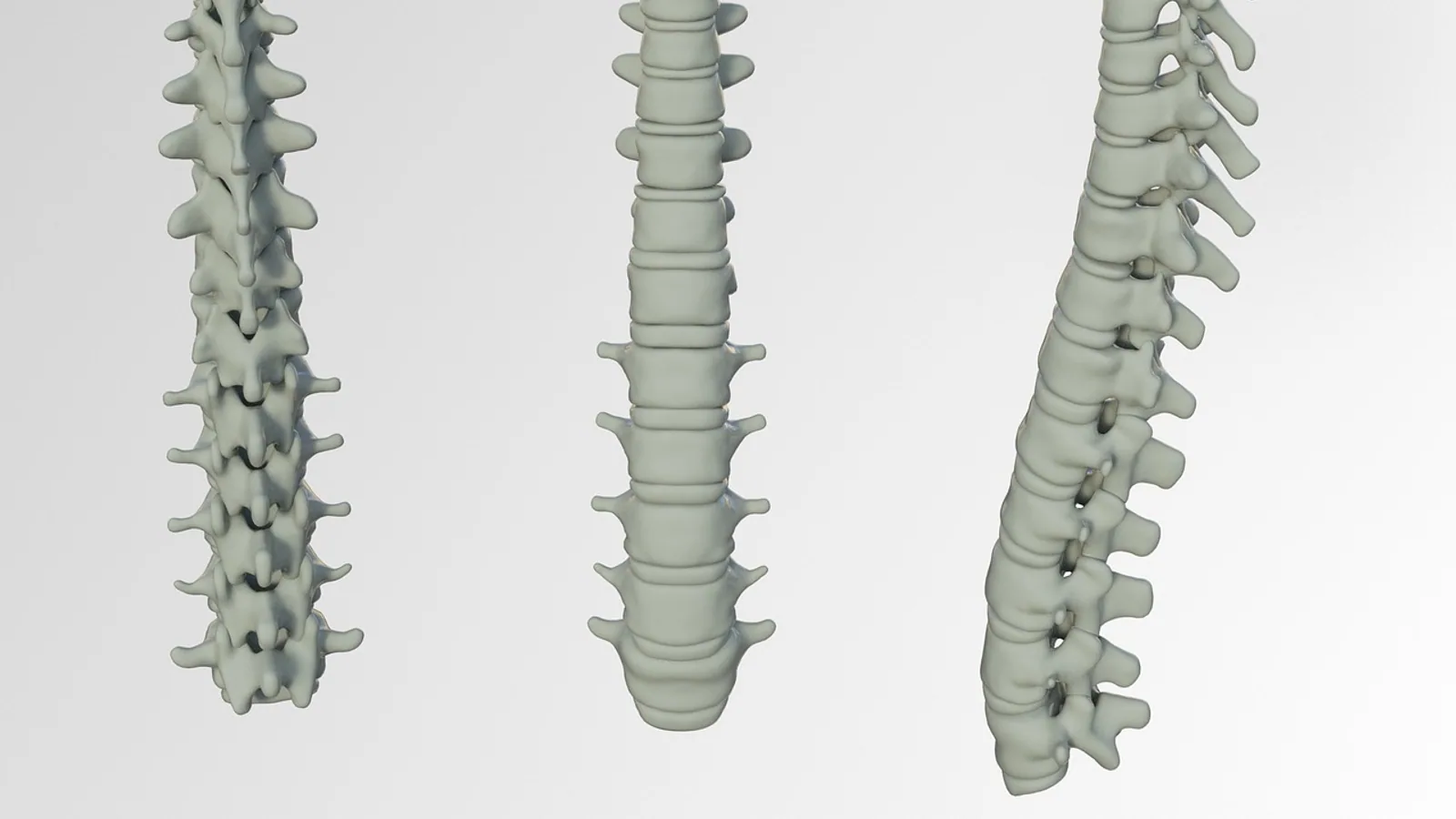
The vertebral artery is the first branch of the subclavian artery that ascends through the neck, passes through the transverse foramina of the cervical vertebrae, loops behind the atlas, and enters the skull to join its partner and form the basilar artery. It is the principal source of blood to the brainstem, cerebellum, and upper spinal cord, and its four named segments (V1 through V4) form the anatomical framework used by surgeons, radiologists, and anatomists to describe its course.
This vessel matters because it is the artery most often injured during cervical spine surgery, the artery most often implicated in posterior circulation stroke, and the artery whose comparative anatomy differs sharply between humans and domestic mammals. A working knowledge of its segments, branches, and variations is required for anyone who reads cervical imaging, places atlantoaxial implants, or interprets brainstem vascular lesions.
Orientation and Terminology
The vertebral artery is a paired vessel. The two vertebral arteries ascend on either side of the cervical spine and unite at the lower border of the pons to form the basilar artery, a single midline trunk. This union is one of only two places in the body where two arteries merge to form a single named vessel [1].
The artery is described in four segments, labeled V1 through V4. This four-segment scheme is the standard used in neurosurgery and radiology, and it is the framework used in photorealistic 3D anatomical models of the V2, V3, and V4 segments [2]. Each segment has a distinct course, a distinct set of branches, and a distinct set of clinical hazards.
A note on laterality. The right and left vertebral arteries are not mirror images in their origin. The right vertebral artery almost always arises from the right subclavian artery. The left vertebral artery arises from the left subclavian artery in most people but arises directly from the aortic arch in a minority. A meta-analysis of 62 studies covering 32,153 vessels found that the aortic arch origin of the left vertebral artery had a prevalence of 4.81%, which is the figure commonly rounded to roughly 5% [3].
The Four Segments
V1: Origin to the C6 Transverse Foramen
The V1 segment, also called the pre-foraminal or ostial segment, begins at the subclavian artery and ends where the artery enters the transverse foramen of the sixth cervical vertebra. The most common site of origin for both vertebral arteries is the subclavian artery [3].
The V1 segment has no named branches of surgical consequence. It runs upward and slightly backward through the root of the neck, passing in front of the transverse process of C7 and behind the inferior cervical ganglion. The artery is relatively mobile in this segment because it is not yet tethered by bone. That mobility is the reason the V1 segment is the target of endovascular procedures and the reason it can be stretched or kinked by neck movement.
Variation in the entry level is common. Ninety-two percent of vertebral arteries enter the transverse foramen of C6, followed in frequency by C5, C7, C4, and least often C3 at 0.1% [3]. The entry level matters because a surgeon who assumes a C6 entry may encounter the artery at an unexpected level during an anterior cervical approach.
V2: Within the Transverse Foramina from C6 to C2
The V2 segment, also called the foraminal or osseous segment, runs inside the transverse foramina of the cervical vertebrae from C6 up to C2. The artery is enclosed by bone on three sides for most of this course, which makes it vulnerable to injury from lateral mass screws, pedicle screws, and any instrumentation that breaches the transverse foramen.
The V2 segment is not a straight tube. It has a characteristic bend, or genu, near the axis vertebra. A cadaveric study of 20 vertebral arteries measured the pre-osseous segment related to the axis (called VAX-1) at 3.8 to 14.5 mm on the right and 4.46 to 10.5 mm on the left, and the osseous segment related to the axis (VAX-2) at 6.82 to 31 mm on the right and 7.35 to 20 mm on the left [4]. The genu of the V2 segment extends toward the midline by a mean of 15.6 mm on the right and 17.5 mm on the left from the midline of the axis vertebral body [4]. These measurements define the safe corridor for atlantoaxial screw placement.
The V2 segment gives off muscular branches to the deep cervical muscles and spinal branches that pass through the intervertebral foramina to supply the vertebral column, the spinal cord, and its meninges. The spinal branches divide into anterior and posterior radicular arteries, which travel along the nerve roots to reach the spinal cord. These radicular arteries are small but functionally important because they contribute to the longitudinal spinal arterial system.
V3: The Atlantoaxial Loop
The V3 segment, also called the atlantic or suboccipital segment, begins as the artery exits the transverse foramen of C2 and ends where it pierces the dura mater. This is the most tortuous part of the vertebral artery. It has three named parts: a vertical part between C2 and C1, a horizontal part that runs along the posterior arch of the atlas, and an exit part that turns upward to enter the skull.
A cadaveric study of 40 vertebral arteries in a South Indian population measured the mean length of the horizontal part at 38.937 mm on the right and 40.237 mm on the left, and the total V3 length at 66.870 mm on the right and 70.350 mm on the left [5]. These values give spine surgeons average measurements for the segment they must avoid during exposure of the craniovertebral junction.
The V3 segment is mobile. It must accommodate rotation of the head, which is why it is looped rather than straight. This mobility is also why the segment is prone to dissection and to kinking during extreme head rotation.
Variations in the V3 course are well documented. A case report described a right vertebral artery that entered the skull through the hypoglossal canal rather than the foramen magnum, a variant attributed to persistence of a primitive hypoglossal artery or partial incorporation of that artery into the vertebral artery [6]. Another case described a right V3 segment that took an anomalous posterosuperior course and penetrated the occipital bone near the jugular foramen, entering the posterior fossa at a higher level than the foramen magnum [7]. These variants are rare but clinically significant because they change the expected location of the artery during skull base surgery.
V4: Intradural Segment to the Basilar Junction
The V4 segment, also called the intradural or intracranial segment, begins where the artery pierces the dura mater and ends where the two vertebral arteries join to form the basilar artery. The artery ascends from the inferolateral to the anterosuperior side of the medulla [8].
The V4 segment gives off the posterior inferior cerebellar artery (PICA), the largest named branch of the vertebral artery. PICA supplies the lateral medulla and the inferior surface of the cerebellum. The V4 segment also gives off small perforating branches to the medulla and contributes to the anterior spinal artery.
The course of the V4 segment over the medullospinal junction is not rare. A study of 162 CT angiograms found that 19.75% of cases had an intradural vertebral artery crossing the ventral side of the medullospinal junction, with an incidence of 17.1% in males and 23.81% in females [8]. When neurological signs of medulla or medullospinal junction compression are present, the course of the vertebral artery should be documented on CT or MRI angiography [8].
Summary Table: Vertebral Artery Segments
| Segment | Course | Branches | Clinical Note |
|---|---|---|---|
| V1 | Subclavian artery to C6 transverse foramen | None of surgical consequence | Origin from aortic arch in 4.81% of left vertebral arteries [3]. Entry level varies, most often C6 [3]. |
| V2 | Within transverse foramina from C6 to C2 | Muscular branches, spinal branches with anterior and posterior radicular arteries | Genu near axis extends 15.6 mm right and 17.5 mm left from midline [4]. At risk during atlantoaxial screw placement. |
| V3 | Exit from C2 foramen to dura, with vertical, horizontal, and exit parts | Muscular branches | Most tortuous segment. Mean total length 66.870 mm right and 70.350 mm left [5]. Rare variants enter through hypoglossal canal [6] or occipital bone [7]. |
| V4 | Intradural ascent to basilar junction | PICA, medullary perforators, anterior spinal artery contribution | Crosses medullospinal junction in 19.75% of cases [8]. PICA supplies lateral medulla and inferior cerebellum. |
Branches in Detail
Muscular Branches
Muscular branches arise from the V2 and V3 segments and supply the deep muscles of the neck, including the longus colli, longus capitis, and semispinalis cervicis. These branches are small and numerous. They are not named individually in standard anatomy texts but are important because they form anastomoses with branches of the occipital artery and the deep cervical artery.
Spinal Branches and Radicular Arteries
Spinal branches arise from the V2 segment and pass through the intervertebral foramina. Each spinal branch divides into an anterior radicular artery and a posterior radicular artery. The anterior radicular artery follows the ventral root to reach the anterior spinal artery. The posterior radicular artery follows the dorsal root to reach the posterior spinal artery.
The radicular arteries are not evenly distributed. Some are larger than others, and the largest anterior radicular artery in the lower cervical and upper thoracic region is traditionally called the artery of Adamkiewicz in human anatomy. In veterinary anatomy, the equivalent vessel is the great radicular artery, which varies in position by species.
The anterior spinal artery is a midline vessel that runs along the ventral surface of the spinal cord. It is formed by contributions from the vertebral arteries at the level of the foramen magnum and reinforced by radicular arteries at intervals along the cord. The anterior spinal artery supplies the anterior two-thirds of the spinal cord, including the corticospinal tracts and the anterior horns.
Posterior Inferior Cerebellar Artery
PICA is the largest branch of the V4 segment. It arises from the vertebral artery near the lower border of the pons, loops around the medulla, and supplies the lateral medulla and the inferior surface of the cerebellum. PICA has the greatest anatomical variability of the cerebellar arteries in terms of origin, course, and branching pattern [9].
PICA can arise from a fenestrated vertebral artery. A case report described a PICA originating from one limb of a large V4 fenestration, an incidental finding on CT angiography performed for sudden headache [9]. Fenestrations of the vertebral artery are more common in the extracranial segment than the intracranial segment [9].
Anterior Spinal Artery Contribution
The vertebral arteries contribute to the anterior spinal artery at the level of the foramen magnum. The anterior spinal artery then descends along the ventral surface of the spinal cord. In humans, the vertebral contribution is significant. In dogs and horses, the contribution is different, as discussed below.
The medullary branches of the vertebral artery have been studied in detail. A microanatomic study of 26 brainstem halves found that perforating medullary arteries averaged 6.7 in number and 0.26 mm in diameter, and most often originated from the anterior spinal artery rather than from the vertebral artery itself. Only 38.5% originated from the vertebral artery, and 11.6% from the basilar artery [10]. These perforators supply the medial medullary region. Lateral medullary arteries, averaging 2.2 in number and 0.31 mm in diameter, usually originated from the vertebral artery and PICA, and supplied the lateral medullary region [10]. Among medullary infarctions, lateral ones were most frequent at 72.8% [10].
Pathway Diagram from Origin to Basilar Artery
The following diagram shows the main pathway of the vertebral artery from its origin to the basilar artery, with the key branch points and clinical decision nodes.
flowchart TD
    A[Subclavian artery] --> B[V1 segment]
    B --> C{Entry level}
    C -->|C6 most common| D[V2 segment]
    C -->|C5 or C7 variant| D
    D --> E[Spinal branches]
    E --> F[Radicular arteries]
    D --> G[V3 segment]
    G --> H{Atlantoaxial loop}
    H -->|Normal| I[V4 segment]
    H -->|Variant course| J[Document on imaging]
    I --> K[PICA]
    I --> L[Anterior spinal artery]
    I --> M[Basilar artery]
Comparative Anatomy: Dogs and Horses
The vertebral artery in domestic mammals is smaller relative to the basilar artery than it is in humans, and the basilar artery is supplied largely by the ventral spinal artery rather than by the vertebral arteries. This difference is important for veterinary students who transfer human anatomy concepts to animal patients.
A comparative study of encephalic arteries in three bird species and eight mammal species found two patterns of brain supply in mammals [11]. In the first pattern, found mostly in ungulates, a carotid rete mirabile supplies the encephalic arteries, the caudal branch of the internal carotid artery is the origin of the basilar artery, and the vertebral artery is only indirectly involved through supply to the rete. In the second pattern, found in humans and some other mammals, the caudal branch of the internal carotid artery becomes the posterior communicating artery, and the basilar artery originates from both vertebral arteries [11].
Dogs fall into a pattern where the vertebral artery is small and the ventral spinal artery is the main source of the basilar artery. The ventral spinal artery in the dog is a prominent midline vessel that runs the length of the spinal cord and continues rostrally to supply the basilar artery. This arrangement means that occlusion of a single vertebral artery in a dog is less likely to cause brainstem ischemia than it would in a human, because the basilar territory is supplied mainly by the ventral spinal artery.
Horses follow the ungulate pattern. The carotid rete mirabile is well developed, and the vertebral artery contributes to the rete rather than directly to the basilar artery. The basilar artery in the horse arises from the caudal branch of the internal carotid artery, and the vertebral artery is indirectly involved through the rete mirabile [11]. This is a fundamental difference from human anatomy and from the anatomy of dogs.
For veterinary students, the practical implication is that the vertebral artery in dogs and horses is not the primary source of basilar artery blood flow. Clinical signs of vertebral artery disease in these species are more likely to reflect spinal cord ischemia than brainstem ischemia.
Clinical Relevance, Limitations and Common Mistakes
Vertebral Artery Hypoplasia
Vertebral artery hypoplasia is defined as a diameter under 2 mm. A hypoplastic vertebral artery is a recognized risk factor for posterior circulation stroke, particularly when associated with a bilateral posterior fetal variant [1]. In a study of unruptured vertebral artery dissections, smaller or hypoplastic vertebral arteries were more likely to occlude [12].
Hypoplasia is not the same as agenesis. Agenesis means the artery is absent. A case report described segmental vertebral artery agenesis with reconstitution by a deep cervical artery, where the left vertebral ostium, V1 segment, and proximal V2 segment were absent and a tortuous deep cervical artery reconstituted the distal V2 segment at the C3-C4 disc level [13]. This variant is rare but demonstrates that collateral pathways can reconstitute flow to the distal vertebral artery.
Vertebral Artery Loops
Vertebral artery loops are focal deviations of the artery's course that can compress nerve roots and increase the risk of iatrogenic injury during cervical spine procedures. A systematic review and meta-analysis of 10 studies covering 7,475 vertebral arteries and 126 loops found a random-effects prevalence of 1.71% [14]. The authors described these loops as a hidden danger whose recognition changes preoperative planning [14].
Dissection and Pearl-and-String Sign
Vertebral artery dissection can present with a pearl-and-string sign on angiography, which consists of a fusiform or bulbous dilatation with stenosis at both ends. A study of 50 unruptured acute vertebral artery dissections with pearl-and-string signs found that 78.4% improved on follow-up. Of these, 48.6% fully recovered, 29.7% retained smooth dilatation, 10.8% developed irregular stenotic segments, 8.1% became occluded, and 2.7% transformed into a saccular lesion [12]. Headaches were linked to subarachnoid hemorrhage in 10 cases with bulbous dilatations, while 50 unruptured cases had fusiform dilatations with no subsequent hemorrhage except for two cases treated preventively [12].
Fenestration and Aneurysm Risk
Vertebral artery fenestration is a duplication of the arterial lumen over a short segment. Fenestrations are associated with aneurysms and arteriovenous malformations [9]. A case report described a pediatric patient with a dissecting aneurysm of a fenestrated V4 segment that caused recurrent subarachnoid hemorrhage and severe vasospasm [15]. The authors noted that dissecting aneurysms in children should be considered in all cases of delayed post-traumatic cranial rebleeding, particularly where anomalous arterial anatomy is present [15].
Basilar Artery Entrapment
The basilar artery can become entrapped in a clival fracture. A case report described a patient with a clival fracture and basilar artery entrapment with severe stenosis, where slab maximum intensity projection images of rotational angiography revealed occlusion of basilar perforators on the right side of the brainstem [16]. Occlusion of bilateral basilar perforators with basilar entrapment can cause brainstem ischemia and unfavorable outcomes [16].
Wallenberg Syndrome
Wallenberg syndrome, or lateral medullary syndrome, results from occlusion of the vertebral artery or PICA. A case report described a patient with untreated hypertension who presented with acute-onset dizziness, visual disturbances, continuous vomiting, difficulty walking, and altered consciousness. MRI and MRA revealed multifocal ischemic lesions consistent with basilar artery thrombosis affecting both vertebral branches and features of Wallenberg syndrome [17]. The lateral medullary region is supplied by lateral medullary arteries that usually originate from the vertebral artery and PICA [10], which explains why vertebral artery occlusion produces this syndrome.
Common Mistakes
The most common mistake is assuming that the vertebral artery enters the transverse foramen at C6 in every patient. It enters at C6 in 92% of cases, but the remaining 8% enter at C5, C7, C4, or C3 [3]. A surgeon who does not check the entry level on preoperative imaging may encounter the artery unexpectedly.
The second common mistake is assuming that the right and left vertebral arteries are symmetric. They are not. The left vertebral artery arises from the aortic arch in 4.81% of cases [3], and the two arteries can differ in diameter, course, and dominance.
The third common mistake is transferring human anatomy to dogs and horses without adjustment. In dogs and horses, the basilar artery is supplied largely by the ventral spinal artery, and the vertebral artery is smaller [11]. A student who assumes the human pattern will misinterpret the vascular anatomy of these species.
The fourth common mistake is overlooking a vertebral artery loop on preoperative imaging. Loops have a prevalence of 1.71% [14], and they are a recognized cause of iatrogenic injury during cervical spine procedures.
This article is educational and is not a substitute for veterinary diagnosis or treatment.
Quick Review
- The vertebral artery has four segments: V1 from origin to C6, V2 within the transverse foramina from C6 to C2, V3 at the atlantoaxial loop, and V4 intradural to the basilar junction.
- The left vertebral artery arises from the aortic arch in 4.81% of cases, which is roughly 5% [3].
- Vertebral artery hypoplasia is defined as a diameter under 2 mm and is a risk factor for posterior circulation stroke [1].
- PICA is the largest branch of the V4 segment and supplies the lateral medulla and inferior cerebellum.
- The V4 segment crosses the medullospinal junction in 19.75% of cases [8].
- In dogs and horses, the basilar artery is supplied largely by the ventral spinal artery, and the vertebral artery is smaller [11].
- Vertebral artery loops have a prevalence of 1.71% and are a hidden danger during cervical spine surgery [14].
Frequently Asked Questions
What are the four segments of the vertebral artery?
The four segments are V1, from the subclavian artery to the C6 transverse foramen, V2, within the transverse foramina from C6 to C2, V3, the atlantoaxial loop from C2 to the dura, and V4, the intradural segment to the basilar junction.
How often does the left vertebral artery arise from the aortic arch?
The left vertebral artery arises directly from the aortic arch in 4.81% of people, which is commonly rounded to roughly 5% [3].
What is vertebral artery hypoplasia?
Vertebral artery hypoplasia is a diameter under 2 mm. It is a risk factor for posterior circulation stroke when associated with a bilateral posterior fetal variant [1].
What does the posterior inferior cerebellar artery supply?
PICA supplies the lateral medulla and the inferior surface of the cerebellum. It is the largest branch of the V4 segment and has the greatest anatomical variability of the cerebellar arteries [9].
How is the vertebral artery different in dogs and horses?
In dogs and horses, the vertebral artery is smaller and the basilar artery is supplied largely by the ventral spinal artery. In horses, the carotid rete mirabile supplies the encephalic arteries and the vertebral artery contributes indirectly through the rete [11].
Why does vertebral artery dissection cause a pearl-and-string sign?
The pearl-and-string sign consists of a fusiform or bulbous dilatation with stenosis at both ends. It is a characteristic finding in vertebral artery dissection and is seen in both ruptured and unruptured cases [12].
Related Articles
- Branches of Biology
- MAPK Pathway: Mechanism, Function, and Clinical Relevance
- Telomere Testing: Methods, Meaning, and Clinical Relevance
- Cellular Signaling: Principles, Pathways, and Clinical Relevance
- RNA Helicase: Mechanisms, Functions, and Clinical Relevance
- DNA Repairsomes: Assembly, Function, and Clinical Relevance
Sources
- Congenital and acquired anomalies of the basilar artery: A pictorial essay.
- Photorealistic 3-Dimensional Models of the Anatomy and Neurosurgical Approaches to the V2, V3, and V4 Segments of the Vertebral Artery.
- The Vertebral Artery: A Systematic Review and a Meta-Analysis of the Current Literature.
- Surgical Anatomy of Vertebral Artery in Relation to Atlantoaxial Instrumentation: A Cadaveric Study.
- Exploring the atlantic part of the vertebral artery in the South Indian population and its implications in spine surgery.
- A typical course of v3 segment of the right vertebral artery: a case study and literature overview.
- V3 segment of the right vertebral artery taking an anomalous posterosuperior course and penetrating occipital bone (wall of the jugular foramen) diagnosed by magnetic resonance angiography.
- Transverse and oblique course of the vertebral artery over the medullospinal junction.
- Posterior inferior cerebellar artery originating from a limb of intracranial vertebral artery fenestration: a Computed Tomography Angiography and Magnetic Resonance Angiography study.
- Medullary branches of the vertebral artery: microsurgical anatomy and clinical significance.
- Composition of encephalic arteries and origin of the basilar artery are different between vertebrates.
- Natural course of the acute unruptured intracranial vertebral artery dissections which show pearl-and-string sign.
- Segmental vertebral artery agenesis with deep cervical reconstitution in a cervicothoracic variant cluster.
- The vertebral artery loops: a systematic review and meta-analysis.
- Paediatric subarachnoid haemorrhage and severe vasospasm secondary to traumatic pseudoaneurysm of a fenestrated vertebral artery: a case report and review of the literature.
- Slab maximum intensity projection images of rotational angiography are useful for evaluation of the perforating branch in a case of basilar artery incarceration secondary to clival fracture.
- Basilar artery thrombosis and Wallenberg syndrome in a patient with uncontrolled hypertension.