Thalamus Function: Relay and Processing
By Dr. Zubair Khalid, DVM, MS, PhD ·
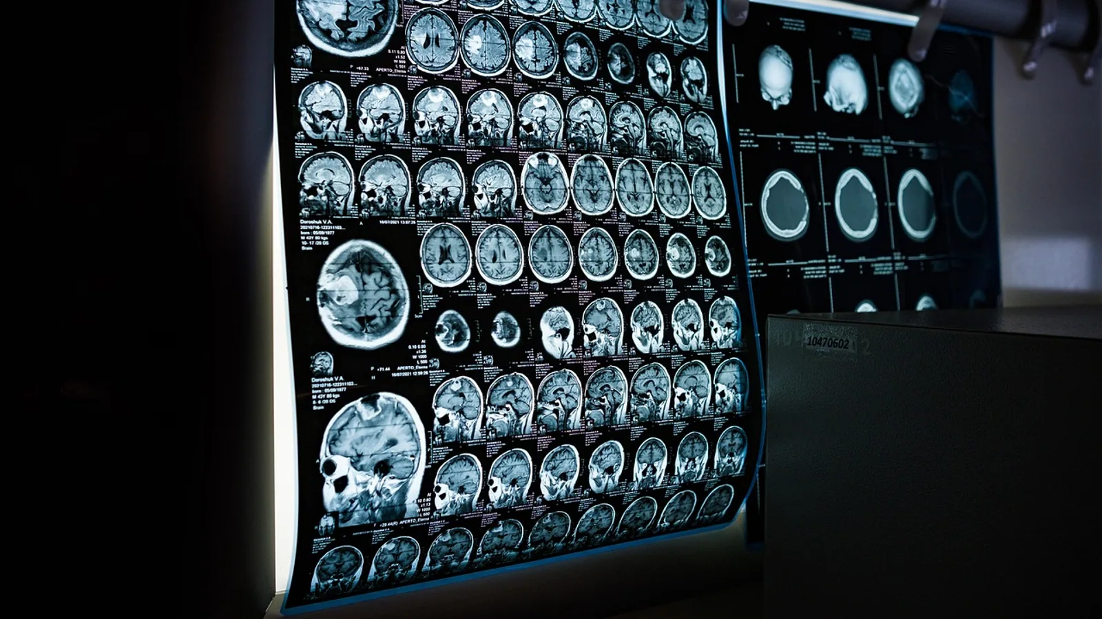
The thalamus is the paired diencephalic structure that receives almost every ascending sensory, motor, and limbic pathway and transmits that information to specific areas of the cerebral cortex through topographically organized thalamocortical projections. Its function is not passive switching: thalamic circuits transform, gate, and synchronize information, and the GABAergic thalamic reticular nucleus (TRN) sits between the thalamus and cortex as an inhibitory gatekeeper [1][2].
Understanding thalamic function matters because it explains how an animal builds a coherent sensory world. A dog tracking a scent, a cat orienting to a mouse under a chair, and a horse steadying itself on uneven ground all depend on thalamic relay circuits that decide which signals reach cortex, with what gain, and in what temporal pattern. When those circuits fail, the result is not blindness or numbness but disordered perception, attention, and arousal. The same circuitry generates sleep spindles and slow oscillations, which is why the thalamus is studied in both sensory physiology and sleep medicine [3][4].
This article is educational and is not a substitute for veterinary diagnosis or treatment.
The Diencephalic Relay Hub: Core Definition
The thalamus, or dorsal thalamus, is a bilateral egg-shaped mass of gray matter on either side of the third ventricle, bounded laterally by the internal capsule and connected to the epithalamus, subthalamus, and hypothalamus. Nearly all sensory pathways that reach the cerebral cortex, with the single exception of olfaction, pass through a thalamic nucleus first [5]. Olfactory information reaches cortex more directly via the piriform cortex, which is why smell is the classic exception in every comparative anatomy course.
Three anatomical facts organize everything else:
- Thalamocortical neurons are glutamatergic and excitatory. They are the projection cells that carry data to cortex.
- The TRN is a thin shell of GABAergic inhibitory neurons wrapped around the lateral thalamus. It is the only source of inhibition to relay cells from outside the nucleus itself and provides inhibitory control over the routing, gain, and timing of thalamocortical communication [1].
- Corticothalamic feedback is massive. Layer 6 of cortex sends more axons back to thalamus than thalamus sends forward, and these feedback axons target both relay cells and TRN [6].
The thalamus therefore sits inside a closed loop rather than at the end of a one-way chain. Biophysically detailed modeling of the mouse whisker pathway shows that closed-loop reciprocal connections between relay neurons in the ventral posteromedial nucleus (VPM) and TRN best reproduce thalamic spiking and local field potential responses in both awake and sleeping states [3].
Specific Versus Nonspecific Nuclei
A useful first cut divides thalamic nuclei into specific and nonspecific groups.
Specific (first-order) nuclei receive their driving input from a subcortical source and project to a single primary cortical area. The ventral posterolateral (VPL) and ventral posteromedial (VPM) nuclei carry somatosensory information from body and face respectively. The lateral geniculate nucleus (LGN) carries vision. The medial geniculate nucleus (MGN) carries audition. Each is a labeled line: the LGN does not send auditory data, and the MGN does not send visual data.
Nonspecific nuclei project more diffusely and carry contextual, arousal, and state information. The intralaminar group, including the centromedian-parafascicular complex (CM-Pf), is the most consistently implicated thalamic nuclear group in consciousness-related functions across the systematic review literature, followed by the mediodorsal and ventral groups [5]. Nonspecific nuclei do not simply add noise. In cortex, they recruit a different inhibitory target than specific nuclei: nonspecific inputs from the rhomboid nucleus powerfully activate slow-responding adapting interneurons, whereas specific ventrobasal inputs preferentially drive fast-spiking interneurons. The result is that specific inputs produce rapid feedforward inhibition that limits response duration, while nonspecific inputs produce delayed inhibition that permits lasting recurrent excitation, allowing sensory and contextual signals to summate [7].
First-Order and Higher-Order Relays
The specific/nonspecific distinction overlaps with, but is not identical to, the first-order/higher-order distinction.
- First-order relays carry simple stimulus properties from the periphery. VPL and VPM are first-order somatosensory relays, LGN is first-order visual, MGN is first-order auditory.
- Higher-order relays carry more complex response properties, provide contextual feedback, and modulate plasticity. The posterior medial nucleus (POm) is the classic higher-order somatosensory relay, and the pulvinar is the higher-order visual relay [8][2].
Higher-order thalamic input to cortex is not a minor add-on. In mouse primary somatosensory cortex, whether a layer 4 neuron receives stronger higher-order input depends on the type of embryonic progenitor it came from, and disrupting this mechanism alters higher-order sensory responses and sensory-evoked plasticity [8]. The functional implication is that the thalamus shapes cortex during development, not only during adult sensation.
The Thalamic Reticular Nucleus as a GABAergic Gate
The TRN deserves its own section because it is the structure students most often reduce to a single sentence and then misuse on exams.
The TRN contains GABAergic neurons that receive collaterals from thalamocortical axons and from corticothalamic axons, and that send inhibition back onto thalamic relay cells. It is functionally and genetically subdivided. In the mouse somatosensory TRN, calbindin-expressing (CB) neurons sit in the central core and connect with the ventral posterior nucleus, the primary somatosensory relay. Somatostatin-expressing (SOM) neurons lie along the surrounding edges and synapse with POm, a higher-order structure carrying both top-down and bottom-up information [2].
This creates two channels. CB neurons inhibit and receive excitation from first-order nuclei. SOM neurons inhibit and receive excitation from higher-order nuclei [9]. Electrical synapses (gap junctions) link TRN neurons both within and across these subtypes, forming homocellular and heterocellular connections, which means the two channels are not fully independent [9]. Modeling work shows that electrical coupling within the TRN cumulatively alters cortical integration of sequential thalamic inputs, changing response rates, duration, and correlation to sensory input [10].
The TRN is also a state-dependent gate. During sleep, TRN neurons participate in distinct oscillatory activities. Reciprocal synaptic circuits between TRN and sensory relay nuclei underlie sleep spindles, and TRN neurons can generate long-lasting plateau potentials and persistent firing that produce sustained inhibition in ventrobasal relay neurons [4]. Sleep spindle density is a measurable readout of this system: roughly a 40 percent reduction in spindle density is reported in a condition linked to weakened TRN-mediated inhibition [1].
Key Nuclei and Their Cortical Targets


The table below summarizes the nuclei a veterinary or biomedical student should be able to name from memory.
| Nucleus | Primary input | Cortical output | Core function |
|---|---|---|---|
| VPL | Contralateral body somatosensory pathways via medial lemniscus and spinothalamic tract | Primary somatosensory cortex, body region | First-order relay of touch, proprioception, nociception from trunk and limbs |
| VPM | Contralateral face somatosensory pathways, including trigeminothalamic | Primary somatosensory cortex, face region | First-order relay from face, oral cavity, and in rodents the whiskers |
| LGN | Retina via optic tract | Primary visual cortex | First-order visual relay, retinotopically organized |
| MGN | Inferior colliculus via brachium | Primary auditory cortex | First-order auditory relay, tonotopically organized |
| VA and VL | Basal ganglia via globus pallidus and substantia nigra, plus cerebellum | Primary motor and premotor cortex | Motor relay and feedback, movement initiation and correction |
| Pulvinar | Superior colliculus, cortex, and other thalamic nuclei | Association visual and parietal cortices | Higher-order visual relay, attention and contextual modulation |
| POm | Somatosensory cortex layer 6, brainstem | Primary and secondary somatosensory cortex | Higher-order somatosensory relay, contextual feedback |
| Intralaminar group, including CM-Pf | Brainstem reticular formation, spinal cord, basal ganglia | Diffuse cortical and striatal projections | Arousal, state setting, consciousness-related signaling |
| Mediodorsal | Amygdala, olfactory and limbic structures, prefrontal cortex | Prefrontal cortex | Limbic and executive relay |
| TRN | Collaterals of thalamocortical and corticothalamic axons | Inhibits thalamic relay nuclei | GABAergic gating of relay output, spindle generation |
Two entries in this table are worth extra attention because they are frequently confused. VA and VL are motor relays, not sensory relays, and they receive their driving input from the basal ganglia and cerebellum rather than from peripheral receptors. The pulvinar is a higher-order visual nucleus, not a first-order one, and it is proportionally much larger in primates than in rodents [11].
Comparative Mammalian Notes
Thalamic organization is conserved in its broad plan and variable in its proportions and lamination.
Lamination. The LGN is the clearest example. In primates it is a six-layered structure with distinct magnocellular and parvocellular laminae, and the layers are separated by koniocellular cells. In carnivores such as the cat, the LGN is also laminated but with a different number and arrangement of layers. In rodents, the LGN is much less obviously laminated. The MGN shows a similar gradient: primates have a distinct parvocellular and magnocellular division, while rodents have a more uniform structure. Students should treat lamination as a primate-and-carnivore specialization rather than a universal thalamic feature.
Relative size of associative nuclei. Foxp2 protein expression across thalamic nuclei in mice, rats, and macaques shows a consistent pattern: expression is highest in midline and intralaminar nuclei, absent in the reticular nucleus and zona incerta, and low and variable in the anterior group. Macaques show broader and in some nuclei more intense expression, particularly in associative regions such as the pulvinar and parts of the ventral group, which fits the greater complexity of primate thalamocortical circuits [11]. In practical terms, the pulvinar is a minor nucleus in a mouse and a major one in a macaque.
The reticular nucleus is conserved in its absence of Foxp2. Across mice, rats, and macaques, Foxp2 is consistently absent from the reticular nucleus and zona incerta [11]. This is a useful marker for identifying TRN in comparative work.
Circuit principles extend beyond mammals. The zebrafish preglomerular complex is a thalamocortical-like pathway that provides the primary visual and vibrational input to the pallium, with sensory-specific and topographically organized responses. Pallial neurons show topographically organized hierarchies from sensory-specific to multimodal and coincidence-detecting responses. The authors conclude that hierarchies of sensory transformations across topographically organized thalamocortical-like circuits reflect a convergent principle across vertebrates [12]. For veterinary students working across fish, amphibian, reptile, bird, and mammal species, this is the key comparative point: the thalamic relay principle is older than the mammalian cortex.
Developmental timing. In developing ferrets, auditory cortex neurons respond to sound before the ears open, and single-unit recordings show responses emerge first in cortical subplate neurons, then in layer 4. Sound-evoked spike latencies are longer in layer 4 than in subplate, consistent with subplate neurons relaying thalamic information to layer 4 [13]. Thalamocortical transmission therefore begins before the mature cortical target layer is even wired.
How Thalamic Function Is Studied
Several methods appear repeatedly in the primary literature, and knowing them helps when reading papers.
Electrophysiology in thalamic slices. Researchers record from TRN and relay neurons in acute slices, often from adult mice, and use pharmacological tools to isolate currents. This approach identified the persistent firing and plateau potentials in TRN neurons that sustain inhibition in ventrobasal relay cells [4].
Optogenetics with Cre driver lines. Three Cre driver lines are characterized for dorsal thalamus: Olig3-Cre expresses broadly including thalamocortical neurons and interneurons, while HDC-Cre and CRH-Cre have restricted patterns within and across sensory relay nuclei. All three express by the time of natural birth. Targeting channelrhodopsin to thalamus with HDC-Cre or CRH-Cre allows optogenetic activation of thalamocortical afferents in primary visual cortex [14]. This is the standard toolkit for dissecting thalamocortical circuitry in mice.
Magnetic resonance spectroscopy and resting-state fMRI. In human work, thalamic GABA and glutamate concentrations can be measured and related to intrinsic functional connectivity between thalamus and somatosensory cortex [15].
Diffusion tractography and connectivity-based parcellation. Thalamic nuclei can be segmented noninvasively by parcellating the thalamus according to its cortical connectivity, and results using modern high-resolution diffusion data remain remarkably similar to the original parcellation work [16].
Biophysical network modeling. Multi-compartmental models of the mouse whisker pathway test whether intrathalamic connections are reciprocal or non-reciprocal. Closed-loop connectivity best reproduces experimental recordings across awake and sleep states and supports spindle oscillations during sleep [3].
Clinical and Comparative Relevance
Thalamic dysfunction produces recognizable patterns across species.
In chronic low back pain patients, four weeks of transcutaneous auricular vagus nerve stimulation or transcutaneous greater auricular nerve stimulation reduced pain intensity, bothersomeness, and interference scores, and both interventions modulated static and dynamic functional connectivity within thalamocortical, limbic, and sensorimotor networks. The vagus stimulation group showed decreased static connectivity and fiber integrity between the mediodorsal nucleus and the left postcentral gyrus [17]. This is a direct demonstration that thalamic relay circuits are modifiable by peripheral nerve stimulation.
In pediatric anti-NMDA receptor encephalitis, total thalamic volumes did not differ from controls, but subnuclear analysis revealed nuclei-specific volumetric changes and structural covariance network reorganization [18]. The lesson for clinicians is that whole-thalamus volume is a blunt instrument. In type 1 diabetes mellitus, by contrast, total thalamic volume is reduced bilaterally by roughly 7.6 percent on the left and 8.0 percent on the right, with the most pronounced atrophy in anterior, medial, and lateral groups [19].
Sensory over-responsivity, the extreme sensitivity to sensory stimuli seen in autism spectrum disorder, anxiety, and ADHD, has been linked to altered thalamic sensory gating. Within the autism group, sensory over-responsivity severity correlated negatively with thalamic GABA and positively with somatosensory cortex glutamate [15]. This is the clearest human evidence that the thalamic excitatory/inhibitory balance is behaviorally meaningful.
In experimental stroke, damage to somatosensory cortex dampens the excitability of surviving thalamocortical circuits. Chronic optogenetic stimulation of thalamocortical axons promotes formation of new and stable thalamocortical synaptic boutons and enhances recovery of somatosensory cortical circuit function and forepaw sensorimotor ability [20]. The thalamus is therefore a therapeutic target in rehabilitation, not only a passive victim of cortical injury.
Quick Review
- All sensory pathways except olfaction synapse in the thalamus before reaching cortex [5].
- Specific nuclei (VPL, VPM, LGN, MGN) carry labeled-line first-order information. Nonspecific nuclei (intralaminar group) carry arousal and contextual signals [5][7].
- Higher-order relays such as POm and pulvinar carry complex properties and contextual feedback, and their cortical targeting is set during development by progenitor type [8].
- The TRN is GABAergic, subdivided into CB and SOM subtypes, and gates first-order and higher-order channels respectively [2][9].
- TRN neurons are electrically coupled and generate sleep spindles and slow oscillations [10][4].
- VA and VL are motor relays from basal ganglia and cerebellum, not sensory relays.
- The pulvinar and other associative nuclei are proportionally larger in primates than in rodents [11].
Clinical Relevance, Limitations and Common Mistakes
Mistake 1: Calling the thalamus a passive relay. The word "relay" invites the idea that the thalamus forwards signals unchanged. It does not. Thalamic circuits transform sensory input, and TRN inhibition controls routing, gain, and timing [1]. Modeling shows that closed-loop connectivity, not simple feedforward transmission, best explains recorded thalamic activity [3].
Mistake 2: Assuming the TRN is one uniform nucleus. The TRN has at least two genetically and topographically distinct subtypes with different targets and different input sources [2][9]. A question that treats the TRN as a single homogeneous inhibitory pool will produce a wrong answer.
Mistake 3: Forgetting the olfactory exception. Students routinely list olfaction as a thalamic relay. It is the exception, and it is the exception in every standard comparative anatomy text.
Mistake 4: Confusing first-order with specific, and higher-order with nonspecific. The two distinctions overlap but are not the same. POm is higher-order but not part of the classic nonspecific intralaminar group. The rhomboid nucleus is nonspecific but is not a first-order relay [7].
Mistake 5: Treating lamination as universal. The six-layered primate LGN is a primate feature. Rodent LGN is far less laminated. Comparative exam questions often hinge on this.
Mistake 6: Using whole-thalamus volume as a proxy for nucleus-specific change. Subnuclear analysis can reveal changes that total volume misses, as shown in pediatric anti-NMDAR encephalitis [18].
Limitations. Most detailed circuit-level work on TRN subtypes, electrical coupling, and closed-loop connectivity comes from rodent models, particularly mice. Extrapolation to dogs, cats, horses, and cattle requires care because thalamic proportions, lamination, and associative nucleus development differ across orders [11]. Individual animals with suspected thalamic or thalamocortical dysfunction need evaluation by a veterinarian, since the clinical signs overlap with cortical, brainstem, and peripheral causes.
A Working Model of Thalamic Relay
The flowchart below traces the main path of a sensory signal from receptor to cortex, including the two points where the TRN can intervene.
flowchart TD
    A[Peripheral receptor] --> B[Ascending sensory pathway]
    B --> C[First order thalamic nucleus]
    C --> D[Thalamocortical neuron]
    D --> E[Cortical layer 4]
    E --> F[Cortical layer 6 feedback]
    F --> G[Thalamic reticular nucleus]
    G --> H[Inhibition of relay]
    F --> C
    I[Higher order thalamic nucleus] --> J[Association cortex]
    J --> I
    G --> I
Frequently Asked Questions
What is the main function of the thalamus?
The thalamus relays almost all sensory and many motor and limbic signals to the cerebral cortex and actively transforms, gates, and synchronizes that information. The GABAergic thalamic reticular nucleus controls the routing, gain, and timing of the relay [1].
Which sensory system does not pass through the thalamus?
Olfaction is the exception. Every other major sensory modality synapses in a thalamic nucleus before reaching cortex [5].
What is the difference between specific and nonspecific thalamic nuclei?
Specific nuclei receive driving input from one subcortical source and project to one primary cortical area, such as VPL to somatosensory cortex. Nonspecific nuclei project more diffusely and carry arousal and contextual information, with the intralaminar group most consistently linked to consciousness-related function [5][7].
What does the thalamic reticular nucleus do?
The TRN is a GABAergic shell that inhibits thalamic relay neurons, gating sensory flow and generating sleep spindles. It contains calbindin-expressing neurons that target first-order relays and somatostatin-expressing neurons that target higher-order relays [2][9].
What are first-order and higher-order thalamic relays?
First-order relays carry simple stimulus properties from the periphery, such as VPL and LGN. Higher-order relays such as POm and pulvinar carry more complex properties and provide contextual feedback to cortex [8].
Do all mammals have the same thalamic organization?
No. The broad plan is conserved, but proportions and lamination differ. Primates have a larger pulvinar and a more distinctly laminated LGN than rodents, and Foxp2 expression in associative thalamic territories is broader in macaques than in mice or rats [11].
Related Articles
- Upstream Processing in Biotechnology: A Practical Guide
- Beeswax Processing and Quality Control
- Downstream Processing: A Practical Guide for Biotech Scientists
- mRNA Processing in Eukaryotes: Steps and Mechanisms
- RNA Processing: Steps, Types, and Mechanisms Explained
- Beef Cattle Arrival Processing Plans
Sources
- How Do Similar Thalamocortical Circuits Produce Psychosis Versus Compulsivity?
- Two dynamically distinct circuits drive inhibition in the sensory thalamus.
- Closed-Loop Connectivity Best Supports Angular Tuning and Sleep Dynamics in a Biophysical Thalamocortical Circuit Model.
- TRPM4 Conductances in Thalamic Reticular Nucleus Neurons Generate Persistent Firing during Slow Oscillations.
- Thalamus and consciousness: a systematic review on thalamic nuclei associated with consciousness.
- Somatosensory control of thalamic relay neurons is regulated by two distinct layer 6 feedback systems.
- Target Interneuron Preference in Thalamocortical Pathways Determines the Temporal Structure of Cortical Responses.
- Higher-order thalamocortical circuits are specified by embryonic cortical progenitor types in the mouse brain.
- Functionally Distinct Circuits Are Linked by Heterocellular Electrical Synapses in the Thalamic Reticular Nucleus.
- Electrical Coupling within Thalamocortical Networks Cumulatively Reduces Cortical Correlation to Sensory Inputs.
- Comparative analysis of Foxp2 expression in the thalamus of mice, rats, and macaques: implications for the evolution of language circuits.
- Hierarchical sensory processing in zebrafish thalamocortical-like circuits.
- Subplate neurons are the first cortical neurons to respond to sensory stimuli.
- Cre driver mouse lines for thalamocortical circuit mapping.
- Sensory over-responsivity is related to GABAergic inhibition in thalamocortical circuits.
- Revisiting the role of structural connectivity-based parcellation in thalamic nuclei segmentation: Benchmarking against recent state-of-the-art methods.
- Effects of transcutaneous auricular nerve stimulation on thalamic relay: A randomized brain imaging study in chronic low back pain patients.
- Thalamic nuclei volumes and structural covariance network in patients with Anti-N-methyl-D-aspartate receptor encephalitis.
- Thalamic Nuclei Atrophy in Type 1 Diabetes Mellitus Across Disease Duration.
- Optogenetic rewiring of thalamocortical circuits to restore function in the stroke injured brain.