Taenia Coli: Anatomy, Structure and Function
By Dr. Zubair Khalid, DVM, MS, PhD ·
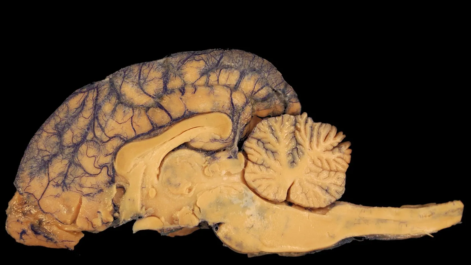
The taenia coli are three narrow bands of longitudinal smooth muscle that run along the outer surface of the colon, separate from the thin sheet of longitudinal muscle that covers the rest of the gut. They are the structural reason the colon looks like a string of pouches rather than a smooth tube, because each band is shorter than the colon it lies on and pulls the wall into segmental folds called haustra.
This matters for veterinary students and clinicians because colonic motility, fecal consistency, and several species-specific digestive strategies depend on how these bands are arranged. A horse, a rabbit, and a dog do not share the same colonic muscle architecture, and those differences shape how each species ferments, stores, and expels ingesta.
This article is educational and is not a substitute for veterinary diagnosis or treatment.
The Three Bands and Their Names
In species that have a fully banded colon, the taeniae are named by their relationship to the mesocolon, the fold of peritoneum that suspends the colon from the dorsal body wall.
- Taenia mesocolica. This band lies at the attachment of the mesocolon. It is the reference point for the other two.
- Taenia omentalis. This band lies where the greater omentum attaches to the colon. In species with a well-developed omentum, it carries the omental apron.
- Taenia libera (antimesocolica). This band lies on the free surface of the colon, opposite the mesocolic attachment and away from the omental attachment. It is the band most easily seen from the abdominal approach during surgery or dissection.
The three bands are spaced roughly evenly around the circumference in the human colon, and the same general arrangement holds in the domestic species that have three taeniae. The names are positional, not structural. Each band has the same basic histology, a compacted strip of longitudinal smooth muscle cells with connective tissue and blood vessels between them.
Why the Names Matter in Practice
During abdominal exploration, the taenia libera is the landmark surgeons and anatomists use to orient the colon. The taenia mesocolica tells you where the mesocolon is attached, which is critical when you are tracing the blood supply. The taenia omentalis tells you where the omentum is anchored. In the horse, where the arrangement is different, the names still apply but the number and distribution of bands change.
Structure: How the Taeniae Are Built
The colonic wall has two muscle layers in the muscularis externa. The inner layer is circular muscle, oriented around the circumference of the tube. The outer layer is longitudinal muscle, oriented along the length of the tube. In most of the small intestine and in parts of the colon, the longitudinal layer is a continuous sheet. In the taeniae, the longitudinal layer is gathered into discrete bands.
Smooth Muscle Orientation
The circular layer and the longitudinal layer work in opposition and in coordination.
- Circular muscle contraction narrows the lumen. When a ring of circular muscle contracts, it creates a segmental narrowing that can pinch off a pouch of ingesta.
- Longitudinal muscle contraction shortens the segment. When the taeniae contract, they pull the colon lengthwise.
The taeniae are not separate muscles. They are thickenings of the longitudinal layer. Between the taeniae, the longitudinal muscle is thin or absent, which is why the wall between bands can bulge outward when the circular muscle contracts. That bulging produces haustra.
The Shorter-Band Principle
The taeniae are shorter than the colon. In the human colon, each taenia is roughly 1 cm wide and runs the length of the colon, but its total length is less than the length of the colonic tube it covers. Because the band cannot stretch to match the tube, it bunches the wall into a series of sacculations. This is the mechanical basis of haustra. The colon is not a smooth cylinder. It is a pleated tube held in that shape by three taut bands.
The same principle applies in the domestic species that have taeniae. The bands are shorter than the colon, so the colon is haustrated. When the bands relax, the haustra can flatten. When the bands contract, the haustra deepen.
Histology of the Taenia
Each taenia is composed of bundles of smooth muscle cells separated by connective tissue. The cells are elongated, spindle-shaped, and arranged in parallel. They contain the usual smooth muscle machinery: actin and myosin filaments, dense bodies, and gap junctions that allow electrical coupling between cells.
A study of the mouse colon using alpha-smooth muscle actin antibodies found that the muscularis propria, the main muscle wall that includes the taeniae, stained uniformly across proximal and distal regions [1]. The muscularis mucosae, a thinner muscle layer deeper in the wall, showed regional differences, with the proximal segment containing a less differentiated smooth muscle population [1]. That finding is about the muscularis mucosae, not the taeniae, but it shows that smooth muscle differentiation varies along the colon.
The taeniae receive autonomic innervation from the enteric nervous system and from extrinsic sympathetic and parasympathetic fibers. They also have interstitial cells of Cajal, the pacemaker cells that generate the slow waves underlying colonic motility.
Function: Haustral Segmentation and Mass Peristalsis
The taeniae do two main things. They help form haustra, and they participate in the two major patterns of colonic motility.
Haustral Segmentation
Haustral segmentation is the mixing motion of the colon. Circular muscle contracts in localized rings, and the taeniae maintain the longitudinal shortening that keeps the wall folded. The result is a series of pouches that slowly churn the ingesta, exposing it to the mucosa for water and electrolyte absorption.
Segmentation is not propulsion. It is mixing. It keeps the colonic contents in contact with the absorptive surface and helps form feces. The taeniae contribute by holding the colon in a shortened, folded state so that circular contractions produce discrete pouches rather than a simple tube.
Mass Peristalsis
Mass peristalsis is the propulsive motion that moves colonic contents over long distances. It is a coordinated wave of contraction that sweeps through a large segment of the colon, often after a meal. The circular muscle contracts behind the bolus, and the longitudinal muscle, including the taeniae, contracts ahead of it to shorten the segment and reduce resistance.
The taeniae are not the sole driver of mass peristalsis. The enteric nervous system, the interstitial cells of Cajal, and the autonomic supply all contribute. But the taeniae provide the longitudinal shortening that makes the wave efficient. Without them, the colon would be a longer, smoother tube, and the propulsive wave would have to work against more resistance.
A study of colonic smooth muscle in a mouse model of functional constipation found that mitochondrial dysfunction in colonic smooth muscle cells was associated with impaired motility [2]. The study linked reduced expression of HSF1 and mitochondrial unfolded protein response components to mitochondrial structural damage and reduced ATP production in smooth muscle cells [2]. ATP is the energy source for smooth muscle contraction, so any condition that reduces ATP in the taeniae or the circular layer will impair both segmentation and peristalsis.
A separate study of slow-transit constipation identified BIN1 and ALDH1B1 as regulators of intestinal motility in human colonic smooth muscle cells [3]. Loss of these proteins impaired ATP-evoked calcium responses and contraction, disrupted mitochondrial architecture, and increased reactive oxygen species [3]. These findings are about human colonic smooth muscle, but the basic mechanism, that smooth muscle contraction depends on mitochondrial energy production, applies across species.
The Role of the Taeniae in Colonic Compliance
The taeniae also affect how the colon stretches. Because they are shorter than the colon, they limit longitudinal expansion. When the colon fills with ingesta, the circular muscle must stretch, but the taeniae resist lengthening. This creates a pressure gradient that helps move contents toward the rectum.
A study of colonic muscle in diverticular disease noted that the colon loses tensile properties throughout life and that the colon operates mostly as a low-pressure system, evacuating only occasionally by mass peristalsis [4]. That description applies to the human colon, but the principle is general: the colon is a storage organ that uses low-pressure mixing and occasional high-pressure propulsion. The taeniae are part of the architecture that makes this possible.
Comparative Anatomy: Species Differences in Colonic Banding
The number and distribution of taeniae vary widely across species. This is one of the most important comparative anatomy points for veterinary students, because the colonic muscle pattern reflects the species' digestive strategy.
Dogs and Cats
Dogs and cats have a relatively simple colon. The longitudinal muscle layer is thin and does not form prominent taeniae in the same way as the human colon. The colon is short and relatively smooth, with less haustration. This matches the carnivore digestive strategy: a short colon that does not need to ferment plant material.
Pigs
Pigs have a moderately banded colon with haustra. The arrangement is closer to the human pattern than the dog or cat pattern, but the bands are less prominent than in the horse.
Horses
The horse has the most complex colonic anatomy of the domestic species. The large colon is sacculated and has four taeniae in some segments. The dorsal and ventral colons have different banding patterns. The small colon has two taeniae. The horse lacks taeniae in some segments, particularly parts of the small colon, where the longitudinal muscle is more uniformly distributed. This variation reflects the horse's role as a hindgut fermenter. The large colon is a fermentation vat, and the taeniae help create the sacculations that increase surface area and slow the passage of ingesta.
Rabbits
Rabbits have a large cecum and a colon with a single prominent taenia. The rabbit colon is sacculated, and the taenia helps form the haustra that mix and ferment ingesta. Rabbits practice cecotrophy, eating a special type of soft feces to recover microbial protein and vitamins. The colonic muscle architecture supports this by allowing separation of soft and hard feces.
Guinea Pigs
Guinea pigs have a large cecum and a colon with taeniae that form haustra. Like rabbits, they are hindgut fermenters and rely on cecal fermentation. The colonic bands help mix ingesta and regulate the passage of fecal material.
Ruminants
Cattle, sheep, and goats have a colon with two taeniae and haustra. The colon is not the primary fermentation site in ruminants, because the rumen does most of the work. The colonic bands are present but less prominent than in the horse.
Species Comparison Table
| Species | Number of taeniae | Haustra present | Notes |
|---|---|---|---|
| Human | 3 (mesocolic, omental, free) | Yes | Taeniae about 1 cm wide, shorter than colon |
| Dog | Thin longitudinal layer, not prominent | Minimal | Short, simple colon |
| Cat | Thin longitudinal layer, not prominent | Minimal | Short, simple colon |
| Pig | 3 | Yes | Moderate haustration |
| Horse | 4 in some segments, 2 in small colon | Yes | Lacks taeniae in some segments |
| Rabbit | 1 prominent | Yes | Large cecum, cecotrophy |
| Guinea pig | Present, number varies | Yes | Hindgut fermenter |
| Ruminant | 2 | Yes | Rumen is primary fermentation site |
How the Taeniae Are Studied and Observed
Gross Anatomy
The taeniae are visible on gross dissection. In the human colon, they appear as three pale bands on the surface of the colon, distinct from the darker, thinner wall between them. In the horse, the bands are visible on the large colon, and their number and distribution can be traced along the length of the organ.
Histology
Histological sections through the colon show the taeniae as thickenings of the longitudinal muscle layer. Standard hematoxylin and eosin staining shows the smooth muscle bundles. Immunohistochemistry with antibodies against alpha-smooth muscle actin, desmin, and other smooth muscle markers can identify the muscle cells and assess their differentiation state [1].
Organ Bath Studies
Organ bath studies measure the contractile response of colonic muscle strips. A strip of colon containing a taenia is mounted in a bath, stretched to a standard tension, and exposed to drugs or electrical stimulation. The force of contraction is recorded. This technique is used to study the effects of neurotransmitters, hormones, and drugs on colonic motility. A study of aryl hydrocarbon receptor-deficient mice used organ bath studies to measure spontaneous peristaltic amplitude and responses to acetylcholine and norepinephrine [5].
Imaging
Radiography, ultrasonography, and computed tomography can show the haustra and the colonic wall. In the horse, radiography and ultrasonography are used to assess the large colon. In small animals, ultrasonography can show the colonic wall layers and the presence of haustra.
Clinical Relevance, Limitations and Common Mistakes
Clinical Relevance
The taeniae are relevant to any condition that affects colonic motility. When colonic smooth muscle function is impaired, as in slow-transit constipation, the taeniae and the circular layer cannot coordinate properly [3]. When mitochondrial function in smooth muscle cells is disrupted, ATP production falls and contraction weakens [2]. These are not diseases of the taeniae specifically, but the taeniae are part of the affected muscle.
A study of MELAS-associated chronic intestinal pseudo-obstruction found marked loss of myofilaments in colonic muscle, along with abnormal mitochondria in smooth muscle cells [6]. The myofilaments are the contractile proteins, and their loss would impair both the taeniae and the circular layer.
A rare case of segmental additional muscle coat in the colon described a sharply defined smooth muscle band between the muscularis mucosae and the muscularis propria [7]. This is not a taenia, but it shows that abnormal smooth muscle bands can cause motility problems. The additional layer was found in the distal colon and was associated with pseudo-obstruction [7].
Limitations
The taeniae are not the only determinant of colonic motility. The enteric nervous system, the interstitial cells of Cajal, the autonomic supply, and the mucosal environment all contribute. A study of aryl hydrocarbon receptor-deficient mice found sex- and age-dependent differences in colonic motility, with reduced spontaneous peristaltic amplitude in females [5]. That study did not focus on the taeniae, but it shows that colonic motility is influenced by factors beyond muscle anatomy.
Species differences mean that findings in one species do not always translate to another. A study of alpha-smooth muscle actin in the mouse colon found regional differences in the muscularis mucosae, but the muscularis propria stained uniformly [1]. That is a mouse-specific finding. The same study would need to be repeated in other species to know whether the pattern holds.
Common Mistakes
- Confusing taeniae with the entire longitudinal muscle layer. The taeniae are thickenings of the longitudinal layer, not the whole layer. Between the bands, the longitudinal muscle is thin or absent.
- Assuming all species have three taeniae. The number varies. Horses have four in some segments and two in others. Rabbits have one prominent band.
- Thinking the taeniae are separate muscles. They are part of the muscularis externa, continuous with the longitudinal layer.
- Believing haustra are fixed structures. Haustra change shape as the muscle contracts and relaxes. They are not permanent anatomical features like ribs or vertebrae.
- Assuming the taeniae are the only cause of haustra. Haustra result from the interaction of the shorter taeniae and the circular muscle. The circular muscle contraction is what actually pinches the wall into pouches.
Quick Review
- The taenia coli are three bands of longitudinal smooth muscle on the colon: mesocolic, omental, and free (antimesocolic).
- The bands are shorter than the colon, which pulls the wall into haustra.
- The circular muscle layer narrows the lumen, and the longitudinal layer shortens the segment.
- Haustral segmentation mixes ingesta, and mass peristalsis propels it.
- Species differ in band number and haustra presence. Horses have four bands in some segments and lack them in others.
- Smooth muscle contraction depends on mitochondrial ATP, so mitochondrial dysfunction impairs colonic motility [2][3].
- The taeniae are part of the muscularis externa, not separate muscles.
Frequently Asked Questions
What are the three taeniae of the colon?
The three taeniae are the taenia mesocolica, the taenia omentalis, and the taenia libera (also called the taenia antimesocolica). They are named by their relationship to the mesocolon and the omentum.
Do all animals have three taeniae?
No. The number varies by species. Humans have three. Horses have four in some segments and two in the small colon. Rabbits have one prominent band. Dogs and cats have a thin longitudinal layer that does not form prominent taeniae.
What causes haustra?
Haustra are caused by the taeniae being shorter than the colon. The bands pull the wall into folds, and circular muscle contractions deepen the pouches. The haustra are not fixed structures. They change shape as the muscle contracts and relaxes.
What is the difference between haustral segmentation and mass peristalsis?
Haustral segmentation is a mixing motion that keeps ingesta in contact with the absorptive surface. Mass peristalsis is a propulsive motion that moves contents over long distances. Both involve the taeniae and the circular muscle.
Why do horses lack taeniae in some colonic segments?
The horse colon has a complex banding pattern that varies by segment. The small colon has two taeniae, and some segments have a more uniform longitudinal muscle layer. This variation reflects the horse's digestive strategy as a hindgut fermenter.
Can the taeniae be seen on ultrasound?
The taeniae themselves are not usually resolved on ultrasound, but the haustra and the colonic wall layers can be seen. The taeniae are best identified on gross dissection or histology.
Related Articles
- Equine Hoof Anatomy: Structures and Function
- Lac Operon: Structure, Function, and Regulation in E. coli
- Ara Operon: Structure, Function, and Regulation in E. coli
- Equine Larynx Anatomy and Function in Respiration
- Canine Lymphatic System: Anatomy and Function
- Canine Liver Anatomy and Lobular Structure
- E. coli UTI: Causes, Symptoms, and Treatment
Sources
- Differential immunoreactivity of alpha-smooth muscle actin in the mouse colon.
- Dysregulation of the HSF1-Mediated UPR(mt) Pathway in Colonic Smooth Muscle Cells Drives Motility Dysfunction in Functional Constipation.
- BIN1 and ALDH1B1 Deficiency in Colonic Smooth Muscle Drives Mitochondrial Dysfunction and Fibrosis in Slow-Transit Constipation.
- Colonic muscle in diverticular disease.
- Aryl hydrocarbon receptor deficiency leads to sex- and age-dependent colonic dysmotility in mice.
- Loss of Myofilaments in Gastrointestinal Smooth Muscle: A Novel Pathological Finding in MELAS-Associated Chronic Intestinal Pseudo-Obstruction.
- Pseudo-Obstruction of the Colon Associated With Segmental Additional Muscle and Fibrous Coat: A Rare Specimen With Novel Quantitative Immunohistochemical Analysis.