Stratum Germinativum: Skin Layer Explained
By Dr. Zubair Khalid, DVM, MS, PhD ·
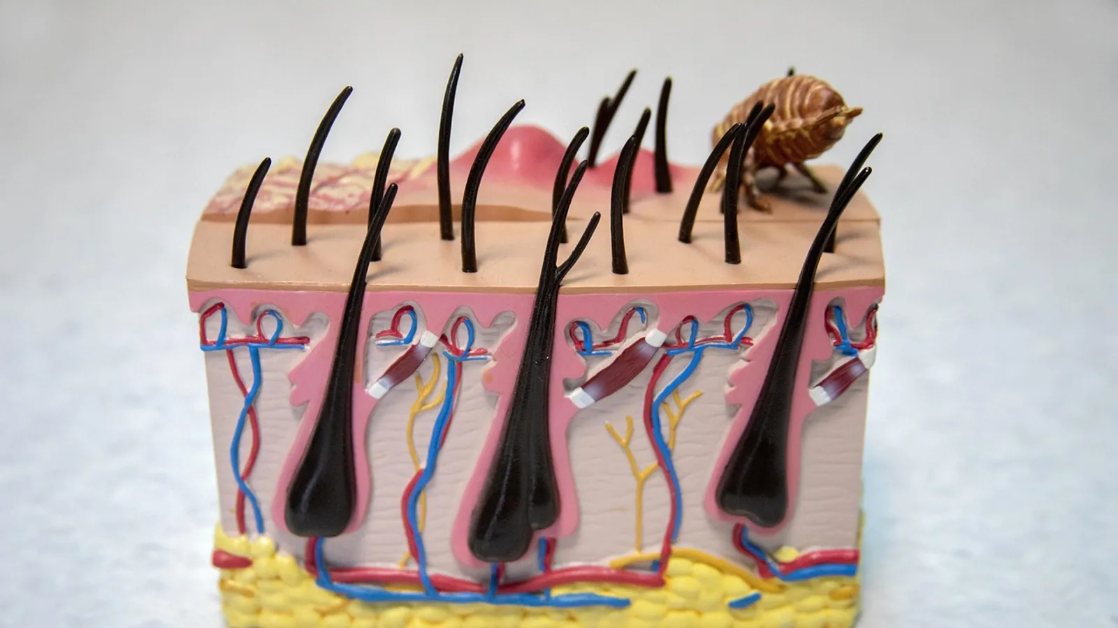
The stratum germinativum is the deepest living layer of the epidermis, a single sheet of cells resting directly on the basement membrane. In modern terminology this layer is called the stratum basale, and the two names refer to the same structure. Older veterinary and human histology texts often used "stratum germinativum" more loosely, applying it to the whole regenerative compartment of the epidermis, including the basal layer and the first one or two suprabasal layers where daughter cells begin to differentiate. That historical usage still appears in comparative anatomy papers today [1][2][3].
This layer matters because every keratinocyte above it was born there. The stratum germinativum is the mitotic engine of the skin, the anchor point for the dermo-epidermal junction, and the site where pigment, touch, and immune functions converge in a single cell-thick sheet.
This article is educational and is not a substitute for veterinary diagnosis or treatment.
What the Stratum Germinativum Is, and What It Is Not
The stratum germinativum is one cell layer thick. Its cells sit on the basement membrane, a specialized extracellular matrix that separates the epidermis from the dermis. Each basal keratinocyte attaches to this membrane through hemidesmosomes, which are adhesion complexes built around integrin beta-4 and laminin-332. Loss of either component weakens that attachment and increases the rate at which basal cells detach and move upward into the differentiating layers, a process called delamination [4][5].
Two clarifications prevent most confusion:
- Stratum germinativum and stratum basale are synonyms. They describe the same single layer. "Germinativum" emphasizes its generative role. "Basale" emphasizes its position at the base.
- Older texts use "germinativum" for a thicker zone. In that convention the term covers the basal layer plus the lower portion of the stratum spinosum, because those cells still retain proliferative capacity. When you read an older paper describing "the stratum germinativum" as several cells thick, check whether the author means the basal layer alone or the regenerative compartment as a whole.
The layer is not uniform across the body surface. It follows the contours of the dermo-epidermal junction, which is flat in thin skin and deeply interdigitated in thick, hairless skin such as the nasal planum or footpads. Those downward projections of epidermis into dermis are rete ridges, and they increase the surface area available for adhesion and nutrient exchange.
The Cells of the Basal Layer
Four cell types reside in or immediately above the stratum germinativum. Keratinocytes dominate numerically. The other three are minority populations with specialized jobs.
Keratinocytes
Keratinocytes make up the vast majority of cells in the basal layer. They are the mitotic cells of the epidermis. A basal keratinocyte divides, and the two daughter cells face different fates. One can remain in the basal layer as a progenitor, while the other detaches from the basement membrane and begins the upward journey of terminal differentiation [6]. Along that journey the cell changes which keratin genes it expresses. Basal cells express keratin 5 and keratin 14. As they enter the suprabasal layers they switch to keratin 1 and keratin 10 [7].
That keratin switch is not cosmetic. In vitro models of primary human keratinocytes show that varicella-zoster virus and herpes simplex virus 1 both replicate poorly when they first infect differentiated keratinocytes, and much more efficiently when they first infect undifferentiated basal cells [7]. The basal layer is the entry point for these viruses because its cells are metabolically active and not yet committed to forming a cornified barrier.
Melanocytes
Melanocytes sit in the basal layer and produce melanin pigment. They are far outnumbered by keratinocytes, on the order of one melanocyte for every ten basal cells. Each melanocyte extends dendritic processes that contact many neighboring keratinocytes, transferring pigment granules to them. The pigment sits over the keratinocyte nucleus and absorbs ultraviolet radiation, which reduces DNA damage.
Melanocytes are visible in comparative skin studies across vertebrate groups. In the flowered racer snake, melanophores and iridophores occupy the dermal layer while pigment cells also influence the appearance of the overlying epidermis [1]. In the shank skin of the Egyptian Balady Duck, melanocytes are found in the stratum basale [2]. The anatomical position is conserved even when the pigment chemistry and the resulting color differ.
Merkel Cells
Merkel cells are mechanoreceptors in the basal layer. They form synapse-like contacts with sensory nerve endings and respond to sustained pressure and texture. They are sparse compared with keratinocytes and are concentrated in touch-sensitive areas such as the snout, paw pads, and lips. In the European hedgehog, ascending nerve fibers run between and through epidermal cells and terminate in the upper layers of the stratum germinativum, which is the anatomical substrate for the animal's acute tactile sensitivity [8].
Langerhans Cells
Langerhans cells are the antigen-presenting cells of the epidermis. They are not born in the basal layer. They migrate into the epidermis from bone marrow precursors and take up residence in the suprabasal region, which older texts include in the stratum germinativum [9]. From there they extend processes between keratinocytes, sampling the environment for foreign antigens.
Quantitative imaging of mouse ear skin found Langerhans cells at a mean depth of 14.9 micrometers, less than 3 micrometers above the dermo-epidermal boundary [9]. The same study measured a Langerhans-to-keratinocyte ratio of about 1:15 in mouse ear skin, compared with about 1:53 in human breast skin [9]. That difference matters for any research that depends on epidermal immune sampling, because a mouse model has roughly three times the relative density of these cells.
| Cell type | Position | Useful marker | Primary function |
|---|---|---|---|
| Keratinocyte | Basal layer, single row on basement membrane | Keratin 5, keratin 14 (basal). CD49f/integrin alpha-6 for progenitor sorting [10] | Mitotic renewal, barrier formation, keratin production |
| Melanocyte | Basal layer, interspersed among keratinocytes | Melanin pigment, tyrosinase-related proteins | Pigment production and transfer to keratinocytes, UV protection |
| Merkel cell | Basal layer, concentrated in touch-sensitive skin | Keratin 20 (in mammals), synaptic contact markers | Light touch and pressure sensation |
| Langerhans cell | Suprabasal, within the regenerative compartment | CD1a, langerin (CD207), MHC class II | Antigen capture and presentation, immune surveillance |
How the Basal Layer Renews the Epidermis
The epidermis is in constant turnover. Cells are lost from the surface and replaced from below. The stratum germinativum is where that replacement starts.
A basal keratinocyte that divides can send one daughter upward while the other stays put. The upward-moving cell leaves the basal layer by delamination, losing its hemidesmosomal attachment and crossing into the stratum spinosum. There it begins producing keratin 1 and keratin 10 and starts assembling the machinery of the cornified envelope [7][4].
The decision to stay or leave is regulated by adhesion and by Notch signaling. When integrin beta-4 is experimentally removed from mouse basal cells, delamination increases. When Notch signaling is artificially boosted, integrin beta-4 expression falls and delamination rises further. When the Notch effector Rbpj is deleted, the opposite happens and delamination decreases [4][5]. This establishes hemidesmosomes as active regulators of differentiation, not just passive anchors.
Cell density and mechanical cues also feed into the decision. A review of epidermal fate coordination describes how basal progenitors respond to the identity, position, and behavior of their neighbors, and how local signaling and mechanical forces control the ordered progression of differentiation in the layers above [6].
The regenerative compartment is not confined to the interfollicular epidermis. The outer root sheath of the hair follicle contains a basal layer with the same general architecture, and cells there can be isolated using integrin alpha-6 (CD49f) as a surface marker. The CD49f-high fraction shows significantly greater proliferative and clonogenic capacity than the CD49f-low fraction or the unsorted population [10]. This confirms that the basal layer is enriched for stem and progenitor cells wherever it occurs.
Anchoring: Hemidesmosomes and the Basement Membrane
Hemidesmosomes are the rivets that hold the basal layer to the basement membrane. They are built from integrin alpha-6 beta-4 paired with laminin-332 in the matrix below, and they link internally to the keratin cytoskeleton. Without them the epidermis separates from the dermis, which is the mechanism behind certain blistering diseases.
The functional evidence is direct. Mosaic loss of integrin beta-4 or its ligand laminin-alpha-3 beta-3 gamma-2 in mouse epidermis does not cause embryonic dermo-epidermal separation, but it does increase delamination and produce mild defects in the orientation of basal cell divisions, with more oblique divisions and altered telophase correction [4][5]. The layer stays attached during development but its cells leave the basal compartment too readily.
The basement membrane is also a signaling platform. Collagen fibrils from the dermis contact it directly, as seen in the apteric skin of ostriches and emus, where numerous collagen fibrils contact the basement membrane of the epidermis [11]. In the trident goby, a few blood capillaries sit just above the basement membrane, shortening the diffusion distance for cutaneous gas exchange in poorly oxygenated water [12]. The basal layer sits at a busy interface.
Species Differences in Thickness and Pigmentation
The stratum germinativum is present in every vertebrate skin examined, but its thickness, the number of suprabasal layers above it, and the pigment arrangement vary widely.
Dogs. The epidermis is relatively thin over most of the body, with a basal layer of cuboidal to columnar cells. Pigment is concentrated in the basal layer and in hair bulbs. Breed variation in coat color reflects differences in melanocyte activity and pigment transfer rather than differences in basal layer architecture.
Cats. Similar to dogs in general architecture, with a thin epidermis and a well-defined single basal row. The nose leather and paw pads have a thicker, more interdigitated epidermis.
Horses. The epidermis is thicker than in dogs and cats in many regions, and the basal layer is correspondingly more prominent. Pigment distribution in the skin and hoof correlates with melanocyte density in the basal layer.
Humans. Human epidermis varies from thin skin with a flat dermo-epidermal junction to thick, glabrous skin with deep rete ridges. The Langerhans-to-keratinocyte ratio in human breast skin is about 1:53, compared with about 1:15 in mouse ear skin [9].
Birds. The scaly shank skin of the Egyptian Balady Duck and the Broad Breasted White Turkey shows a defined basal cell layer within the stratum germinativum. Langerhans cells are found within that basal layer, and melanocytes are present in the stratum basale of the duck [2]. The shank skin color ranges from yellow to black in the duck and is creamy-white in the turkey, reflecting differences in pigment cell activity rather than a different basal layer structure.
Reptiles. In the flowered racer snake, the epidermis includes a basal stratum germinativum beneath alpha-keratin, a mesos layer, a beta-layer, and the outer Oberhautchen [1]. In three other reptile species, the outer epidermis again shows a stratum germinativum beneath the keratin layers, with marked variation in pigment cell types among species [3].
Ratites. In ostriches and emus, the dorsal epidermis has a columnar basal layer, three to five intermediate suprabasal layers, and a thick corneous layer. The emu neck epidermis is much thinner, with a flat basal layer and only two to three narrow cell layers above it [11]. Same layer, very different proportions.
Amphibians and fish. In adult bullfrog skin, alpha-ENaC mRNA is expressed in the stratum germinativum as well as the granulosum and spinosum, and ouabain-binding studies in frog skin localize the majority of sodium pump sites deep to the outer living cell layer, in the stratum spinosum and stratum germinativum [13][14]. In the trident goby, the stratum germinativum consists of stratified columnar cells at the base of a thick epidermis adapted for cutaneous respiration [12].
Practical Implications for Owners and Keepers
The basal layer is the reason superficial skin injuries can heal without scarring. As long as the basement membrane and the basal layer remain intact, the epidermis can rebuild itself from below. When a wound is deep enough to destroy the basal layer, healing depends on migration of cells from the wound edges and from hair follicle reservoirs.
Several practical points follow from the anatomy:
- Abrasions that remove only the surface layers heal quickly. The basal layer is preserved and supplies new cells.
- Blisters indicate separation at or near the basement membrane. The fluid collects between the basal layer and the dermis, or within the basal layer itself. This is a deeper injury than a scrape.
- Pigment changes can signal basal layer disturbance. Because melanocytes sit in the basal layer, conditions that damage or stimulate that layer can change skin color.
- Topical products penetrate differently depending on the layer. A dry electrode designed for ECG recording uses microneedles that penetrate only to the stratum germinativum, bypassing the electrically insulating stratum corneum [15]. The same principle applies to transdermal drug delivery. The basal layer is a target, not a barrier.
- Bacterial penetration depends on epidermal integrity. In turkey skin, conventional defeathering left a thin layer of epidermis including about 0.3 micrometers of stratum germinativum on the surface, and that residual layer acted as an effective physical shield against Salmonella typhimurium. Steam-spray defeathering removed all epidermis and allowed the deepest bacterial penetration, beyond 200 micrometers [16].
Clinical Relevance, Limitations and Common Mistakes
The stratum germinativum is the target of immune attack in several skin diseases. In cutaneous lupus erythematosus, cytotoxic lymphocytes invade the basal layer of the epidermis and induce keratinocyte cell death, producing the interface dermatitis that defines the condition [17]. This is a direct demonstration that the basal layer is an immunological battleground, not just a structural foundation.
The basal layer also participates in barrier regulation. High-molecular-weight hyaluronan is produced in large amounts in the epidermal basal layer, and treatment of HaCaT keratinocytes with high-molecular-weight hyaluronan decreases expression of claudin-1, a tight junction protein. Knockdown of the hyaluronan receptor CD44 blocks that effect, and knockdown of hyaluronidase 1 also decreases claudin-1 expression [18]. The basal layer is metabolically active in ways that reach beyond simple cell production.
Vitamin D receptor and calcium-sensing receptor signaling in keratinocytes regulate proliferation and differentiation. Deleting both receptors from keratin 14-expressing epidermal keratinocytes in mice causes alopecia, delays wound re-epithelialization, and leads to spontaneous squamous cell carcinoma with age. The same cells show reduced expression of oxidative stress response genes and DNA repair genes, including Xpc and Gadd45a [19]. This links basal layer signaling to long-term epidermal health.
Common mistakes in reading histology and in clinical reasoning:
- Treating germinativum and basale as different layers. They are the same layer. The confusion arises from older usage that extended the term to include suprabasal cells.
- Assuming the basal layer is always one flat sheet. It follows rete ridges and is columnar in some species and regions, cuboidal in others [11][12].
- Forgetting that Langerhans cells are suprabasal. They reside in the regenerative compartment but not in the basal row itself [9].
- Assuming melanocyte density is the same across species. The ratio of Langerhans cells to keratinocytes alone varies more than threefold between mouse and human skin, and pigment cell distribution varies even more across vertebrate groups [9][1][2][3].
- Overlooking the basement membrane. The basal layer's behavior depends on its attachment. Hemidesmosome loss changes differentiation dynamics even without visible blistering [4][5].
What remains uncertain is the precise regulation of the stay-or-leave decision in different species and body sites. Most mechanistic work on delamination and Notch signaling comes from mouse models, and the extent to which those findings translate to dogs, cats, and horses is not fully established. Individual animals vary, and any skin lesion that concerns an owner should be evaluated by a veterinarian.
Frequently Asked Questions
Is the stratum germinativum the same as the stratum basale?
Yes. They are two names for the same single basal layer of the epidermis. "Germinativum" refers to its role in generating new cells, and "basale" refers to its position at the base.
Why do some textbooks describe the stratum germinativum as multiple layers?
Older texts used the term for the entire regenerative compartment, which includes the basal layer and the lower suprabasal cells that still retain proliferative capacity. In modern usage the term refers only to the single basal row.
What holds the stratum germinativum to the dermis?
Hemidesmosomes anchor basal keratinocytes to the basement membrane. These structures are built around integrin beta-4 and laminin-332, and they link to the keratin cytoskeleton inside the cell.
How many melanocytes are in the basal layer?
Roughly one melanocyte for every ten basal cells. Each melanocyte contacts many surrounding keratinocytes and transfers pigment to them.
Where are Langerhans cells located?
Langerhans cells sit in the suprabasal region, within the regenerative compartment but above the basal row. They extend processes between keratinocytes to sample for antigens.
Do all animals have a stratum germinativum?
Yes. A basal epidermal layer is present across vertebrates, from fish and amphibians to reptiles, birds, and mammals. Its thickness and the number of layers above it vary widely.
Can the epidermis regrow if the basal layer is destroyed?
Regrowth depends on surviving basal cells at the wound edges and on stem cells in hair follicles. If the basal layer and its reservoirs are completely destroyed over a large area, the epidermis cannot rebuild itself from that site.
Why does the basal layer matter for skin infections?
Many pathogens enter through the basal layer because its cells are undifferentiated and metabolically active. Viruses such as varicella-zoster and herpes simplex 1 replicate more efficiently when they first infect basal keratinocytes than when they infect differentiated cells above.
Related Articles
- Canine Integumentary System: Skin Layers and Adnexal Structures
- Layers of a Veterinary Bandage: A Practical Guide
- Aviary Layer System Management
- Layer Hen Lighting Programs
- Starting a Commercial Layer Farm
- Feeder and Waterer Placement for Layer Flocks
Sources
- Dorso-ventral skin of flowered racer snake (coluber flourulentus, perreti schatti, 1988): Histological, Keratin immunohistochemical, and ultrastructural characterization.
- Unique insights into morphological characterization and functional adaptation of the scaly shank skin in aquatic and terrestrial birds.
- Comparative ultrastructural-functional characterizations of the skin in three reptile species; Chalcides ocellatus, Uromastyx aegyptia aegyptia, and Psammophis schokari aegyptia (FORSKAL, 1775): Adaptive strategies to their habitat.
- Hemidesmosomes and Notch signaling regulate epidermal differentiation via delamination.
- Hemidesmosomes regulate epidermal differentiation during embryogenesis.
- Keeping up with the neighbors: Coordinating cell fate within and across epidermal layers.
- Comparative analysis of varicella-zoster virus and herpes simplex virus 1 interaction with epidermal terminal differentiation in primary human keratinocytes models of differentiation.
- Localisation and structure of the receptors in the snout skin of the European hedgehog (Erinaceus europaeus) at the level of light and electron microscopy.
- Multiphoton high-resolution 3D imaging of Langerhans cells and keratinocytes in the mouse skin model adopted for epidermal powdered immunization.
- Isolation of Keratinocytes from Human Hair Follicles and Selection of a Population Enriched in Epithelial Stem and Progenitor Cells by Flow Cytometry.
- Ultrastructure and immunohistochemistry of apteric skin in ratites and its epidermal soft cornification.
- Histology and morphometry of the skin of the trident goby Tridentiger brevispinis (Perciformes, Gobiidae).
- Localization of ENaC subunit mRNAs in adult bullfrog skin.
- Localization of Na+-pump sites in frog skin.
- Skin-Potential Variation Insensitive Dry Electrodes for ECG Recording.
- Penetration of Salmonella typhimurium into Turkey Skin.
- Anifrolumab has a direct immunoregulatory effect on inflamed keratinocytes: implications for the treatment of lupus erythematosus skin lesions.
- Role of HYAL1 on hyaluronan metabolism and the regulation of epidermal tight junction component CLDN1 in HaCaT cells.
- Deletion of vitamin D receptor with calcium sensing receptor in keratinocytes promotes epidermal tumorigenesis by limiting dna repair and oxidative stress response genes.