Splenius Capitis Muscle: Anatomy and Function
By Dr. Zubair Khalid, DVM, MS, PhD ·
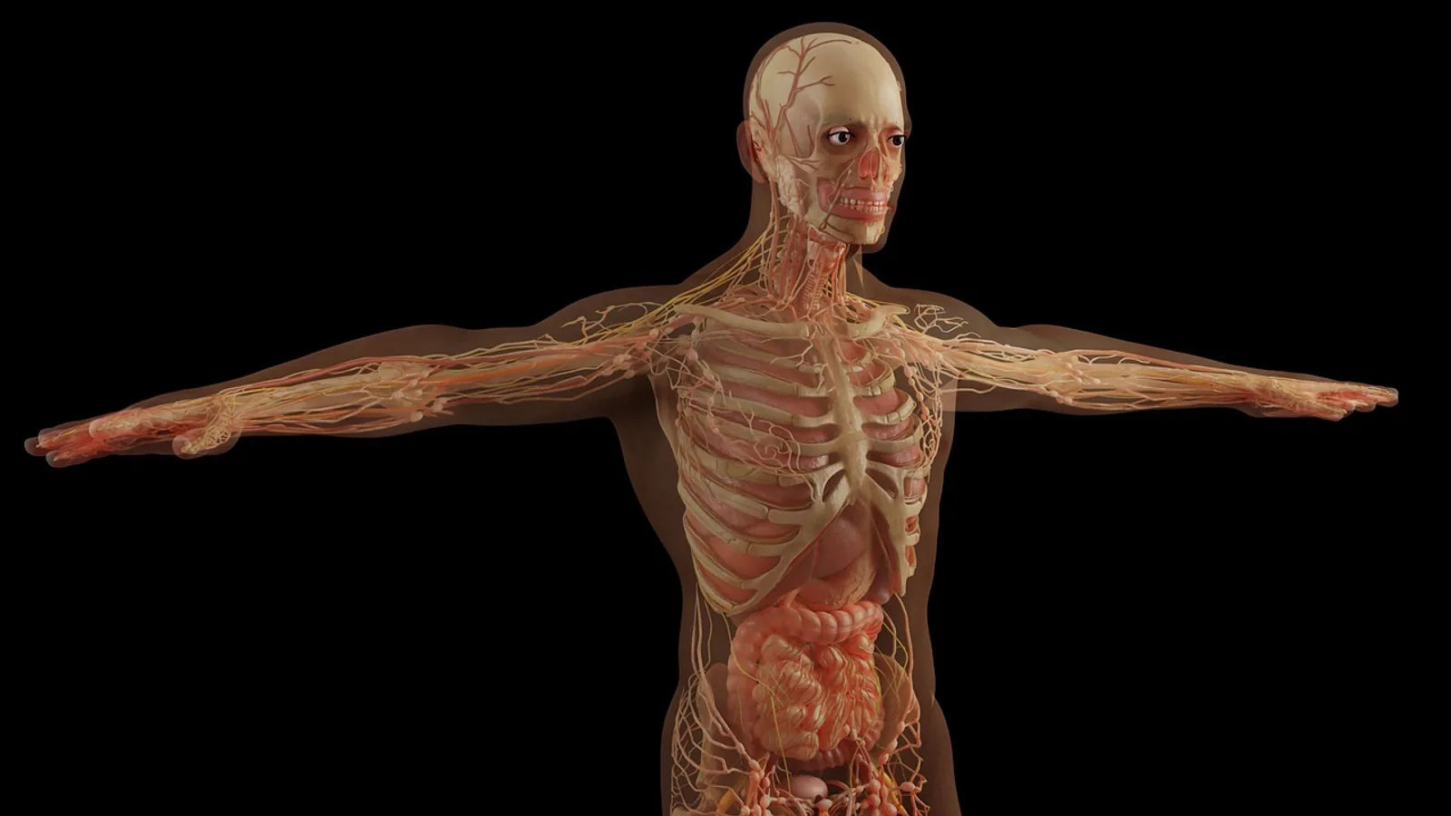
The splenius capitis muscle is a flat, strap-like dorsal neck muscle that arises from the nuchal ligament and the spinous processes of the cranial thoracic vertebrae and inserts on the occipital bone and mastoid region of the skull. Its contraction extends the head and neck and rotates the head toward the same side (ipsilateral rotation), a function that is especially prominent in quadrupedal mammals, where the splenius acts as a major head elevator rather than a secondary postural stabilizer.
Understanding the splenius capitis matters because it sits at the crossroads of anatomy, surgery, and rehabilitation. It is one of the most frequently injected muscles in cervical dystonia, a target for botulinum neurotoxin therapy, and a landmark for surgeons harvesting the occipital artery [1][2][3]. In veterinary patients, it is the principal extensor of a head that must stay level against gravity while the animal walks, eats, and looks up. A student who can reconstruct this muscle from its attachments alone can predict its action, its innervation, and its clinical behavior without memorizing a list.
This article is educational and is not a substitute for veterinary diagnosis or treatment.
Why the Splenius Capitis Matters
Movement of the head is a balance between small, deep muscles that fine-tune position and large superficial muscles that carry load. The splenius capitis belongs to the second group. In a standing dog, horse, or cow, the muscle fires continuously during quiet standing to keep the head from dropping. That endurance demand leaves a structural signature. In samples of the splenius capitis muscle from young men, the muscle contained 51.2% type 1 fibers compared with 39.7% in the vastus lateralis, and its fibers were smaller, measuring roughly 35.5 to 40.9 micrometers versus 47 to 56.1 micrometers [4]. Fiber type and fiber size follow function, and the splenius is built for sustained postural work.
Size and activity also link this muscle to disease. In people with chronic neck pain, the splenius capitis including the splenius cervicis shows altered muscle volume and fat infiltration on MRI, and in whiplash-associated disorder the fat infiltration of the splenius capitis is significantly greater than in controls [5][6]. In cervical dystonia, the splenius capitis is the most commonly injected neck muscle among seven international movement disorder centers, used in 84.9% of 305 patients [3]. Veterinary students should read those numbers as a reminder that a muscle's anatomical position determines where needles and blades go, and that mistakes carry consequences.
Anatomical Position and Attachments


The splenius capitis lies in the superficial layer of the dorsal neck, deep to the trapezius and the rhomboid muscles, and superficial to the semispinalis capitis and longissimus capitis [7][8]. In quadrupeds, it covers the dorsolateral surface of the cranial cervical region and blends rostrally with the fascia over the poll. A useful first step in dissection is to remove the trapezius and rhomboid muscles to expose the superficial neck layer that contains the splenius group.
Historically, the splenius group is treated as two parts. The splenius capitis inserts on the skull, and the splenius cervicis inserts on the cranial cervical vertebrae. In many quadrupeds these two parts merge into one sheet, which is why the term splenius capitis muscles is sometimes used to describe a composite muscle. The two parts can also be considered separately by their attachments, which is how surgeons localize them for injection or dissection.
Origin
The splenius capitis arises from the nuchal ligament and the spinous processes of the cranial thoracic vertebrae. In people, the muscle takes origin from the lower half of the ligamentum nuchae and from the spinous processes of C7 to T3 or T4. Comparative anatomists recognize the same pattern in the domestic species, with the exact number of thoracic spinous processes varying by the length of the neck. In a horse or a cow, with a long cervical column, the thoracic contribution runs further caudal than in a dog or cat, but the principle is constant: the muscle anchors on the midline dorsal axial skeleton and fans laterally as it travels cranially.
The nuchal ligament deserves separate emphasis. In the horse and ox, it is a powerful elastic structure that supports the head with minimal muscular effort, and the splenius capitis uses it as a stable origin. In the dog and cat, the nuchal ligament is less prominent, so the spinous processes of the cranial thoracic vertebrae carry most of the origin.
Insertion
The splenius capitis inserts on the occipital bone and the mastoid region of the skull. The exact footprint includes the lateral part of the occipital squama and the mastoid process of the temporal bone. The mastoid process is a small bony prominence just caudal to the external acoustic meatus, and it is the landmark that identifies the muscle from the surface in many species. In the horse and ruminant, the mastoid region is represented by the mastoid part of the temporal bone, which provides the same attachment function.
An important reinforcement for surgeons is that the cranial part of the longissimus capitis muscle contains an intramuscular tendon that inserts on the mastoid process, which serves as a reliable ultrasound landmark for injection of that muscle [9]. The splenius capitis itself attaches immediately adjacent to this region, so the occipital and mastoid area collects several distinct muscles within a small space. In the posterior neck of the dog, the splenius capitis forms the superomedial border of a muscular triangle that also contains the rhomboid minor inferomedially and the levator scapulae laterally, a region where the proximal brachial plexus can be approached by a muscle-sparing route [10].
Table 1. Splenius Capitis Key Facts
| Feature | Description |
|---|---|
| Origin | Nuchal ligament and spinous processes of cranial thoracic vertebrae |
| Insertion | Occipital bone and mastoid region of the temporal bone |
| Innervation | Dorsal rami of cervical spinal nerves |
| Primary action | Extension of the head and neck |
| Secondary action | Ipsilateral rotation of the head |
| Layer | Superficial dorsal neck, deep to trapezius |
| Fiber profile | Predominantly type 1 (approximately 51% type 1 fibers in a sampled human cohort) [4] |
| Common clinical role | Target for botulinum toxin injection in cervical dystonia [3] |
| Vascular relationship | Occipital artery passes near or through the muscle [2] |
Comparative Anatomy Across Species
The splenius capitis is conserved in position and function but varies in how it is divided and in how much fascia binds it to its neighbors. Recognizing species differences is essential for dissection, surgery, and imaging interpretation.
Dog
In the dog, the splenius capitis and splenius cervicis are distinct but closely related muscles. The splenius capitis is a thick, triangular sheet that covers the dorsolateral neck from the nuchal ligament and cranial thoracic spinous processes to the mastoid region and the occipital bone. It lies superficial to the semispinalis capitis and is covered by the trapezius and the rhomboid muscles. The dog is a brachycephalic or dolichocephalic species depending on breed, and the muscle's length and thickness track skull shape and neck length. Because the dog carries its head forward and low during locomotion, the splenius capitis works against a substantial gravitational moment arm.
Cat
The cat resembles the dog but has a more flexible neck and a more uniform segmentation of the splenius group. The splenius capitis is a thin, flat muscle, and its boundary with the splenius cervicis is sometimes difficult to define by gross inspection alone. In this species, the insertion on the mastoid region is small, so more of the insertion concentrates on the occipital bone. Grooming and predatory pouncing both demand fast extension and rotation of the head, activities that recruit the splenius capitis together with the rectus capitis dorsalis group.
Horse
In the horse, the splenius capitis is a large, powerful sheet and one of the principal elevators of the head and neck. It runs from the nuchal ligament and the spinous processes of the cranial thoracic vertebrae to the occipital bone and the mastoid part of the temporal bone. The equine neck has an extensive nuchal ligament, and the splenius capitis lies superficial to the semispinalis capitis and the longissimus system. The muscle is important in the horse's ability to flex and extend the poll, and its fascial connections to the surrounding neck muscles make it part of a functional chain rather than an isolated unit.
Ruminants
In cattle, sheep, and goats, the splenius capitis is thick and broad. It arises from the nuchal ligament and the thoracic spinous processes and inserts on the occipital and mastoid region. In cattle, the muscle is often described together with the splenius cervicis as a single sheet, because the division between the capitis and cervicis parts is less obvious than in the dog. The horns and the heavy skull of the bull increase the load on the muscle, and the attachments reflect that demand. In horned breeds, extension of the head during head-to-head conflict is powered largely by the splenius and semispinalis system.
The general rule is that the more the species depends on a raised head for vision, fighting, or feeding from tall vegetation, the more robust the splenius capitis becomes. The dog and cat, which hold the head forward, have thinner muscles that are nonetheless active throughout the day because the head is cantilevered on a flexible neck.
Innervation and Neuromuscular Control
The splenius capitis is innervated by dorsal rami of the cervical spinal nerves. Dorsal rami are the posterior branches of the spinal nerves, and they supply the epaxial muscles, the muscles that sit above the transverse processes of the vertebrae. The epaxial group includes the splenius, longissimus, and semispinalis systems, all of which share this dorsal ramus supply.
The specific cervical levels vary by species and by the segmental patterning of the neck. In general, the branches correspond to the mid and caudal cervical segments, so the muscle receives several small nerve branches rather than a single named nerve. The dorsal rami also supply the skin and fascia of the dorsal neck, which is why damage to these branches can produce both weakness and altered sensation.
Control of the splenius capitis is closely tied to the trigeminocervical reflex. In human neurophysiology, electrical stimulation of trigeminal nerve branches such as the supraorbital and infraorbital nerves produces short-latency reflex responses not only in the sternocleidomastoid and trapezius but also in the splenius capitis, on the ipsilateral and contralateral sides [11]. This reflex loop links the face and the neck, allowing head orientation to be adjusted rapidly when the face encounters a stimulus. In quadrupeds, the equivalent circuitry coordinates head position with the visual and trigeminal fields as the animal moves.
Proprioceptive input from the splenius capitis travels back through the same dorsal rami. Muscle spindles are concentrated in particular regions of the muscle. In a study of the splenius muscles in 24 adult individuals, the center of the region of highest spindle abundance was localized to a specific point relative to the external occipital protuberance and the spinous process of the third thoracic vertebra, and the depth of that center was also measured [12]. That kind of mapping matters because it tells clinicians where the muscle's sensory machinery is densest, which is not always the same as the motor point or the nerve-dense region.
Action and Biomechanics
The splenius capitis has two actions that can be understood from its line of pull. Because it runs from the dorsal midline to the lateral skull, it crosses the atlanto-occipital and intervertebral joints obliquely.
Extension
Bilateral contraction of the splenius capitis extends the head and neck. Extension means the head lifts and the nose moves dorsally, or in the case of a quadruped, the head rises. In a standing horse or cow, this action counteracts the weight of the head, which is substantial. The muscle works together with the semispinalis capitis, the rectus capitis dorsalis group, and the longissimus capitis to produce a smooth lift rather than a jerky motion.
Unilateral contraction contributes to extension as well, because the muscle's line of pull on the same side still has a dorsal component. This is a common point of confusion for students who assume that a muscle with a rotatory action cannot also extend.
Ipsilateral Rotation
Unilateral contraction rotates the head toward the same side as the contracting muscle. Ipsilateral rotation means that if the right splenius capitis contracts, the head turns to the right. This occurs because the muscle pulls the mastoid and occipital region caudolaterally and dorsally, and the atlantoaxial joint allows most of the rotation. In the dog and cat, head turning during scent tracking and visual scanning depends on this action, balanced by the contralateral splenius and by muscles such as the sternocleidomastoid, which produces contralateral rotation.
Head Elevation in Quadrupeds
Veterinary students should avoid the human-centered framing that treats the splenius capitis as a minor postural muscle. In quadrupeds, the head is carried at the end of a horizontal or oblique lever, and the splenius capitis is a primary head elevator. A dog lifting its head from a down position, a horse raising its head to look over a fence, and a cow lifting its head from grazing all depend on bilateral splenius activity. The muscle is also active during locomotion, when the head must remain stable as the trunk moves up and down.
The biomechanics explain the fiber-type data. A muscle that must hold a load for hours on end needs a high proportion of fatigue-resistant type 1 fibers, which is exactly what the sampled human splenius capitis showed [4]. The same logic applies to the domestic species, even though the specific fiber percentages have not been measured in every species.
Vascular Relations
The occipital artery runs near or through the splenius capitis. In a cadaveric study of 33 sides, the uppermost fibers of the splenius capitis attached to the superior nuchal line, and the occipital artery passed through the muscle and crossed the superior nuchal line at defined points [2]. In the anterolateral approach for jugular foramen tumors, the occipital artery is described as passing over the longissimus capitis muscle and then into the external carotid artery [7]. The relationship between the splenius capitis and the occipital artery is not incidental. It is the reason the muscle is used as a surgical landmark and the reason harvest procedures can damage the artery if the plane is not respected.
How the Splenius Capitis Is Studied and Observed
Several methods are used to identify and evaluate this muscle in living animals and in cadavers.
Gross Dissection
Dissection begins with removal of the skin and the platysma, followed by reflection of the trapezius and rhomboid muscles. The splenius capitis is then visible as a flat sheet with a midline origin and a lateral skull insertion. In the dog, the superomedial border of the muscle helps define a surgical triangle in the posterior neck that also contains the rhomboid minor and the levator scapulae [10]. Tension applied to the muscle while observing the skull confirms the extension and ipsilateral rotation actions.
Ultrasonography
Ultrasound can identify the splenius capitis and adjacent muscles by their surface landmarks. For the longissimus capitis, the intramuscular tendon is a reliable landmark, and injection of that muscle can be performed in its cranial portion between the tendon and the mastoid insertion [9]. The splenius cervicis can be localized using the spinous processes of C4 and C5 and the body of the levator scapulae as reference points, with the needle advanced laterally through the levator scapulae [9]. Ultrasound is also used to check the accuracy of injections into the splenius capitis, where anatomy-guided needle placement is imprecise. In a study of 332 injections in 56 patients, the overall accuracy was 76.6%, and the lowest accuracy, 67.9%, was observed for the splenius capitis muscle [13].
Magnetic Resonance Imaging
MRI provides cross-sectional measurements of muscle volume and fat infiltration. In a study of chronic neck pain, the splenius capitis including the splenius cervicis was segmented from C3 through T1 alongside other muscles, and women with chronic idiopathic neck pain showed differences in muscle volume and fat infiltration compared with controls [5]. In whiplash-associated disorder, the splenius capitis showed significantly greater fat infiltration than in healthy controls [6]. These measurements are used in research and increasingly in clinical assessment of muscle health.
Electromyography and Reflex Testing
Electromyography can record the electrical activity of the splenius capitis during movement and during reflex testing. The trigeminocervical reflex can be recorded from the splenius capitis after stimulation of the supraorbital and infraorbital nerves, although the response in the splenius is less reliable than in the ipsilateral sternocleidomastoid [11]. For veterinarians, electromyography of the epaxial muscles is used in the workup of neck pain and in the evaluation of animals with suspected neuromuscular disease, though the technique requires sedation or anesthesia and careful needle placement.
Clinical Relevance, Limitations and Common Mistakes
The splenius capitis has three main clinical roles. It is a target for injection in cervical dystonia, where pooled data from seven movement disorder centers showed that the splenius capitis was the most commonly injected muscle, used in 84.9% of patients, and received the highest doses in the reported series [3]. It is a surgical landmark for the occipital artery. The intersection between the sternocleidomastoid and the splenius capitis forms the occipital artery triangle, which marks the proximal end of the transitional segment of the artery and allows safe distal-to-proximal harvest [1]. In the far lateral approach, the splenius capitis is used as a landmark to find the occipital artery as it crosses the superior nuchal line [2].
The muscle is also part of the paraspinal muscle group used to assess cervical spine health. In posterior cervical fusion, preoperative morphometry of the splenius capitis and other paraspinal muscles is being evaluated as a predictor of mechanical failure [14]. In chronic neck pain and whiplash, the muscle shows fat infiltration and stiffness changes [5][6][15].
Several limitations deserve mention. Most of the quantitative morphometric data come from human studies, and the extrapolation to dogs, cats, horses, and ruminants is based on comparative anatomy rather than direct measurement in every species. Fiber-type percentages, capillary density, and spindle distribution have not been measured in every domestic species. The anatomy of the splenius is also variable. A previously undescribed variant muscle connecting the longissimus capitis and the semispinalis capitis has been reported in a cadaveric dissection, and the authors suggested it arose from incomplete or abnormal columnar segregation during development [16]. Awareness of such variants matters during surgery.
Common mistakes in veterinary anatomy and practice include confusing the splenius capitis with the splenius cervicis, assuming the splenius capitis is a pure extensor that cannot rotate the head, and treating the human anatomy as the template for quadrupeds. Another frequent error is to ignore the relationship between the splenius capitis and the occipital artery during dissection of the caudal skull. Finally, anatomy-guided injections into the splenius capitis are less accurate than many clinicians assume, and imaging guidance improves accuracy [13].
Individual cases require evaluation by a veterinarian. This article is educational and does not replace a clinical examination.
Quick Review
- The splenius capitis originates on the nuchal ligament and cranial thoracic spinous processes.
- It inserts on the occipital bone and mastoid region of the temporal bone.
- It is innervated by dorsal rami of cervical spinal nerves.
- Its actions are head and neck extension and ipsilateral rotation.
- In quadrupeds, it is a primary head elevator because the head is cantilevered at the end of the neck.
- It lies superficial to the semispinalis capitis and deep to the trapezius.
- It is a landmark for the occipital artery and a common target for botulinum toxin injection in cervical dystonia.
Frequently Asked Questions
What does the splenius capitis muscle do?
The splenius capitis extends the head and neck when both sides contract, and rotates the head toward the same side when one side contracts. In quadrupeds, the extension action is a primary means of lifting the head against gravity.
Where does the splenius capitis attach?
It originates on the nuchal ligament and the spinous processes of the cranial thoracic vertebrae, and it inserts on the occipital bone and the mastoid region of the temporal bone.
What nerve supplies the splenius capitis?
The muscle is supplied by dorsal rami of the cervical spinal nerves, the posterior branches that also serve the other epaxial muscles of the neck.
Is the splenius capitis the same in dogs, cats, horses, and cattle?
The muscle is conserved in position and function, but its division from the splenius cervicis differs. The dog and cat have relatively distinct capitis and cervicis parts, while in horses and ruminants the two parts are more extensively fused into a broad sheet.
Why is the splenius capitis important in cervical dystonia?
It is the most commonly injected neck muscle for cervical dystonia and receives the highest doses in pooled clinical series, which makes its anatomy and injection accuracy directly relevant to treatment planning.
How does the splenius capitis relate to the occipital artery?
The occipital artery passes near or through the splenius capitis, and the intersection of the sternocleidomastoid and splenius capitis forms a surgical triangle used to locate the artery during harvest.
Related Articles
- Equine Muscular System: Major Muscle Groups and Function
- Equine Larynx Anatomy and Function in Respiration
- Canine Lymphatic System: Anatomy and Function
- Equine Hoof Anatomy: Structures and Function
- Fish Gills: Anatomy, Function, and Common Health Issues
- Muscle Protein Synthesis
Sources
- The Intersection Between the Sternocleidomastoid and Splenius Capitis as the Anatomical Landmark to Facilitate Occipital Artery Harvest: A Retrospective Clinical Study.
- Topographic Anatomy and Step-Wised Harvest of Intermuscular Occipital Artery in Far Lateral Approach.
- Dose per muscle in cervical dystonia: pooled data from seven movement disorder centres.
- Fiber-type composition and 3D capillary analysis of the human splenius capitis muscle.
- Greater muscle volume and muscle fat infiltrate in the deep cervical spine extensor muscles (multifidus with semispinalis cervicis) in individuals with chronic idiopathic neck pain compared to age and sex-matched asymptomatic controls: a cross-sectional study.
- Changes in Muscle Morphology in Female Chronic Neck Pain Patients Using Magnetic Resonance Imaging.
- The Anterolateral Approach, Revisited for Benign Jugular Foramen Tumors With Predominant Extracranial Extension: Microsurgical Anatomy and Case Series (SevEN-012).
- Surgical anatomy of the nuchal muscles in the posterior cervicothoracic junction: significance of the preservation of the C7 spinous process in cervical laminoplasty.
- Anatomo-sonographic identification of the longissimus capitis and splenius cervicis muscles: principles for possible application to ultrasound-guided botulinum toxin injections in cervical dystonia.
- Surgical Anatomy of the Posterior Intermuscular Approach to the Brachial Plexus
- Short-Latency Trigeminocervical Reflex Obtained Without Muscle Activation: Topographic Distribution and Methodological Approach.
- Optimal target localization for botulinum toxin A in treating splenius muscles dystonia based on the distribution of intramuscular nerves and spindles.
- Anatomy-guided injections of botulinum neurotoxin in neck muscles: how accurate is needle placement?
- Cervical Paraspinal Muscle Morphometry and Mechanical Failure: A Propensity-Matched Analysis.
- Can measuring passive neck muscle stiffness in whiplash injury patients help detect false whiplash claims?
- Previously undescribed variant muscle connecting longissimus and semispinalis capitis muscles.