Spermatic Cord: Anatomy, Contents, and Function
By Dr. Zubair Khalid, DVM, MS, PhD ·
The spermatic cord is the composite soft tissue pedicle that suspends the testis from the abdominal wall and carries every vessel, duct, and nerve that serves the testis and epididymis. In domestic mammals it consists of the testicular artery, the testicular vein and pampiniform plexus, lymphatic vessels, the ductus deferens (vas deferens), the cremaster muscle, and autonomic nerves, all wrapped within layers of the vaginal tunic.
The cord matters because it is the physical and physiological link between the gonad and the body. It holds the testis outside the thermal core of the abdomen, it cools arterial blood before that blood reaches the heat-sensitive seminiferous tubules, and it provides the anatomical landmark that surgeons, sonographers, and andrologists use every day. A student who understands the cord can predict why a varicocele damages sperm, why a retained testis is a cancer risk, why a stallion with a twisted cord is a surgical emergency, and why the inguinal canal feels different in a dog, a bull, and a horse.
What the Spermatic Cord Is
The spermatic cord is a funiculus, meaning a cord-like bundle. It begins at the internal inguinal ring, where its contents leave the abdominal cavity, and ends at the dorsal pole of the testis and the head of the epididymis. Along that course it passes through the inguinal canal and emerges at the external inguinal ring into the scrotum.
Two features define the cord in every domestic species. First, it is a conduit. Blood, lymph, sperm, and nerve signals all travel through it. Second, it is a heat exchanger. The testis requires a temperature a few degrees below core body temperature for normal spermatogenesis, and the cord is the structure that makes that gradient possible.
The cord is not the same as the testis. The testis is the gonad. The epididymis is the coiled duct that matures sperm. The cord is the suspensory and vascular pedicle that connects the testis and epididymis to the body wall. Students often blur these three structures, and that blurring causes errors in examination and in surgery.
Layers of the Cord
The cord is wrapped in coverings that are continuous with the layers of the abdominal wall. These layers are derived from the same embryonic tissues that form the inguinal canal, which is why the cord and the canal share a fascial architecture.
The outermost covering is the external spermatic fascia, derived from the external oblique aponeurosis. Deep to it lies the cremaster muscle and its fascia, derived from the internal oblique muscle. The innermost covering is the internal spermatic fascia, derived from the transversalis fascia. Inside these layers sits the vaginal tunic, a serous sac of peritoneum that has been dragged into the scrotum during testicular descent.
The vaginal tunic has two parts. The parietal layer lines the scrotal cavity. The visceral layer covers the testis, epididymis, and the contents of the cord. The potential space between them is the vaginal cavity, which normally contains only a small amount of serous fluid. In the horse and the dog this cavity is large and clinically important. In the ruminant it is smaller.
Contents of the Spermatic Cord
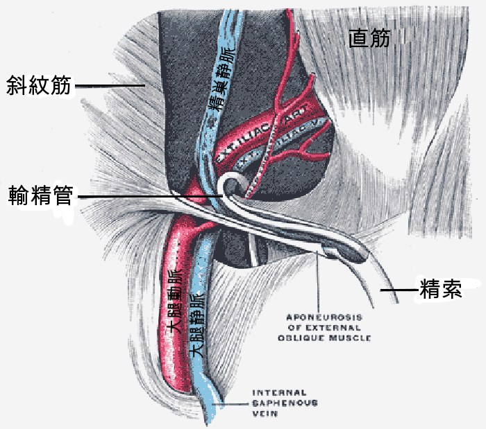

The contents of the cord are best learned as a list with origins and functions. The table below summarizes them.
| Structure | Origin | Function |
|---|---|---|
| Testicular artery | Abdominal aorta, just caudal to the renal artery | Supplies oxygenated blood to the testis and epididymis |
| Testicular vein and pampiniform plexus | Testis and epididymis, draining to the caudal vena cava (right) or renal vein (left) | Returns blood and cools arterial blood by countercurrent exchange |
| Lymphatic vessels | Testis, epididymis, and cord | Drain interstitial fluid and immune cells to the lumbar lymph nodes |
| Ductus deferens (vas deferens) | Tail of the epididymis | Transports mature sperm to the pelvic urethra |
| Cremaster muscle | Internal oblique muscle of the abdomen | Elevates and retracts the testis, adjusts scrotal position |
| Autonomic nerves | Sympathetic fibers from the testicular plexus, parasympathetic fibers from the pelvic plexus | Regulate blood flow, smooth muscle tone, and duct motility |
Testicular Artery
The testicular artery arises from the abdominal aorta. It is a long, slender vessel that becomes increasingly tortuous as it descends through the cord. In the ram, the artery shows a clear tortuous pattern along its course toward the testis, though it is less tortuous near the inguinal ring [1]. The wall of the artery thins progressively as it approaches the testis, and the vessel gives off one or two branches to the head of the epididymis and the ductus deferens in the bull [2].
The tortuosity is not decorative. It increases the surface area available for heat exchange with the surrounding veins and slows blood flow, which allows more time for thermal equilibration.
Testicular Vein and Pampiniform Plexus
The pampiniform plexus is a network of veins that surrounds the testicular artery in the cord. In the bull, the plexus is formed from intratunical veins, and its veins are smaller in diameter but more numerous than the intratunical veins [2]. The density of plexus veins in the bovine cord averages 9.37 veins per square millimeter [2]. The testicular vein proper begins 90 to 121 mm above the superior pole of the testis in the bull, and in about 2.9 percent of specimens it is doubled [2].
The plexus is the core of the cord's cooling system. It is a countercurrent heat exchanger. Warm arterial blood descending toward the testis passes through a coil of cooler venous blood returning from the testis. Heat moves from the artery to the vein, so the blood that reaches the testis is cooler than core arterial blood. The same arrangement allows the returning venous blood to be warmed before it re-enters the body, which reduces heat loss from the animal.
The relationship between the artery and the plexus is not a set of direct anastomoses. In the human cord, microscopic casts show a narrow space between the testicular artery and the plexus veins, and that space contains a capillary network that connects the two [3]. The same indirect connection through the vasa vasorum of the testicular artery has been demonstrated in the bull [2]. This means the artery and vein do not simply touch. They communicate through a fine vascular net that likely contributes to both heat exchange and local blood flow regulation.
Lymphatic Vessels
Lymphatic vessels run in the cord alongside the blood vessels. They drain the testis, epididymis, and cord to the lumbar lymph nodes. These vessels are clinically important because they are the route by which testicular tumors and infections spread, and because they must be preserved during microsurgical procedures on the cord. In microsurgical denervation of the spermatic cord for chronic orchialgia, the surgeon protects the testicular arteries and several lymphatic vessels while dividing the fibrous and neural tissue [4].
Ductus Deferens
The ductus deferens, also called the vas deferens, is the muscular tube that carries sperm from the tail of the epididymis to the pelvic urethra. It is the most rigid and palpable structure in the cord. In the bull and ram it is thick-walled and easy to identify on palpation. In the dog it is smaller but still distinct. The ductus deferens has its own blood supply from the deferential artery, a branch of the umbilical artery, and it is accompanied by autonomic nerves.
Cremaster Muscle
The cremaster muscle is a thin sheet of skeletal muscle derived from the internal oblique muscle of the abdomen. It forms a sling around the cord and testis. When it contracts, it pulls the testis toward the inguinal canal. This is the muscle responsible for the cremasteric reflex, the retraction of the testis that occurs with cold, touch, or fear.
The cremaster varies in thickness and strength across species. In ruminants it is strong and well developed, which reflects the need to retract the testis in response to environmental temperature and handling. In the dog it is thinner. In humans, a hyperactive cremaster reflex can cause testicular retraction severe enough to produce chronic pain, and men with this condition have a mean cremaster muscle thickness of 3.9 mm compared with 1.0 mm in controls [5]. Surgical release of the muscle resolves retraction in essentially all cases and resolves pain in most [6].
The cremaster is also a useful experimental model. Because it is a thin, accessible skeletal muscle with a well-defined vascular pedicle, it has been used in microcirculation research, including studies of ischemia-reperfusion injury [7] and radiation-induced changes in vascular permeability [8].
Autonomic Nerves
The cord carries sympathetic and parasympathetic nerve fibers. Sympathetic fibers arise from the testicular plexus and travel with the testicular artery. They regulate blood flow through the testicular and deferential vessels and influence smooth muscle tone in the ductus deferens. Parasympathetic fibers from the pelvic plexus contribute to duct motility and glandular secretion.
These nerves are the target of microsurgical denervation procedures for chronic orchialgia. The goal is to divide the pain-carrying fibers while preserving the arteries and lymphatics [4].
The Pampiniform Plexus as a Countercurrent Heat Exchanger
The countercurrent mechanism deserves a step-by-step explanation because it is the single most important physiological function of the cord.
- Blood leaves the abdomen through the testicular artery at core body temperature, roughly 38 to 39 degrees Celsius in most domestic mammals.
- As the artery descends through the cord, it is surrounded by the pampiniform plexus, which contains venous blood that has already passed through the testis and lost heat to the scrotal surface.
- Heat flows down the temperature gradient from the warmer arterial blood to the cooler venous blood.
- By the time the arterial blood reaches the testis, its temperature has fallen by several degrees.
- The warmed venous blood returns to the body, which conserves heat.
This arrangement maintains the testis at a temperature below core. The scrotum, with its thin skin and sparse hair, provides the surface for heat loss. The cord provides the internal heat exchange that makes the gradient possible.
Doppler studies in animals confirm the hemodynamic consequences of this anatomy. In jackasses, blood flow decreases between the level of the pampiniform plexus and the capsular artery, which helps reduce testis temperature and oxygen pressure [9]. In Assaf rams, peak systolic velocity, pulsatility index, and resistive index are higher in the proximal supratesticular artery than in the middle and distal regions, while end-diastolic velocity is higher distally [1]. Total arterial blood flow increases progressively along the cord toward the testis [1]. These findings show that the cord is not a passive conduit. It actively modifies the pressure and flow characteristics of the blood that reaches the gonad.
Comparative Anatomy Across Species
The spermatic cord differs among domestic species in length, inguinal canal diameter, and cremaster strength. These differences have practical consequences for examination, diagnosis, and surgery.
Dog
In the dog the spermatic cord is short and the inguinal canal is narrow. The external inguinal ring is small and not easily palpable as a distinct opening. The vaginal tunic is large relative to the cord, and the vaginal cavity is roomy. These features make the dog cord relatively protected from torsion but also make it more difficult to palpate the cord structures through the scrotum.
Spermatic cord tumors do occur in dogs. A case report describes a 13-year-old intact male mixed-breed dog with a B-cell lymphoma of the spermatic cord found incidentally on abdominal ultrasound, which later relapsed to the brain [10]. This is a reminder that the cord is a site of primary neoplasia, not just a conduit.
Horse
In the horse the spermatic cord is long and the external inguinal ring is palpable. The inguinal canal is wide, which is why inguinal herniation and spermatic cord torsion are more common in stallions than in dogs. The vaginal cavity is large, and the cord is mobile within it.
Spermatic cord torsion is a recognized emergency in stallions. A 180-degree torsion alters testicular arterial blood flow, and Doppler ultrasonography can detect the change in resistance and pulsatility indices in the supratesticular and capsular arteries [11]. The long cord and wide canal also make the horse prone to other cord pathology. A 19-year-old stallion with recurrent colic was found to have a leiomyoma of the spermatic cord, which was treated by hemicastration [12]. A liposarcoma of the spermatic cord extending through the internal inguinal ring into the retroperitoneal cavity has also been reported, treated with laparoscopic-assisted resection [13].
Ruminants
In ruminants the spermatic cord is relatively short and the cremaster muscle is strong. The strong cremaster allows the bull and ram to retract the testis toward the body in cold weather or during handling. The short cord and strong cremaster also make the testis less mobile than in the horse, which reduces the risk of torsion.
The bovine cord has been studied in detail. The testicular artery forms a cone-like structure with its base at the proximal part of the gonad [2]. The pampiniform plexus is dense and highly anastomosed [2]. Doppler studies in bulls show that testicular artery hemodynamics change with age. In 12-month-old bulls, pulsatility and resistivity indices are higher than in 24-month-old bulls, and diastolic velocity is lower [14]. These changes reflect the maturation of the testicular vascular bed.
In young bulls, ultrasonographic evaluation of the pampiniform plexus is used to assess testicular development under different environmental conditions [15]. The plexus is visible on B-mode and color Doppler, and its perfusion characteristics can be measured over time.
Boar
The boar has been used as a model for studying the heritability of pampiniform plexus vessel size and varicocele. In a study of 327 maternal-line boars, varicocele was diagnosed in 23.17 percent by one method and 15.1 percent by another, and heritability estimates for average plexus vessel area were 0.52 on the right and 0.46 on the left [16]. These estimates suggest that vessel size and varicocele could be selected against in breeding programs to improve semen quality [16].
How the Cord Is Examined in Practice
Veterinarians assess the spermatic cord by palpation, ultrasonography, and Doppler imaging.
Palpation is the first step. In the horse, the external inguinal ring can be palpated, and the cord can be traced from the ring to the testis. In the bull and ram, the ductus deferens is palpable as a firm, cord-like structure. In the dog, the cord is short and less accessible, but the testis and epididymis can be evaluated through the scrotal skin.
Ultrasonography provides more detail. B-mode imaging can show the testicular parenchyma, the mediastinum, and the pampiniform plexus. In young bulls, B-mode and color Doppler are used to evaluate testicular echogenicity, heterogeneity, and plexus perfusion over time [15]. In boars, ultrasonography can measure plexus vessel area and detect varicocele earlier than palpation [16].
Doppler imaging measures blood flow. Spectral Doppler of the supratesticular artery provides values for peak systolic velocity, end-diastolic velocity, resistance index, and pulsatility index. In stallions, these values differ between normal testes and testes with 180-degree torsion [11]. In rams, Doppler velocimetry shows a progressive decline in flow velocity, pulsatility, and resistivity as the artery enters the testis [1]. In jackasses, blood flow at the capsular artery correlates most strongly with semen production [9].
These techniques are not just research tools. They are used clinically to evaluate testicular viability, to diagnose torsion, and to monitor recovery after injury or surgery.
Clinical Relevance, Limitations and Common Mistakes
The spermatic cord is involved in several clinically important conditions.
Varicocele is an abnormal tortuosity and dilatation of the pampiniform plexus veins. It is associated with impaired spermatogenesis. In men with varicocele, testicular arterial blood flow is significantly decreased compared with controls, and sperm concentration correlates positively with left testicular artery blood flow [17]. The mechanism likely involves impaired microcirculation and defective heat exchange. The smooth muscle layer of spermatic veins is degraded in varicocele, and the longitudinal smooth muscle layer is absent in grade III varicocele [18]. This suggests that the vein wall itself is part of the disease process, not just a passive dilated tube.
Spermatic cord torsion is a twisting of the cord that compromises blood flow. In stallions, 180-degree torsion changes the Doppler indices of the supratesticular and capsular arteries [11]. Torsion is an emergency because prolonged ischemia damages the testis.
Spermatic cord tumors are rare but important. Liposarcoma, leiomyoma, and lymphoma have all been reported in the cord [13][12][10]. These tumors can extend through the inguinal ring into the retroperitoneal cavity, which complicates surgical planning [13].
Chronic orchialgia related to a hyperactive cremaster reflex is a condition in which the testis retracts into the inguinal canal and causes pain. Microsurgical cremaster muscle release resolves retraction in all reported cases and resolves pain in most [6]. The cremaster muscle is thicker in affected individuals [5].
Common mistakes in learning the cord include confusing the cord with the testis, forgetting that the vaginal tunic is a serous membrane rather than a fascial layer, and assuming that the pampiniform plexus is a simple vein rather than a complex countercurrent exchanger. Another common error is to think that the testicular artery and vein anastomose directly. They do not. They communicate through a capillary net and the vasa vasorum [3][2].
This article is educational and is not a substitute for veterinary diagnosis or treatment.
Quick Review
- The spermatic cord contains the testicular artery, testicular vein and pampiniform plexus, lymphatic vessels, ductus deferens, cremaster muscle, and autonomic nerves.
- All of these structures lie within the vaginal tunic, which is a serous sac derived from peritoneum.
- The pampiniform plexus is a countercurrent heat exchanger that cools arterial blood before it reaches the testis.
- The testicular artery and plexus veins do not anastomose directly. They connect through a capillary net and vasa vasorum.
- The cremaster muscle retracts the testis and varies in strength across species, being strong in ruminants and thinner in dogs.
- The cord is short in the dog, long in the horse, and relatively short in ruminants.
- Doppler ultrasonography of the cord is used clinically to assess testicular blood flow and to diagnose torsion.
Frequently Asked Questions
What are the contents of the spermatic cord?
The spermatic cord contains the testicular artery, the testicular vein and pampiniform plexus, lymphatic vessels, the ductus deferens, the cremaster muscle, and autonomic nerves. All of these structures are enclosed within the vaginal tunic.
What does the pampiniform plexus do?
The pampiniform plexus cools arterial blood before it reaches the testis. It acts as a countercurrent heat exchanger, transferring heat from the warm descending arterial blood to the cooler ascending venous blood.
How does the spermatic cord differ between dogs and horses?
In dogs the cord is short and the inguinal canal is narrow. In horses the cord is long and the external inguinal ring is palpable. The horse also has a wider inguinal canal, which makes torsion and herniation more likely.
Why is the cremaster muscle important?
The cremaster muscle elevates and retracts the testis. It adjusts scrotal position in response to temperature and touch. In ruminants it is strong and well developed.
Can the spermatic cord develop tumors?
Yes. Liposarcoma, leiomyoma, and lymphoma have all been reported in the spermatic cord of animals. These tumors can extend through the inguinal ring and require surgical removal.
What is a varicocele?
A varicocele is an abnormal dilatation and tortuosity of the pampiniform plexus veins. It is associated with decreased testicular arterial blood flow and impaired spermatogenesis.
Related Articles
- Canine Spinal Cord Anatomy and Segmental Innervation
- Equine Larynx Anatomy and Function in Respiration
- Canine Lymphatic System: Anatomy and Function
- Equine Hoof Anatomy: Structures and Function
- Fish Gills: Anatomy, Function, and Common Health Issues
- Equine Muscular System: Major Muscle Groups and Function
Sources
- Morphological and ultrasonographic characterization of the three zones of supratesticular region of testicular artery in Assaf rams.
- Angioarchitecture of the bovine spermatic cord.
- The human testicular artery and the pampiniform plexus--where is the connection?
- Microvascular Doppler Assisted Microsurgical Denervation of the Spermatic Cord in Treating Chronic Orchialgia.
- Cremaster muscle thickening: the anatomic difference in men with testicular retraction due to hyperactive cremaster muscle reflex.
- Microsurgical subinguinal cremaster muscle release for chronic orchialgia secondary to hyperactive cremaster muscle reflex in adults.
- Protective effects of immunosuppressants and steroids against ischemia-reperfusion injury in cremaster muscle flap at microcirculatory level.
- An intravital microscopy study of radiation-induced changes in permeability and leukocyte-endothelial cell interactions in the microvessels of the rat pia mater and cremaster muscle.
- Examination of jackass (Equus asinus) accessory sex glands by B-mode ultrasound and of testicular artery blood flow by colour pulsed-wave Doppler ultrasound: Correlations with semen production.
- A case of spermatic cord B-cell lymphoma relapsing to the brain in a dog.
- Doppler assessment of testicular arterial blood flow in stallions: influence of age and 180° spermatic cord torsion.
- [[Unilateral leiomyoma in the spermatic cord - a reason for intermittent colic in stallions?].](https://pubmed.ncbi.nlm.nih.gov/40897169/)
- Laparoscopic-assisted surgery for liposarcoma of the spermatic cord extending to the retroperitoneal cavity through the internal inguinal ring.
- Testicular shape, scrotal skin thickness and testicular artery blood flow changes in bulls of different ages.
- Ultrasonographic evaluation of testicular and Pampiniform plexus characteristics in young bulls under different microclimatic conditions in a tropical environment.
- The heritability of pampiniform plexus vessel size and varicocoele in boars.
- Effect of varicocele on testicular artery blood flow in men--color Doppler investigation.
- The complex structure of the smooth muscle layer of spermatic veins and its potential role in the development of varicocele testis.