Simple Cuboidal Epithelium: Function and Locations
By Dr. Zubair Khalid, DVM, MS, PhD ·
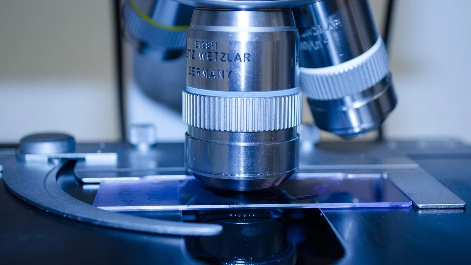
Simple cuboidal epithelium is a single layer of cube-shaped cells, each with a round nucleus near its center, that lines small ducts, kidney tubules, thyroid follicles, and the surface of the ovary. Its main jobs are secretion, absorption, and the active transport of ions, water, and small molecules across the cell layer.
This article covers what simple cuboidal epithelium looks like under the microscope, where it is found in domestic animals, how it differs from other epithelial types, and why veterinarians and pathology labs care about it. It is written for veterinary and biomedical students, technicians, and pet owners who want a clear, source-grounded reference.
This article is educational and is not a substitute for veterinary diagnosis or treatment.
What Simple Cuboidal Epithelium Is
Epithelium is one of the four basic tissue types in animals, alongside connective tissue, muscle, and nervous tissue. Epithelia cover surfaces, line cavities and tubes, and form glands. They are classified by two features: the number of cell layers and the shape of the cells in the outermost layer.
Simple means one layer. Every cell sits on the basement membrane, a thin sheet of extracellular matrix that anchors the epithelium to the tissue below. Cuboidal means the cells are roughly as tall as they are wide, like small cubes. In cross-section, a simple cuboidal epithelium looks like a single row of boxes with a round nucleus in the middle of each box.
The round, centrally placed nucleus is the single most useful feature for identifying simple cuboidal epithelium on a slide. In a simple squamous epithelium, the cells are flat and the nucleus is flattened and often off-center. In a simple columnar epithelium, the cells are taller than they are wide and the nucleus usually sits in the basal third of the cell.
Simple cuboidal cells are polarized. The apical surface faces the lumen or the outside world. The basal surface rests on the basement membrane. The lateral surfaces contact neighboring cells and are joined by junctional complexes, including tight junctions that seal the space between cells and control what passes through the layer. This polarity is what allows the sheet to move fluid and solutes in one direction rather than leaking in both directions.
The retinal pigment epithelium is a well-studied example of this architecture. In the human eye, the pigment epithelium begins as a pseudostratified columnar layer with apical tight junctions and differentiates into a simple cuboidal epithelium with melanosomes and mitochondria distributed randomly through the cytoplasm. As development proceeds, the cells enlarge, grow long apical microvilli, and form apicolateral junctional complexes. By 36 weeks of gestation, the basal cell membrane develops infoldings that increase surface area for transport [1]. That sequence, from a multilayered precursor to a mature single cuboidal layer with a transport-ready basal surface, captures what simple cuboidal epithelium is built to do.
Function of Simple Cuboidal Epithelium
The function of simple cuboidal epithelium falls into three broad categories: secretion, absorption, and transport. Many cuboidal epithelia do more than one of these at once.
Secretion
Cuboidal cells contain the organelles needed for protein synthesis and export. They have a well-developed rough endoplasmic reticulum, a prominent Golgi apparatus, and secretory vesicles. In glandular ducts, cuboidal cells secrete ions, water, and mucus that modify the primary secretion produced deeper in the gland. In the thyroid gland, cuboidal follicular cells synthesize the protein thyroglobulin and secrete it into the follicle lumen, where it is stored and later reclaimed to make thyroid hormone.
The lining of a thymic cyst illustrates how cuboidal epithelium can appear in a secretory or lining role outside the classic glandular organs. In a series of 36 resected primary thymic cysts, 18 were classified as squamous or cuboidal type, and these cuboidal-lined cysts were more often multilocular than the ciliated columnar type [2]. The point is not that the thymus is a normal site for cuboidal secretion, but that cuboidal epithelium is a common default lining for small cystic and ductal spaces throughout the body.
Absorption
Absorption requires a large surface area and a high density of membrane transporters. Cuboidal cells in the proximal kidney tubule are packed with mitochondria that supply ATP for sodium-potassium pumps, and their apical surface carries a brush border of microvilli that multiplies the area available for reabsorption. The same principle applies in smaller ducts, where cuboidal cells reclaim water and ions from the fluid passing through.
Transport and Barrier Control
Transport is the movement of specific molecules across the cell layer, either from lumen to blood or from blood to lumen. Tight junctions between cuboidal cells make the layer a controlled barrier rather than a leaky sieve. This matters in the retinal pigment epithelium, where the layer sits between the photoreceptors and the choroidal blood supply. The pigment epithelium must move nutrients inward, carry waste outward, and physically separate two compartments with very different chemical environments. The appearance of basal infoldings late in development is a structural sign that the cell is expanding its transport capacity [1].
In the kidney, transport is the dominant function. Cuboidal cells of the proximal tubule reabsorb the bulk of filtered sodium, water, glucose, and amino acids. Cells of the distal tubule and collecting duct fine-tune sodium, potassium, and acid-base balance under hormonal control. The cuboidal shape gives each cell enough cytoplasmic volume to house the mitochondria and transporter proteins this work requires.
Simple Cuboidal Epithelium Locations
Simple cuboidal epithelium is found in a limited but important set of sites. The list below covers the locations most relevant to veterinary anatomy and histology.
- Kidney tubules. The proximal and distal convoluted tubules, and parts of the collecting duct system, are lined by simple cuboidal epithelium. The proximal tubule has a brush border and eosinophilic cytoplasm. The distal tubule lacks a brush border and has a clearer lumen.
- Ducts of glands. Small intralobular and interlobular ducts of the salivary glands, pancreas, mammary gland, sweat glands, and lacrimal glands are lined by simple cuboidal cells that modify the secretion as it travels toward the surface.
- Thyroid follicles. The follicular cells that line each thyroid follicle are simple cuboidal in the resting gland. They become flatter when the gland is inactive and taller when it is actively secreting.
- Ovary surface. The ovarian surface epithelium is a simple cuboidal layer in many species, resting on a dense connective tissue layer called the tunica albuginea. In the bovine fetus, the surface of the ovarian base or hilum is cuboidal from the earliest stages examined, and by mid-development the whole ovarian surface is covered by either a stratified or simple epithelium of cuboidal cells. Later in fetal life, most of the ovarian surface is covered by a simple cuboidal epithelium [3].
- Retinal pigment epithelium. This single cuboidal layer sits between the photoreceptors and the choroid in the eye [1].
- Choroid plexus. The epithelial layer that produces cerebrospinal fluid is cuboidal and carries apical microvilli and cilia in many species.
- Small collecting ducts and some respiratory bronchioles. Simple cuboidal epithelium appears in the smallest airways and duct systems, where cilia may be present on the apical surface.
A useful contrast comes from the female reproductive tract of the Arabian oryx. The ovarian surface is covered by a squamous to cuboidal germinal epithelium over the tunica albuginea, but the uterine tube mucosa is lined by a pseudostratified epithelium with ciliated cells [4]. That single animal shows both patterns side by side, which is why species and organ context matter when you read a histology slide.
Table: Organ, Function, and Notable Features
| Organ or site | Primary function | Notable histologic features |
|---|---|---|
| Kidney proximal tubule | Absorption and transport | Brush border of microvilli, abundant mitochondria, eosinophilic cytoplasm |
| Kidney distal tubule | Ion transport and acid-base regulation | No brush border, clearer lumen, fewer microvilli |
| Collecting duct | Water and electrolyte transport | Cuboidal to columnar cells, principal and intercalated cell types |
| Salivary, pancreatic, mammary ducts | Secretion and modification of glandular fluid | Single layer, round central nuclei, often surrounded by connective tissue |
| Thyroid follicle | Synthesis and storage of thyroglobulin | Cell height varies with gland activity, colloid fills the lumen |
| Ovarian surface | Barrier and transport at the ovary surface | Simple cuboidal over tunica albuginea, cell shape varies by region and stage [3] |
| Retinal pigment epithelium | Transport, barrier, phagocytosis of photoreceptor tips | Melanosomes, apical microvilli, basal infoldings [1] |
| Choroid plexus | Cerebrospinal fluid production | Apical microvilli, cilia in many species, fenestrated underlying capillaries |
| Small ducts and bronchioles | Transport of fluid and mucus | Cilia may be present on the apical surface |
How to Recognize Simple Cuboidal Epithelium on a Slide


Start with the nucleus. In simple cuboidal epithelium, each cell has one round nucleus that sits near the center of the cell. The nuclei line up in a single row at roughly the same height, which gives the layer a regular, tiled appearance.
Next, check the cell height. Cuboidal cells are about as tall as they are wide. If the cells are clearly taller than wide, you are looking at simple columnar epithelium. If they are flat and the nuclei are elongated, you are looking at simple squamous epithelium.
Then, confirm that there is only one layer. If you can see nuclei at two or more levels within the same epithelium, the tissue is stratified or pseudostratified, not simple. Pseudostratified epithelium looks layered because the nuclei sit at different heights, but every cell still touches the basement membrane. That is a different tissue type with different functions, and it should not be confused with simple cuboidal epithelium.
Finally, look at the apical surface. A brush border indicates an absorptive cell, as in the proximal tubule. Cilia indicate a transport function, as in some small airways and the choroid plexus. A smooth apical surface with secretory vesicles suggests a secretory role.
Species Notes: Dog, Cat, and Horse
The basic histology of simple cuboidal epithelium is conserved across domestic mammals. A proximal tubule cell in a dog looks much like a proximal tubule cell in a cat or a horse: same brush border, same central nucleus, same mitochondrial density. What varies between species is the distribution and relative prominence of cuboidal-lined structures, not the fundamental cell design.
Kidney. The kidney tubule histology of the dog, cat, and horse is similar in its essentials. All three species have proximal tubules with a prominent brush border and distal tubules without one. Species differences show up mainly in the relative length of the loops of Henle and in the concentrating ability of the medulla, which reflects the animal's natural water economy rather than a difference in the cuboidal cells themselves. Dogs and cats have well-developed urinary concentrating ability. Horses produce more dilute urine and have a different medullary architecture, but the cuboidal epithelium of the cortical tubules follows the same plan.
Glandular ducts. The distribution of cuboidal-lined ducts varies more than the kidney does. Salivary gland duct systems differ in complexity between species, and the relative amount of cuboidal versus columnar epithelium in the larger ducts is not identical across dogs, cats, and horses. The mammary gland duct system also differs with reproductive status and species. In all three, the smallest ducts are the most consistently cuboidal.
Thyroid. The thyroid follicle is lined by cuboidal cells in the resting state in all domestic mammals. Follicular cell height is a dynamic feature that reflects gland activity, so a pathologist reads thyroid histology with the animal's metabolic status in mind rather than treating cuboidal height as a fixed species trait.
Ovary. The ovarian surface epithelium is simple cuboidal in many species, but its thickness and continuity vary with age, reproductive cycle, and region of the ovary. In the bovine fetus, the surface is cuboidal at the hilum from the earliest stages and becomes a complete simple cuboidal layer later in development [3]. In the mouse, the ovarian surface is described as simple squamous to simple cuboidal, resting on a distinct basal lamina, and oocytes migrate through this layer during neonatal development [5]. In the human ovary, E-cadherin expression, a marker of cell-to-cell adhesion, is most prominent in columnar surface cells, variable in cuboidal cells, and absent in flat cells [6]. That finding shows that cuboidal ovarian surface cells sit in an intermediate state of adhesion and differentiation, which is relevant to how ovarian surface lesions develop.
Eye. The retinal pigment epithelium is a simple cuboidal layer in the ferret, with basal folds, apical microvilli, and pigment granules at various stages of maturation [7]. The same general architecture is present across mammals, including dogs and cats.
Other species context. Comparative work in non-mammalian species confirms that cuboidal epithelium is a widespread building block. The rainbow trout esophagus shows a gradual transition from stratified cuboidal epithelium anteriorly to simple columnar epithelium distally, with mucous cells distributed along the length [8]. In insects, the hindgut of the gall wasp larva is built from cuboidal cells, and the follicular epithelium of the earwig ovary includes a cuboidal subpopulation covering the oocyte [9][10]. These examples are outside veterinary clinical practice, but they show that the cuboidal cell shape is an ancient and flexible solution for lining and transporting across a surface.
Simple Cuboidal Versus Other Epithelia
Students often mix up the simple epithelia. The table below separates them by structure and typical location.
| Epithelium | Cell shape | Layers | Typical locations |
|---|---|---|---|
| Simple squamous | Flat, nucleus flattened | One | Alveoli, endothelium, mesothelium |
| Simple cuboidal | Cube-shaped, round central nucleus | One | Kidney tubules, gland ducts, thyroid, ovary surface |
| Simple columnar | Taller than wide, basal nucleus | One | Stomach, intestine, gallbladder |
| Pseudostratified columnar | Tall cells, nuclei at multiple levels | One, but looks layered | Trachea, bronchi, epididymis |
| Stratified squamous | Flat cells at surface, multiple layers | Many | Skin, oral cavity, esophagus, vagina |
The key distinction is that simple cuboidal epithelium has one layer and cube-shaped cells. Pseudostratified and stratified epithelia are different tissues with different functions and should not be grouped with it.
Clinical Relevance, Limitations and Common Mistakes
Simple cuboidal epithelium matters in veterinary practice for three reasons.
First, it is the site of a large share of kidney pathology. Because the proximal tubule is metabolically active and takes up filtered toxins, it is vulnerable to damage from drugs, heavy metals, and ischemic injury. When a nephrotoxic insult kills proximal tubule cells, the cuboidal lining is lost and replaced by flattened cells or by scar tissue, and the kidney loses concentrating and reabsorptive capacity. Recognizing normal cuboidal tubule histology is the baseline for recognizing this damage.
Second, cuboidal epithelium is the tissue of origin for several tumors. Renal tubular adenomas and carcinomas, thyroid follicular tumors, and ovarian surface epithelial tumors all arise from cuboidal cell populations. The ovarian surface epithelium is the source of common epithelial ovarian carcinomas in humans, and it acquires complex epithelial characteristics, including E-cadherin expression, during carcinogenesis [6]. The ovarian surface epithelium and the adjacent peritoneal mesothelium meet at a junction near the ovarian hilum, and this junction is a site of interest in ovarian cancer research because epithelial junctions are hotspots for abnormal cell growth [11]. In animals, similar tumors occur, and histologic grading depends on recognizing the normal cuboidal pattern.
Third, cuboidal epithelium appears in cystic and ductal lesions. A tracheal duplication cyst lined by cuboidal epithelium with sebaceous gland nests has been reported in a human patient, an unusual overlap of endodermal and ectodermal development [12]. In veterinary medicine, cysts and dilated ducts lined by cuboidal cells are common incidental findings, and the pathologist's job is to decide whether the lining is normal, hyperplastic, or neoplastic.
Common mistakes to avoid:
- Calling any small cell "cuboidal." Cell shape must be judged in a well-oriented section. A tangentially cut columnar epithelium can look cuboidal.
- Confusing simple cuboidal with pseudostratified. If nuclei appear at more than one level, look for whether every cell reaches the basement membrane before deciding.
- Assuming cuboidal means inactive. Cuboidal cells are often highly active. The proximal tubule is one of the most metabolically demanding epithelia in the body.
- Treating ovarian surface epithelium as fixed. Its cell shape varies with location, age, and reproductive status, and it can be squamous, cuboidal, or columnar in the same animal [6][5].
- Forgetting the basement membrane. Simple cuboidal epithelium always sits on a basement membrane. Loss of that attachment is a feature of invasive disease.
This article covers general principles. Individual animals need a veterinarian for diagnosis and treatment.
Frequently Asked Questions
What is simple cuboidal epithelium?
Simple cuboidal epithelium is a single layer of cube-shaped cells, each with a round nucleus near the center, that lines small ducts, kidney tubules, thyroid follicles, and the ovarian surface.
What is the main function of simple cuboidal epithelium?
Its main functions are secretion, absorption, and the active transport of ions, water, and small molecules across the cell layer.
Where is simple cuboidal epithelium found in the body?
It is found in kidney tubules, the ducts of glands such as salivary and pancreatic ducts, thyroid follicles, the ovarian surface, the retinal pigment epithelium, and the choroid plexus.
How do you identify simple cuboidal epithelium on a slide?
Look for a single row of cells that are about as tall as they are wide, each with one round nucleus near the center of the cell.
Is simple cuboidal epithelium the same as simple columnar epithelium?
No. Cuboidal cells are about as tall as they are wide, while columnar cells are taller than they are wide and usually have a nucleus in the basal third of the cell.
Does simple cuboidal epithelium have cilia?
Cilia can be present on simple cuboidal cells in specific locations such as the choroid plexus and some small airways and ducts, but most simple cuboidal epithelia do not have cilia.
Why does the thyroid follicle change shape?
Thyroid follicular cells are cuboidal when the gland is resting and become taller or flatter depending on how actively they are producing and storing thyroglobulin.
Are kidney tubules the same in dogs, cats, and horses?
The cuboidal epithelium of the kidney tubules is similar across these species. Differences between them are mainly in medullary architecture and urine concentrating ability, not in the basic cuboidal cell design.
Related Articles
- RNA Location: Types, Functions, and Cellular Distribution
- Z-DNA Found in Biology: Location, Function, and Detection
- Prokaryotic DNA: Location, Structure, and Function
- Bioinformatics Salary by Location
- Swine Barn Location and Site Selection
- CRISPR Explained: A Simple Guide to Gene Editing
- Simple Columnar Epithelium: Function and Locations
- Transitional Epithelium: Structure and Function
- Stratified Epithelium: Types, Locations, and Function
Sources
- Autophagy and Exocytosis of Lipofuscin Into the Basolateral Extracellular Space of Human Retinal Pigment Epithelium From Fetal Development to Adolescence.
- Thymic cyst: A clinicopathological, immunohistochemical, and molecular study of 36 cases focusing on squamous- and ciliated-type epithelium.
- Formation of the Bovine Ovarian Surface Epithelium during Fetal Development.
- Histology of the internal reproductive organs of the female Arabian oryx (Oryx leucoryx).
- Ultrastructure of oocyte migration through the mouse ovarian surface epithelium during neonatal development.
- Increased E-cadherin expression in ovarian surface epithelium: an early step in metaplasia and dysplasia?
- Histology of the ferret retina.
- Light and electron microscopic studies on the oesophageal epithelium of the rainbow trout, Salmo gairdneri.
- Integration of Morphology, Histology, and Ultrastructure of the Gall-Inducing Andricus quercustozae (Cynipidae) Larval Alimentary Canal.
- A very simple mode of follicular cell diversification in Euborellia fulviceps (Dermaptera, Anisolabididae) involves actively migrating cells.
- Characterization of High-grade Serous Carcinoma Involving the Ovarian Surface Epithelium-peritoneal Junction.
- A case report of tracheal duplication cyst with steatocystomatous histology: a rare embryological overlap.