Rib Anatomy: Features, Landmarks, and Labeling
By Dr. Zubair Khalid, DVM, MS, PhD ·
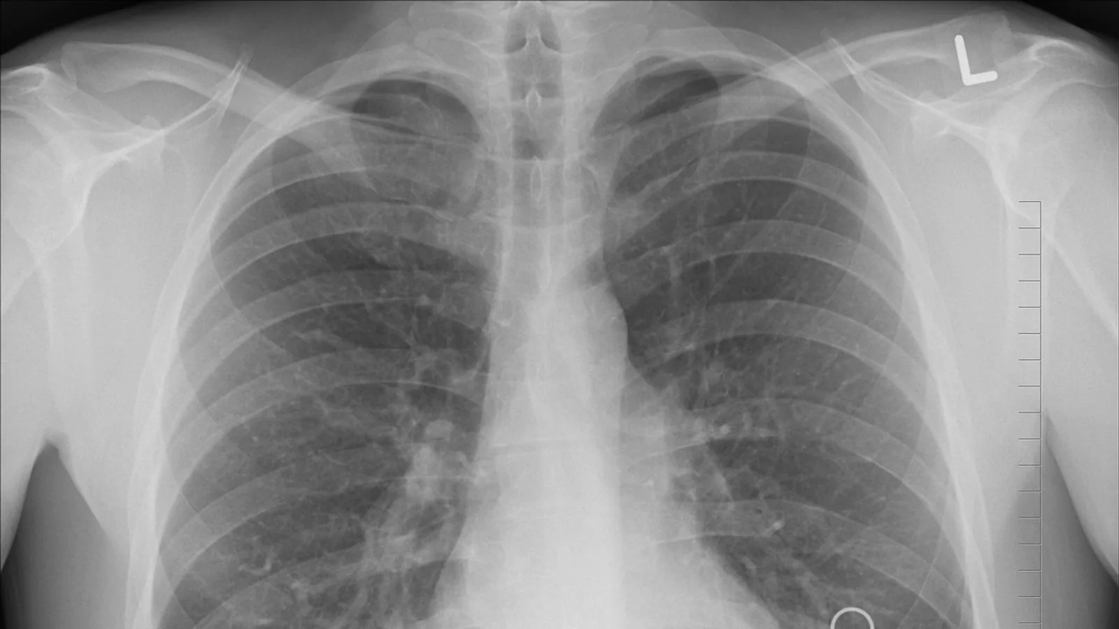
A rib is a paired, curved, dermal bone of the thoracic wall that articulates with the vertebral column and, in most cases, with the sternum through a costal cartilage. Rib anatomy covers the features found on each bone (head, neck, tubercle, angle, costal groove, and shaft) plus the way ribs are grouped into true, false, and floating categories.
Rib labeling matters because the thorax is where auscultation, thoracic radiography, thoracocentesis, and rib fracture management all begin. A clinician who can name rib 9 in a dog or rib 14 in a horse can localize a lung field, place a chest tube, or interpret a fracture line with far more confidence. This guide walks through the anatomy of a rib bone by bone, compares rib counts across species, and flags the labeling errors students make most often.
This article is educational and is not a substitute for veterinary diagnosis or treatment.
What a Rib Is and Where It Sits


A rib (Latin costa) is one of a series of paired bones that form the lateral and ventral walls of the thorax. Each rib articulates dorsally with a thoracic vertebra at the costovertebral joint and curves ventrally and downward to enclose the thoracic cavity. In mammals, most ribs reach the sternum through a segment of hyaline cartilage called the costal cartilage. The costal cartilage is not bone. It is a separate cartilaginous segment that calcifies with age in some species, which is why it can look bright and bony on radiographs of older animals.
The rib cage protects the heart and lungs, provides attachment for the muscles of respiration, and acts as a mechanical bellows. When the external intercostal muscles contract, they rotate the ribs cranially and outward, increasing thoracic volume and drawing air into the lungs. Rib morphology therefore reflects both protection and ventilation. Comparative work on fossil hominins has shown that rib cross-sectional shape at midshaft varies between groups, with Neanderthal ribs tending toward rounder cross-sections than recent human ribs, a difference that likely reflects both body form and mechanical loading [1]. Rib cross-sectional mineralized area also follows a U-shaped trend along the costal series in modern humans, being lower at the extremes and higher in the middle thoracic ribs, and this pattern differs in some fossil hominins [2]. These findings reinforce a core principle for veterinary students: rib shape is not arbitrary. It tracks function, loading, and phylogeny.
The Features Found on a Typical Rib
A typical rib is a single bone with a defined set of landmarks. When a question asks you to check all that are features found on ribs, these are the structures that belong on the list.
Head (Capitulum)
The head is the rounded, club-shaped proximal end of the rib. It carries one or two articular facets that meet the corresponding facets on the bodies of adjacent thoracic vertebrae. In most domestic mammals, the head of a typical rib articulates with two vertebrae, the one of the same number and the one cranial to it. This creates a stable, overlapping articulation that resists rotational forces during breathing.
Neck
The neck is the short, constricted segment between the head and the tubercle. It is narrower than both and serves as the attachment site for the costal ligaments that stabilize the costovertebral joint. The neck is a useful landmark in dissection because it separates the articular head from the more lateral tubercle.
Tubercle
The tubercle is a raised prominence on the caudal or lateral aspect of the rib, just distal to the neck. It bears a small articular facet that contacts the transverse process of the corresponding thoracic vertebra, forming the costotransverse joint. The tubercle is present on typical ribs but is reduced or absent on the most caudal ribs, which do not articulate with transverse processes. In the dog and cat, the tubercle is well developed on the cranial ribs and progressively smaller toward the last ribs.
Angle
The angle is the bend or change in curvature along the rib, located a short distance distal to the tubercle. It marks the point where the rib turns from its vertebral portion, which is relatively straight, to its sternal portion, which curves ventrally. The angle is a consistent landmark for measuring rib length and for describing fracture location.
Costal Groove
The costal groove is a shallow furrow running along the caudal or medial surface of the rib shaft. It accommodates the intercostal vessels and nerve. The groove is deepest in the middle third of the rib and shallows toward both ends. In surgical anatomy, the groove defines the neurovascular bundle's protected position: the intercostal nerve, artery, and vein lie in the groove, which is why needle decompression and thoracocentesis are performed along the cranial border of the rib below the target space, avoiding the groove.
Shaft (Body)
The shaft is the long, curved main portion of the rib between the angle and the sternal end. It is flattened and has an outer convex surface and an inner concave surface. The shaft's cross-sectional shape varies by species and by position along the costal series. In humans and other hominins, midshaft cross-sectional shape has been used to infer mechanical loading and growth patterns, and mineralized area at midshaft follows a predictable distribution along the rib series [2]. In domestic mammals, the shaft is generally more oval or flattened than in humans, reflecting different thoracic mechanics.
Sternal End and Costal Cartilage
The sternal end is the ventral tip of the rib. In true ribs, it joins the costal cartilage, which in turn articulates with the sternum. The costal cartilage is hyaline cartilage, not bone. It provides flexibility to the thoracic wall, allowing the rib cage to expand during respiration and to absorb impact. In older animals, the costal cartilage may calcify, which can mimic bone on radiographs. Students should be able to state clearly that the costal cartilage is a separate structure from the rib bone and that the two are joined at the costochondral junction.
True Ribs, False Ribs, and Floating Ribs
Ribs are classified by their ventral attachments. This classification is one of the most commonly tested concepts in comparative anatomy.
True Ribs (Vertebrosternal Ribs)
True ribs are those that connect directly to the sternum through their own costal cartilage. In the dog, the first nine ribs are true ribs. In the horse, the first eight are true ribs. In humans, the first seven are true ribs. The term vertebrosternal reflects the two articulations: vertebral dorsally and sternal ventrally.
False Ribs (Vertebrochondral Ribs)
False ribs are ribs whose costal cartilages do not reach the sternum directly. Instead, they join the cartilage of the rib above them, forming a continuous costal arch. In the dog, ribs 10, 11, and 12 are false ribs. In humans, ribs 8, 9, and 10 are false ribs. The term vertebrochondral describes the vertebral attachment and the chondral (cartilage) connection to the arch rather than to the sternum.
Floating Ribs
Floating ribs are the most caudal ribs. Their ventral ends do not attach to the sternum or to the costal arch. They end free in the abdominal musculature. In the dog, rib 13 is a floating rib. In humans, ribs 11 and 12 are floating ribs. Floating ribs still articulate with the vertebral column dorsally, so they are not free-floating in the literal sense. They are free only at their ventral ends. This is a frequent source of confusion: a floating rib is not unattached. It is unattached to the sternum.
A useful way to remember the three categories is to ask two questions. Does the rib's own cartilage reach the sternum? If yes, it is a true rib. If no, does its cartilage join the costal arch? If yes, it is a false rib. If no, it is a floating rib.
Summary Table: Rib Features and Classification
| Feature or category | Definition | Present on typical ribs? | Notes |
|---|---|---|---|
| Head (capitulum) | Rounded proximal end with articular facets | Yes | Articulates with two adjacent vertebrae in most species |
| Neck | Constricted segment between head and tubercle | Yes | Attachment site for costal ligaments |
| Tubercle | Raised prominence with facet for transverse process | Yes | Reduced or absent on caudal ribs |
| Angle | Bend between vertebral and sternal portions | Yes | Landmark for rib length and fracture location |
| Costal groove | Furrow for intercostal vessels and nerve | Yes | Deepest in middle third of shaft |
| Shaft (body) | Long curved main portion | Yes | Cross-sectional shape varies by species and position |
| Sternal end | Ventral tip joining costal cartilage | Yes | Costal cartilage is not bone |
| Costal cartilage | Hyaline cartilage segment | Yes, on most ribs | May calcify with age |
| True rib | Direct sternal attachment via own cartilage | Not applicable | Vertebrosternal |
| False rib | Cartilage joins costal arch | Not applicable | Vertebrochondral |
| Floating rib | No sternal or arch attachment | Not applicable | Still articulates with vertebrae dorsally |
Comparative Rib Counts Across Species
Rib counts differ across species, and students should not assume a single number applies to all mammals. The table below summarizes typical counts for the species most commonly seen in veterinary practice.
| Species | Typical rib pairs | True ribs | False ribs | Floating ribs |
|---|---|---|---|---|
| Human | 12 | 7 | 3 | 2 |
| Dog | 13 | 9 | 3 | 1 |
| Cat | 13 | 9 | 3 | 1 |
| Horse | 18 | 8 | 10 | 0 |
| Cattle | 13 | 8 | 5 | 0 |
| Pig | 14 to 15 | 7 | 7 to 8 | 0 |
| Bird | Varies by species | Varies | Varies | Varies |
Several points deserve emphasis. First, the horse has the largest rib count among common domestic mammals, with 18 pairs. The horse also has a long thoracic sling and a relatively rigid thorax, which is relevant to respiratory mechanics and to the interpretation of thoracic radiographs. Second, the dog and cat both have 13 pairs, but the distribution differs slightly in labeling conventions. Third, cattle have 13 pairs, with 8 true ribs and 5 false ribs. Fourth, pigs typically have 14 to 15 pairs, which is more than the dog or cow. Fifth, birds do not follow the mammalian pattern. Avian ribs vary in number by species, and many birds have uncinate processes, which are small bony projections that overlap the rib behind them and stiffen the thoracic wall for flight. The uncinate process is not a feature of mammalian ribs, so it should not appear on a checklist of typical mammalian rib features.
Rib counts are also not perfectly uniform within a species. Individual variation occurs, and vertebral formula can differ by one segment in some animals. This is why radiographic rib counting should be confirmed against vertebral landmarks rather than assumed from a textbook number.
How Rib Anatomy Is Observed and Tested
Gross Anatomy and Dissection
In the dissection lab, ribs are identified by palpating the head, neck, and tubercle at the costovertebral junction, then following the shaft ventrally to the costochondral junction. The costal groove is best seen on the medial surface of the shaft. The angle is easiest to locate by running a finger along the outer surface until the curvature changes. Labeling exercises typically ask students to identify these features on a disarticulated rib, on a mounted skeleton, and on a radiograph.
Radiography and Cross-Sectional Imaging
On thoracic radiographs, ribs appear as curved, radiopaque bands that cross the lung field. The costal cartilage is usually not visible unless it has calcified. The costochondral junction can be identified as a slight change in opacity and contour. On computed tomography, rib cross-sections can be measured at midshaft, and this approach has been used in comparative anatomy to quantify mineralized area and shape [2]. In clinical practice, CT is the reference standard for rib fracture localization, and mixed reality systems have been piloted to project patient-specific 3D holograms onto the body surface to mark fracture sites before surgery [3]. In that pilot study, mixed reality identified 54 rib fractures compared with 30 identified by ultrasound, with a mean marking time of about 9 minutes for mixed reality and about 10 minutes for ultrasound [3]. These numbers illustrate how imaging modality affects rib labeling accuracy, though the study was small and focused on human trauma patients.
Palpation and Surface Landmarks
In live animals, ribs are palpable through the skin and muscle. The first rib is short and largely hidden by the shoulder. The last rib is often the most mobile and is a useful landmark for identifying the caudal thoracic boundary. In dogs and cats, the costal arch formed by the false ribs is easily palpated caudal to the last true rib. In horses, the long rib cage extends further caudally, and the last rib may overlap the flank.
Clinical Relevance, Limitations and Common Mistakes
Rib anatomy has direct clinical consequences. Rib fractures are painful and can impair ventilation. In human trauma patients, analgesia for rib fractures has been compared between erector spinae plane block and thoracic epidural analgesia, with one retrospective study reporting that erector spinae plane catheters were associated with a greater increase in incentive spirometry volume and lower pain scores, though the study was limited by its retrospective design and single-center setting [4]. Veterinary patients with rib fractures are managed along similar principles: pain control, monitoring of respiratory function, and treatment of concurrent injuries.
Thoracocentesis and chest tube placement depend on accurate rib labeling. The intercostal vessels and nerve run in the costal groove along the caudal border of each rib, so needles and tubes are placed just cranial to the rib below the target intercostal space. Mistaking the groove for a safe entry point risks neurovascular injury.
Common mistakes students make include the following.
- Confusing costal cartilage with bone. The costal cartilage is hyaline cartilage. It is not part of the rib bone, even when it calcifies with age.
- Assuming all ribs attach to the sternum. Floating ribs do not, and false ribs attach indirectly through the costal arch.
- Forgetting that floating ribs still articulate with vertebrae. They are free only at the ventral end.
- Applying a single rib count to all species. Dogs and cats have 13 pairs, horses have 18, cattle have 13, and pigs have 14 to 15.
- Listing uncinate processes as a feature of mammalian ribs. They are avian structures.
- Mislabeling the costal groove as a cranial feature. It runs along the caudal or medial surface of the shaft.
Limitations of this material are straightforward. Individual animals can vary in rib count and in the shape of specific ribs. Imaging artifacts, age-related cartilage calcification, and positioning can all affect how ribs appear on radiographs. A veterinarian should evaluate any specific animal with a suspected rib injury or thoracic abnormality.
Quick Review
- A typical rib has six named features: head, neck, tubercle, angle, costal groove, and shaft.
- True ribs attach directly to the sternum through their own costal cartilage.
- False ribs attach to the costal arch, not directly to the sternum.
- Floating ribs have no ventral attachment to the sternum or arch but still articulate with vertebrae dorsally.
- Costal cartilage is cartilage, not bone, and may calcify with age.
- Rib counts vary: humans 12, dogs 13, cats 13, horses 18, cattle 13, pigs 14 to 15, birds variable with uncinate processes.
- The intercostal vessels and nerve run in the costal groove, which guides safe needle placement.
Frequently Asked Questions
What are the main features found on a typical rib?
The main features are the head, neck, tubercle, angle, costal groove, and shaft. The sternal end and costal cartilage are also part of the rib complex, though the cartilage is a separate tissue.
How do true ribs differ from false ribs?
True ribs connect directly to the sternum through their own costal cartilage. False ribs connect to the costal arch formed by the cartilage of the ribs above them.
Do floating ribs attach to the sternum?
No. Floating ribs have no ventral attachment to the sternum or costal arch. They still articulate with the vertebral column dorsally.
How many ribs do dogs and cats have?
Dogs and cats typically have 13 pairs of ribs. In both species, the first nine pairs are true ribs, three pairs are false ribs, and the last pair is a floating rib.
How many ribs does a horse have?
Horses typically have 18 pairs of ribs. The first eight pairs are true ribs, and the remaining pairs are false ribs.
Are bird ribs the same as mammal ribs?
No. Bird ribs vary in number by species, and many birds have uncinate processes, which are bony projections that overlap the adjacent rib. Mammalian ribs do not have uncinate processes.
Related Articles
- Bovine Heart Anatomy and Auscultation Landmarks
- Plasmid Backbones 101: Anatomy of a Recombinant DNA Vector
- Operon Found in Prokaryotes: Gene Regulation Explained
- Z-DNA Found in Biology: Location, Function, and Detection
- Why Is My Cat Sneezing All The Time
- Why Is My Cat Sneezing All Of The Sudden
Further Reading
Sources
- Over 100 years of Krapina: New insights into the Neanderthal thorax from the study of rib cross-sectional morphology.
- Rib Cross-Sectional Mineralized Area in Early Pleistocene Hominins: Insights From the Homo antecessor and H. erectus s. l. Fossil Record.
- Mixed Reality for Localisation of Rib Fractures Before Surgical Stabilization: A Pilot Study of an Innovative Tool.
- Who Hits the Target? Erector Spinae Plane Block Versus Epidural Analgesia in Traumatic Rib Fractures.