Purkinje System: Cardiac Conduction Explained
By Dr. Zubair Khalid, DVM, MS, PhD ·
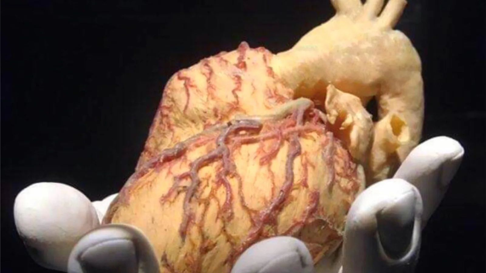
The Purkinje system is the network of specialized, fast-conducting cardiomyocytes that spreads electrical excitation through the ventricular walls so that ventricular muscle contracts as a coordinated unit rather than a patchwork of independent twitches. It is the distal half of the cardiac conduction system, formed by the bundle of His, the right and left bundle branches, and the free-running Purkinje fibers that terminate at the Purkinje-myocardial junctions.
A heart that beats is not automatically a heart that pumps. The ventricles must squeeze from apex to base in a single coordinated wave, and they must do so after the atria have finished emptying into them. Both requirements are timing problems, and the Purkinje system is the timing solution. When it fails, the result is a widened QRS complex, loss of synchronized contraction, reduced stroke volume, and a substrate for ventricular arrhythmia. In humans, ventricular arrhythmias often originate within the Purkinje system, and although these cells are a tiny fraction of ventricular mass, their pathogenic role is disproportionate [1]. The same principle applies across domestic species, which is why the Purkinje network sits at the center of veterinary electrocardiography.
This article is educational and is not a substitute for veterinary diagnosis or treatment.
The Conduction Pathway From Start to Finish
Step 1: The Sinoatrial Node Fires
The sinoatrial node is the primary pacemaker. It sits subepicardially in the right atrial wall near the junction with the superior vena cava [2]. Its cells depolarize spontaneously because their membrane potential drifts upward during diastole until it reaches threshold. The node is stabilized by fibrous insulation, cellular heterogeneity, and discrete exit pathways that regulate how the impulse leaves and enters atrial muscle [3]. In the dog, the sinoatrial node receives blood from the sinoatrial nodal artery, a branch of the right coronary artery in roughly 60% of individuals and the left circumflex coronary artery in the remaining 40% [2]. Ischemic damage to that vessel can silence the pacemaker.
Step 2: Atrial Activation
From the sinoatrial node, the impulse spreads through atrial myocardium at roughly 0.3 to 0.5 m/s. Interatrial conduction occurs through aligned atrial muscle, with the interatrial bundle and posterior connections supporting rapid activation without strict insulation [3]. This is why the right and left atria depolarize almost simultaneously, producing the P wave on the surface ECG. Note that atrial conduction is not a dedicated cable system in most species. It is ordinary working myocardium doing an extraordinary job of speed.
Step 3: The Atrioventricular Node and the Delay
The atrioventricular node lies subendocardially within the triangle of Koch, bordered posteriorly by the coronary sinus ostium, superiorly by the tendon of Todaro, and anteriorly by the septal leaflet of the tricuspid valve [2]. Conduction velocity here falls to about 0.05 m/s, roughly one tenth that of atrial muscle. This is the slowest point in the entire pathway, and the slowness is deliberate.
The node achieves its delay through slow, safe conduction shaped by specialized cellular architecture, connexin gradients, and dual-pathway organization [3]. Because gap junctions are sparse and the cells are small, current flows poorly from cell to cell. The result is an AV nodal delay of roughly 0.1 seconds. That tenth of a second allows ventricular filling. While the ventricles are electrically quiet, the atria have already contracted and delivered their volume. The AV node also acts as a filter, protecting the ventricles from very fast atrial rates such as those seen in atrial fibrillation.
The AV node is the most frequency-sensitive part of the conduction system. Shortening the cycle length prolongs intranodal conduction time, and autonomic blockade shifts conduction velocity less here than at any other level [4]. That sensitivity is why AV nodal conduction is the target of so many clinical decisions.
Step 4: Bundle of His and Bundle Branches
The AV node tapers into the bundle of His, which bifurcates into right and left bundle branches coursing along the interventricular septum [2]. These structures are part of the His-Purkinje system, which delivers rapid, synchronous ventricular activation through fibrous-insulated pathways [3]. The insulation matters. Because the bundles are electrically shielded from surrounding myocardium, the impulse travels down them without prematurely exciting the muscle it passes.
The His bundle and bundle branches are histologically distinct from nodal tissue. They show high expression of the cardiac sodium channel Na(v)1.5 and are HCN4-positive and Cx43-negative, whereas the sinoatrial and atrioventricular nodes show low Na(v)1.5 expression [5]. High sodium channel density is the molecular basis of fast conduction, because the upstroke of the action potential depends on rapid sodium influx.
Step 5: Purkinje Fibers and the Purkinje-Myocardial Junction
The bundle branches give rise to Purkinje fibers in the subendocardial regions of the ventricles [2]. Conduction velocity in Purkinje fibers reaches 1 to 4 m/s, the fastest in the heart. This is roughly ten times faster than ventricular muscle, which conducts at 0.3 to 0.5 m/s. The Purkinje network therefore activates the entire endocardial surface of both ventricles almost simultaneously, and the depolarization wave then sweeps outward through the ventricular wall from endocardium to epicardium.
At the terminal arborizations, the Purkinje fiber meets ventricular muscle at the Purkinje-myocardial junction. This junction is an inherent source-sink discontinuity. A small Purkinje cell must deliver enough current to depolarize a much larger mass of ventricular myocytes, and the mismatch supports reliable excitation while also creating a site prone to conduction block [3]. Structural imaging of sheep hearts shows that free-running Purkinje fibers, the Purkinje-myocardial junction, and surrounding myocardium differ in collagen content, adipocytes, and cell composition, which is why they can be distinguished by specialized MRI contrast [6].
The Purkinje network is not a passive wire. Purkinje cells have their own calcium handling machinery. In a canine model of coronary ligation, Purkinje cells 48 hours after occlusion showed increased SERCA2 pump expression, suggesting enhanced sarcoplasmic reticulum calcium sequestration that can drive abnormal calcium-dependent depolarizations [7]. This is one mechanism by which the terminal conduction system becomes an arrhythmia source during myocardial infarction.
flowchart TD
    A[Sinoatrial node fires] --> B[Atrial muscle]
    B --> C[Atrioventricular node]
    C --> D[AV nodal delay]
    D --> E[Bundle of His]
    E --> F[Right bundle branch]
    E --> G[Left bundle branch]
    F --> H[Purkinje fibers]
    G --> H
    H --> I[Purkinje myocardial junction]
    I --> J[Ventricular myocardium]
    J --> K[Coordinated contraction]
Conduction Velocity and ECG Correlation
The table below summarizes the pathway, the approximate conduction velocity at each level, and the surface ECG event that corresponds to it. Velocities are standard values taught in cardiac physiology. ECG correlates refer to the standard limb lead tracing.
| Structure | Conduction velocity | ECG correlate |
|---|---|---|
| Sinoatrial node | Slow, spontaneous depolarization | Onset of P wave |
| Atrial myocardium | 0.3 to 0.5 m/s | P wave |
| Atrioventricular node | About 0.05 m/s | PR segment |
| Bundle of His | Fast, 1 to 4 m/s | PR segment to onset of QRS |
| Bundle branches | Fast, 1 to 4 m/s | QRS complex |
| Purkinje fibers | 1 to 4 m/s | QRS complex |
| Ventricular myocardium | 0.3 to 0.5 m/s | QRS complex to end of T wave |
The PR interval on the ECG measures the time from the start of atrial depolarization to the start of ventricular depolarization. Most of that interval is AV nodal delay. The QRS duration measures how long the ventricles take to depolarize, and it is therefore a direct readout of His-Purkinje function. A narrow QRS means the Purkinje system is delivering the impulse quickly and synchronously. A wide QRS means conduction is slow somewhere in the ventricular pathway, or that the impulse is traveling through working myocardium instead of the fast network.
The PR interval and QRS duration are the two ECG numbers that matter most when you are assessing the conduction system. In a longitudinal study of mdx mice, a model of Duchenne muscular dystrophy, ECG recordings showed a progressive increase in PR interval over time and a prolonged QRS compared with wild-type mice [8]. Those changes occurred even though the morphology and maturation of the Purkinje fiber network appeared normal, which is a useful reminder that a normal-looking network can still conduct abnormally.
Species Differences in Purkinje Distribution
The Purkinje system is not built to one blueprint. Comparative anatomy reveals meaningful differences in how deep the network penetrates the ventricular wall, and those differences change the direction of ventricular activation and the shape of the ECG.
Ungulates and Other Species With Intramural Purkinje Networks
In the pig, the Purkinje fiber network is found in the subendocardium and extends deep into the ventricular walls [9]. This intramural distribution produces ventricular electrical activation in an apex-to-base direction. The mean electrical axis is cranially oriented, and it is influenced more by the presence of the intramural Purkinje network than by left ventricular mass, which is the dominant determinant in humans [9]. This is a critical point for anyone using the pig as a translational model, because action potential shape, activation pattern, and ion channel composition also differ between porcine and human hearts [9].
The common hippopotamus shows a similar pattern. Its Purkinje fiber strands extend deep into the ventricular walls and consist of large, ovoid cells. Orthogonal ECG recordings revealed a mean electrical axis pointing toward the neck, again indicating apex-to-base activation [10]. Sheep also have free-running Purkinje fibers that can be imaged ex vivo, and these fibers show distinct structural composition compared with the Purkinje-myocardial junction and surrounding myocardium [6].
Ungulates are therefore the group to remember when the question is about deep intramural Purkinje penetration. In these species, the fast-conducting network is not confined to a thin subendocardial shell. It reaches into the mid-wall and even toward the epicardium, which shortens transmural conduction time and changes the sequence of activation recorded at the body surface.
Carnivores and Humans
In dogs and humans, the Purkinje network is predominantly subendocardial, and the depolarization wave moves from endocardium to epicardium across the ventricular wall. The intramural Purkinje fibers described in pigs are not the rule. This is one reason why the pig is a useful but imperfect model for human arrhythmia research, and why findings in one species should be translated with care.
A Transmural Gradient in Sodium Channel Expression
Even within a single ventricle, conduction is not uniform. In the mouse, Na(v)1.5 labeling intensity is lower in the subepicardium than in the subendocardium, and maximal action potential upstroke velocity is significantly lower in subepicardial myocytes (mean 309 V/s) compared with subendocardial myocytes (mean 394 V/s) [5]. This transmural heterogeneity in sodium channel availability means that the outer wall conducts more slowly than the inner wall, which contributes to the normal dispersion of repolarization. It also means that the same Purkinje impulse arrives at different layers of the wall with different safety margins.
How the System Is Studied and Observed
Electrocardiography
The surface ECG is the first-line tool. PR interval and QRS duration give indirect but reliable information about AV nodal and His-Purkinje conduction. In the mdx mouse study, ECG recordings detected progressive PR prolongation, QRS prolongation, and a greater prevalence of premature ventricular complexes after beta-adrenergic stimulation, all without visible Purkinje degeneration [8]. This shows that ECG changes can precede or occur independently of structural change.
His Bundle Recording
His bundle electrocardiography, performed with endocavitary catheters, separates the PR interval into its components. The A-H interval measures AV nodal conduction, and the H-V interval measures His-Purkinje conduction. In anesthetized dogs, mild hyperkalemia under acetylcholine infusion decreased AV nodal conduction time by 20% and AV nodal effective refractory period by 17%, while His-Purkinje conduction time (H-V interval) showed no variation or only slight, occasionally biphasic changes [11]. The H-V interval is therefore a relatively stable measure of the distal conduction system, less swayed by autonomic tone than the A-H interval.
Imaging
High-resolution imaging is changing how the conduction system is visualized. Contrast-enhanced micro-CT of human hearts allows 3D reconstruction of cardiac conduction system components alongside surrounding structures [12]. Inhomogeneous magnetization transfer MRI can distinguish free-running Purkinje fibers from the Purkinje-myocardial junction and surrounding myocardium in fixed sheep hearts, with higher ihMTR values in free-running fibers compared with the junction (11.5 ± 1.5% versus 9.0 ± 2.9%) [6]. These techniques are research tools today but point toward better anatomical mapping for education, simulation, and procedural planning.
Computational Modeling
Computer models couple the Purkinje network to ventricular muscle to simulate activation. An open-source GPU-based solver using the monodomain equation coupled with a Purkinje network has been used to run 512 simulations concurrently on 128 compute nodes, completing coarse biventricular mesh simulations in under 24 minutes [13]. Calibration of the Purkinje-muscle junction is a key step in these models, which confirms how much the junction matters to the overall activation pattern.
Clinical Relevance, Limitations and Common Mistakes
Why the Purkinje System Matters Clinically
Damage to the His-Purkinje axis has direct procedural consequences. In transcatheter aortic valve replacement, pacemaker rates remain near 16% in contemporary multicenter practice, and the odds of pacing rise 25% per millimeter of implantation below the estimated His bundle [14]. Depth of implant relative to the membranous septum is the dominant modifiable factor, and single-center strategies that interpret depth against membranous septum length have reached 3 to 5% pacing rates [14]. Conduction-system pacing outcomes also depend on proximity to the left bundle branch origin, with each millimeter closer adding a 0.25% absolute gain in ejection fraction and each millimeter closer to the left ventricular endocardium adding 0.63%, chiefly in patients with depressed baseline function [14]. A CT-based framework for left bundle branch area pacing shows that fixed His-to-lead distances ignore anatomical variance, and that targets cluster at approximately one-third of the horizontal His distance-to-right ventricular apex at 20 mm from the tricuspid annulus [15].
After myocardial infarction, conduction system damage contributes to arrhythmia. In neonatal mice, conduction system restoration during heart regeneration prevented arrhythmias, whereas non-regenerative stages showed disorganized bundling of conduction fibers and global His-Purkinje disruption [16]. Modeling in the infarcted human heart implicated the non-regenerative phenotype as causative for heart block [16]. This links structural Purkinje injury to a clinical syndrome veterinarians recognize as high-grade atrioventricular block.
Limitations
Conduction velocities are population values. Individual animals vary with heart rate, autonomic tone, electrolytes, temperature, and disease. The AV nodal delay of roughly 0.1 seconds is a normal resting value and shortens with sympathetic stimulation and lengthens with vagal tone. A single ECG or a single measurement cannot characterize the conduction system in a patient with intermittent signs. Interpretation requires clinical context, serial recordings, and often advanced diagnostics. Any animal with syncope, exercise intolerance, or an unexplained arrhythmia needs a veterinarian, not a textbook.
Common Mistakes
The most frequent error is treating the Purkinje system as a simple wire. It is an active tissue with its own ion channels, calcium handling, and arrhythmogenic potential. Purkinje cells can generate abnormal depolarizations after ischemia, and their calcium sequestration machinery changes within 48 hours of coronary occlusion [7].
A second mistake is assuming that a normal-appearing Purkinje network guarantees normal conduction. In mdx mice, conduction defects and arrhythmias occurred with no defects in the morphology and maturation of the Purkinje fiber network, though the hearts were larger and showed fibrosis and hypertrophy [8]. Structure and function can dissociate.
A third mistake is applying human activation patterns to all species. The pig and hippopotamus have intramural Purkinje networks and apex-to-base activation, which changes the mean electrical axis [9][10]. A veterinarian or researcher who reads a porcine ECG with human assumptions will misjudge the axis.
A fourth mistake is confusing the AV nodal delay with a conduction block. A PR interval at the upper end of normal is not the same as first-degree atrioventricular block, and the distinction depends on the reference range for the species and the clinical picture.
Quick Review
- The Purkinje system is the fast-conducting distal conduction pathway: bundle of His, bundle branches, and Purkinje fibers.
- Conduction velocity is about 0.05 m/s in the AV node, 1 to 4 m/s in Purkinje fibers, and 0.3 to 0.5 m/s in ventricular muscle.
- The AV nodal delay of roughly 0.1 seconds allows ventricular filling before the ventricles contract.
- The PR interval reflects atrial depolarization plus AV nodal delay. The QRS duration reflects His-Purkinje and ventricular conduction.
- Purkinje fibers terminate at the Purkinje-myocardial junction, a source-sink discontinuity that supports reliable excitation but can also block.
- Ungulates such as pigs and hippopotamuses have intramural Purkinje networks that produce apex-to-base activation and a cranially oriented mean electrical axis.
- Purkinje cells are arrhythmogenic. They can generate abnormal calcium-dependent depolarizations after ischemia.
Frequently Asked Questions
What is the Purkinje system?
The Purkinje system is the network of specialized fast-conducting cardiomyocytes that carries the electrical impulse from the bundle of His through the bundle branches and Purkinje fibers to the ventricular myocardium. It ensures that the ventricles depolarize and contract as a synchronized unit.
Why does the impulse slow down at the AV node?
Conduction velocity in the AV node is about 0.05 m/s because gap junctions are sparse and the cells are small, so current flows poorly from cell to cell. This creates a delay of roughly 0.1 seconds that allows the atria to finish filling the ventricles before ventricular contraction begins.
How fast do Purkinje fibers conduct compared with ventricular muscle?
Purkinje fibers conduct at 1 to 4 m/s, roughly ten times faster than ventricular muscle at 0.3 to 0.5 m/s. The speed comes from high expression of the cardiac sodium channel Na(v)1.5 in the His-Purkinje system.
What does a wide QRS complex mean?
A wide QRS complex means ventricular depolarization is taking longer than normal, usually because conduction through the His-Purkinje system is slowed or blocked. The impulse then spreads through working myocardium, which conducts much more slowly.
Do all animals have the same Purkinje distribution?
No. Pigs and hippopotamuses have Purkinje fibers that extend deep into the ventricular walls, producing apex-to-base activation and a cranially oriented mean electrical axis. Dogs and humans have predominantly subendocardial Purkinje networks with endocardium-to-epicardium activation.
Can the Purkinje system cause arrhythmias?
Yes. Purkinje cells can generate abnormal calcium-dependent depolarizations, especially after myocardial ischemia, and they are a common origin site for ventricular arrhythmias. Conduction system damage after infarction is also linked to heart block.
Related Articles
- Canine Cardiac Physiology: Electrical Conduction and Arrhythmias
- Canine Heart Anatomy: Chambers, Valves, and Conduction System
- Equine Cardiac Conditions: Diagnosis and Management
- Why Is My Dog collapsing suddenly? Cardiac Arrest Fainting and Hypoglycemia
- Veterinary Cardiopulmonary Resuscitation: Post-Cardiac Arrest Care
- Pimobendan Vetmedin Dose: Cardiac Inotrope Dose and MVD Timing
- Myocardium and Heart Layers: Cardiac Muscle Anatomy
- Cardiac Muscle: Structure and Function Explained
- Function of Nerves: How the Nervous System Works
Sources
- [[Development, morphogenesis, and regeneration of cardiac Purkinje fibers].](https://pubmed.ncbi.nlm.nih.gov/42466687/)
- Accelerating Transmural Conduction: The Role of Intramural Purkinje Fibers in the Pig Heart.
- The cardiac conduction system: A narrative review.
- [[The influence of frequency and blockade of the autonomic nervous system on the functional behaviour of the human conduction system. Part A: Conduction velocity (author's transl)].](https://pubmed.ncbi.nlm.nih.gov/65170/)
- The cardiac sodium channel displays differential distribution in the conduction system and transmural heterogeneity in the murine ventricular myocardium.
- Structural Characterization of Cardiac Free-Running Purkinje Fibers Using Inhomogeneous Magnetization Transfer (ihMT): A Proof of Concept MRI-Histology Approach.
- Increased Ca(2+) Sequestration by the Sarco-/Endoplasmic Reticulum in Cardiac Purkinje Cells After Myocardial Infarction.
- Conduction defects and arrhythmias in mdx mice are not associated with a degeneration of the cardiac Purkinje network.
- Characterization of the Porcine Cardiac Conduction System and Electrocardiogram: Implications for Using the Pig in Translational Arrhythmia Research.
- Cardiac conduction system and the electrocardiogram of the common hippopotamus (Hippopotamus amphibius).
- Effects of mild hyperkalemia on conduction velocity and effective refractory period in various parts of the dog heart in situ.
- High-resolution 3D visualization of human hearts with emphases on the cardiac conduction system components-a new platform for medical education, mix/virtual reality, computational simulation.
- Toward cardiac electrophysiology digital twins with an efficient open source scalable solver on GPU clusters.
- Avoid It or Reach It? The Cardiac Conduction Axis as a Common yet Divergent Determinant of Patient Outcomes in Transcatheter Aortic Valve Replacement and Conduction-System Pacing.
- Anatomical computed tomography-based framework for optimizing left bundle branch area pacing of the human cardiac conduction system.
- Cardiac conduction system regeneration prevents arrhythmias after myocardial infarction.