Pulmonary Arteries and Lung Vessels: Anatomy Guide
By Dr. Zubair Khalid, DVM, MS, PhD ·
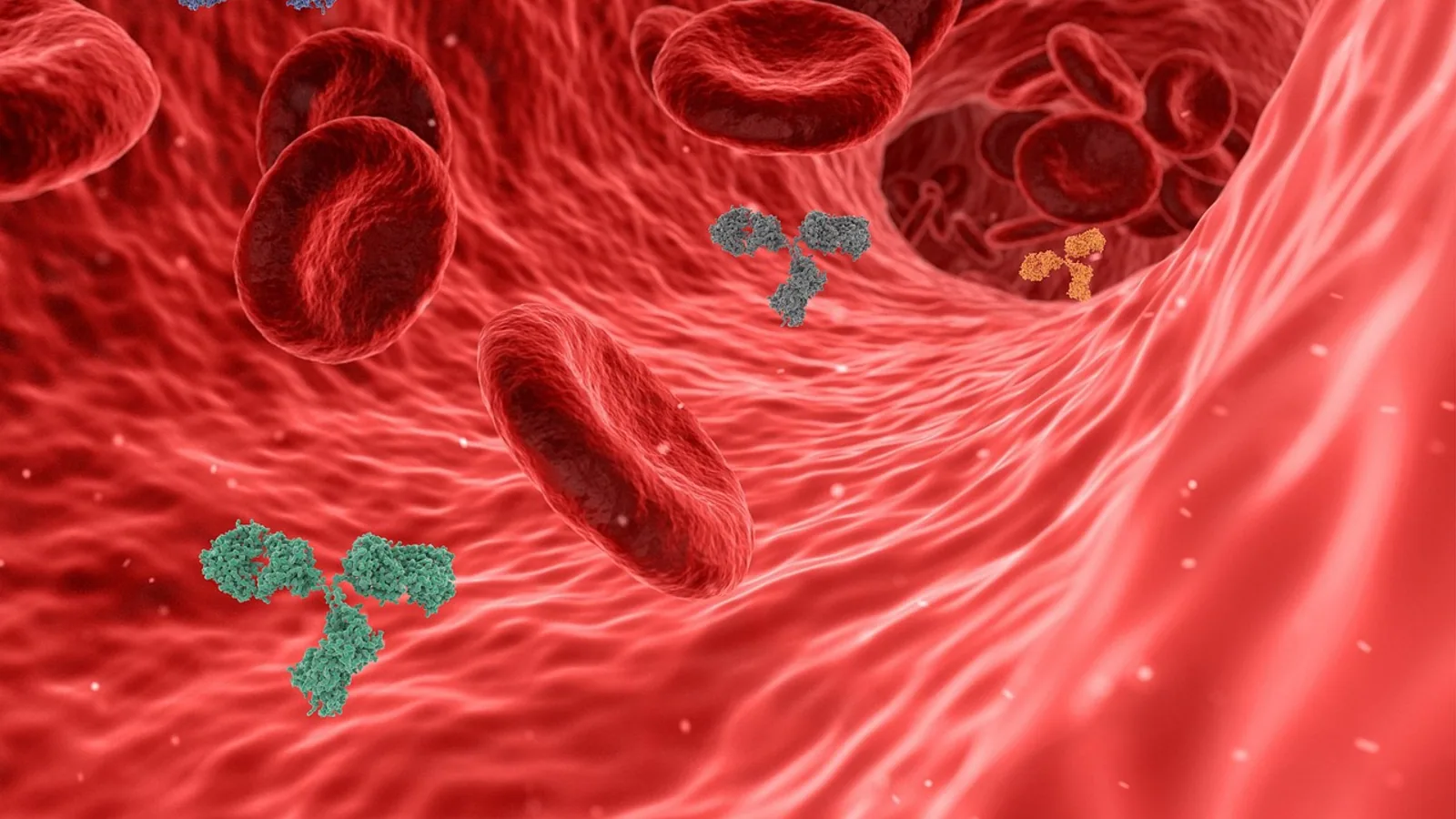
The pulmonary arteries are the great vessels that carry deoxygenated blood from the right ventricle of the heart to the lungs, where the blood is oxygenated and returned to the left atrium by the pulmonary veins. Together these vessels form the pulmonary circulation, a low-pressure circuit that is anatomically and functionally distinct from the higher-pressure bronchial circulation that supplies the airway walls themselves.
This anatomy matters in every species a veterinarian handles. Congenital heart disease in puppies and kittens often turns on the size and branching pattern of the pulmonary arteries. Thoracic radiography, computed tomography, and echocardiography all depend on knowing where the pulmonary trunk sits, how it divides, and how many pulmonary veins enter the left atrium. Species differences in lobar artery origins and trunk length change what you see on an angiogram or a CT study. The pulmonary circulation is also the site of clinically important disease, including pulmonary hypertension and pulmonary thromboembolism, so a working knowledge of its normal anatomy is the foundation for interpreting abnormal findings. Congenital anomalies of the pulmonary arteries and tracheobronchial tree, such as pulmonary artery sling with long-segment tracheal stenosis, are surgically corrected in children and have been studied with an emphasis on tracheal arborization and lung hypoplasia as predictors of outcome [1].
This article is educational and is not a substitute for veterinary diagnosis or treatment.
The Pulmonary Trunk and Its Bifurcation


Origin from the right ventricle
The pulmonary trunk (also called the main pulmonary artery) arises from the right ventricle at the pulmonic valve. It is a single, short, wide vessel in most domestic mammals, and it is the first major branch of the right ventricular outflow tract. The pulmonary trunk carries deoxygenated blood away from the heart, in contrast to the aorta, which carries oxygenated blood away from the left ventricle. This reversal of the usual "artery carries oxygenated blood" rule is the single most common point of confusion for beginning students.
The pulmonary trunk passes craniodorsally from the right ventricle, crosses ventral to the aorta in most species, and divides into the right and left pulmonary arteries. In some species the trunk is notably longer than in others. The horse is the classic example of an unusually long pulmonary artery, a feature that is relevant during cardiac catheterization and when interpreting angiographic studies of the equine heart.
Imaging studies in animals and in people have characterized the elastic properties of the pulmonary trunk and its branches. Using 640-slice volume computed tomography in subjects free of cardiovascular disease, researchers found that aortic and pulmonary artery distensibility and compliance decreased in the order main pulmonary artery, right pulmonary artery, then left pulmonary artery, and that these elasticity measures correlated with age [2]. This kind of data is useful when interpreting vessel behavior in disease, but the working anatomy for a clinician is simpler: the trunk leaves the right ventricle, then splits.
Bifurcation into right and left pulmonary arteries
The bifurcation of the pulmonary trunk produces the right pulmonary artery and the left pulmonary artery. Each then divides further into lobar arteries that follow the bronchi into the individual lung lobes. Because the airways and the pulmonary arteries branch together, the lobar arteries are named for the lobes they supply: the right cranial, middle, caudal, and accessory lobar arteries on the right, and the left cranial and caudal lobar arteries on the left. (In dogs and cats the left lung has cranial and caudal lobes, with the cranial lobe often subdivided into cranial and caudal parts, so the left lobar arteries may be described in corresponding subdivisions.)
Blood flow is not evenly split between the two lungs. In a study of children and young adults with congenital heart disease, threshold-based CT volumetry found that right pulmonary vascular volume averaged 66.3 percent, while right lung perfusion measured by scintigraphy averaged 69.1 percent [3]. The two methods agreed closely enough to be clinically comparable. The takeaway is that the right lung normally receives more than half of pulmonary blood flow, a fact that becomes relevant when disease or surgery changes the balance between the two lungs.
Pulmonary artery growth and pathology
The pulmonary arteries are dynamic structures. They grow with the animal, and their caliber changes in response to disease. When a lobe is removed, the remaining pulmonary vasculature adapts. A retrospective study of patients who underwent lung resection found that lobectomy produced a greater increase in the pulmonary artery to aorta diameter ratio than sublobar resection, with the lobectomy group showing a significantly higher ratio three years after surgery [4]. Because the pulmonary artery to aorta ratio correlates with pulmonary artery pressure, this finding links surgical removal of lung tissue to a measurable increase in the load on the right side of the heart.
The pulmonary arteries can also be narrowed. Pulmonary artery stenosis and stenting are well studied in animal models, and a swine model of congenital heart disease has been used to compare imaging modalities for measuring pulmonary artery diameters, including stenosis and stents [5]. In that model, three-dimensional rotational angiography agreed closely with conventional catheter angiography and multi-slice CT, while phase-contrast magnetic resonance angiography could not reliably visualize stents and distal vessels [5]. These details matter for anyone who plans or interprets vascular imaging in small animals.
Pulmonary Veins and Return to the Left Atrium
Four pulmonary veins
In the dog and cat, and in domestic mammals generally, the lungs are drained by pulmonary veins that return oxygenated blood to the left atrium. The standard veterinary anatomy is that four pulmonary veins enter the left atrium: one from each of the major lobar drainage territories, typically paired on the right and left. The right pulmonary veins drain the right cranial, middle, caudal, and accessory lobes, and the left pulmonary veins drain the left cranial and caudal lobes. The precise number and pattern of ostia can vary, but the function is constant: all oxygenated pulmonary blood returns to the left atrium.
Pulmonary veins are the only veins in the body that carry oxygenated blood. Like the pulmonary arteries, this reverses the usual rule. Students who remember "veins carry deoxygenated blood" will mislabel the pulmonary veins on an exam.
Clinical relevance of pulmonary vein anatomy
Pulmonary vein anatomy is central to some cardiac procedures. In people, cardiac computed tomography before radiofrequency catheter ablation of atrial fibrillation is performed specifically to evaluate pulmonary vein anatomy, and a study of 606 consecutive patients found that routine cardiac CT scans obtained for this purpose could also evaluate coronary artery lesions in 93 percent of patients [6]. This is a human clinical example, but it illustrates a general principle: the pulmonary veins are anatomically consistent enough to be mapped, and mapping them is standard practice in interventional cardiology. In veterinary medicine, pulmonary vein anatomy is assessed during echocardiography and during CT angiography of the thorax.
Pulmonary Versus Bronchial Circulation
Two separate vascular systems supply the lung. Confusing them is a common error.
Pulmonary circulation
The pulmonary arteries carry deoxygenated blood from the right ventricle to the lungs at low pressure. The pulmonary circuit is a low-resistance, high-flow system. The capillaries of the pulmonary circulation are thin-walled and optimized for gas exchange across the blood-gas barrier. The pulmonary veins then return oxygenated blood to the left atrium.
Bronchial circulation
The bronchial arteries carry oxygenated blood at systemic pressure to supply the walls of the bronchi, the supporting connective tissue of the lung, and the nerves and vasa vasorum of the pulmonary arteries themselves. The bronchial circulation is a nutrient supply system, not a gas exchange system. According to a comparative review of the tracheobronchial circulation, the subgross anatomy of the sheep, cow, pig, and horse appears similar to that of humans, with a major bronchial artery in a consistent location [7]. That same review notes that the vasa vasorum of the pulmonary arterial system derive from the bronchial arteries, so the composition of bronchial artery blood can modulate pulmonary vasomotor responses [7]. In pathological situations, the bronchial circulation of the sheep behaves similarly to that of humans, which is one reason sheep are used as models [7].
A second comparative study of bronchial artery distribution across ten mammalian species and humans found marked subgross differences in how the bronchial artery reaches the pleura, interlobular septa, distal airways, and alveoli, and in the presence of bronchial artery to pulmonary artery anastomoses [8]. The bronchial artery is considered the nutrient artery to the lung, and its distribution depends on the amount of supportive, non-gas-exchanging tissue present, which varies greatly between species [8]. This is a reminder that species selection matters when animal data are meant to inform understanding of another species.
Summary comparison
| Vessel | Origin | Destination | Blood oxygenation status | Pressure |
|---|---|---|---|---|
| Pulmonary trunk | Right ventricle | Bifurcates into right and left pulmonary arteries | Deoxygenated | Low |
| Right and left pulmonary arteries | Pulmonary trunk | Lung lobes via lobar arteries | Deoxygenated | Low |
| Lobar pulmonary arteries | Right or left pulmonary artery | Individual lung lobes | Deoxygenated | Low |
| Pulmonary veins | Lung lobar drainage | Left atrium via four veins | Oxygenated | Low |
| Bronchial arteries | Aorta or intercostal arteries | Bronchial walls, lung connective tissue, vasa vasorum | Oxygenated | Systemic |
| Bronchial veins | Bronchial walls | Azygos or other systemic veins | Deoxygenated | Systemic |
This table captures the single most testable fact in pulmonary vascular anatomy: pulmonary arteries carry deoxygenated blood, pulmonary veins carry oxygenated blood, and the bronchial arteries carry oxygenated blood at systemic pressure to nourish the airways.
Step-by-Step Flow Path from Heart to Lungs
The path of blood through the pulmonary circulation can be traced in a fixed sequence. This is the flow path a student should be able to recite.
- Deoxygenated blood enters the right atrium from the cranial and caudal venae cavae and from the coronary sinus.
- Blood passes through the right atrioventricular (tricuspid) valve into the right ventricle.
- The right ventricle contracts and ejects blood through the pulmonic valve into the pulmonary trunk.
- The pulmonary trunk divides into the right and left pulmonary arteries.
- Each pulmonary artery divides into lobar arteries that follow the bronchi into the lung lobes.
- Lobar arteries branch into progressively smaller arteries and then into the pulmonary capillary bed, where gas exchange occurs across the blood-gas barrier.
- Oxygenated blood is collected by venules and then by pulmonary veins.
- Pulmonary veins return blood to the left atrium, typically via four ostia.
- Blood passes through the left atrioventricular (mitral) valve into the left ventricle.
- The left ventricle ejects blood into the aorta for systemic distribution.
The bronchial circulation runs in parallel but separately. Bronchial arteries arise from the aorta or its branches, carry oxygenated blood at systemic pressure, and supply the walls of the airways and the supporting tissues of the lung [7][8].
The following flowchart summarizes the main flow path and the parallel bronchial supply.
flowchart TD
    A[Right ventricle] --> B[Pulmonary trunk]
    B --> C[Right pulmonary artery]
    B --> D[Left pulmonary artery]
    C --> E[Lobar arteries]
    D --> E
    E --> F[Pulmonary capillaries]
    F --> G[Pulmonary veins]
    G --> H[Left atrium]
    I[Aorta] --> J[Bronchial arteries]
    J --> K[Airway walls and vasa vasorum]
    K --> L[Bronchial veins]
Comparative Anatomy Across Common Species
Lung lobation and lobar arteries
The number of lung lobes varies by species, and the lobar arteries follow the lobar bronchi. Dogs and cats have well-developed lobation, with the right lung typically having four lobes (cranial, middle, caudal, and accessory) and the left lung having two or three depending on how the cranial lobe is subdivided. Ruminants and pigs have a distinct lobar pattern as well, and pigs are commonly used as models for pulmonary artery imaging because their anatomy is tractable for catheterization and CT [5].
Separate origin of the right cranial lobar artery
In ruminants and pigs, the right cranial lobar artery often arises from a separate branch rather than from the right pulmonary artery in the same pattern seen in dogs. This means the right cranial lobar artery may take origin directly from the pulmonary trunk or from a distinct early branch. The practical consequence is that when you cannulate or image the right pulmonary artery in a calf or a pig, you may not see the right cranial lobar artery where you would expect it in a dog. The separate origin is a normal variant in these species, not an anomaly.
The horse: long pulmonary artery
The horse has an unusually long pulmonary artery. The pulmonary trunk and its main branches are elongated, which changes the geometry of the right ventricular outflow tract and affects how catheters and imaging probes are positioned. Horses are also prone to exercise-induced pulmonary hemorrhage, and the pulmonary capillary bed has been studied in this context. A comparative study of pulmonary capillary strength in rabbit, dog, and horse measured blood-gas barrier thickness and capillary radius and found that the barrier was thinnest in the rabbit and intermediate in the dog, with the horse having the thickest barrier of the three [9]. The same study noted that the capillary pressure for failure in the horse was estimated from the mean of pulmonary arterial and left atrial pressures observed in galloping Thoroughbreds known to develop exercise-induced pulmonary hemorrhage [9]. This is a species-specific anatomical and physiological consideration that has clinical implications for equine athletes.
Species differences in the bronchial circulation
The bronchial circulation varies more between species than the pulmonary circulation does. The distribution of the bronchial artery to the pleura, interlobular septa, distal airways, and alveoli differs markedly across mammals, as does the presence of bronchial artery to pulmonary artery anastomoses [8]. Sheep, cow, pig, and horse share a subgross pattern similar to humans with a consistent major bronchial artery, which makes these species useful for studies of the human bronchial circulation [7]. Dogs and cats have their own patterns, and caution is warranted when extrapolating findings from one species to another [8].
Imaging the pulmonary arteries across species
Modern imaging has made comparative pulmonary artery anatomy more accessible. In a swine model of congenital heart disease, three-dimensional rotational angiography, multi-slice CT, and conventional catheter angiography all provided accurate measurements of pulmonary artery diameters, while phase-contrast magnetic resonance angiography was less reliable for stents and distal vessels [5]. In human medicine, three-dimensional BFFE and TRANCE magnetic resonance techniques have been compared for non-contrast pulmonary artery imaging, with TRANCE showing higher contrast-to-noise ratios at several regions of interest [10]. These technical comparisons are useful background for understanding how pulmonary artery anatomy is documented in practice, though the specific protocols are not directly transferable to veterinary patients without validation.
Clinical Relevance, Limitations and Common Mistakes
Pulmonary hypertension
Pulmonary hypertension is a sustained increase in pressure in the pulmonary arteries. It increases the workload on the right ventricle, which can lead to right ventricular hypertrophy and eventually right heart failure. The pulmonary artery to aorta diameter ratio on CT correlates with pulmonary artery pressure and is a simple tool for estimating the load on the right side of the heart [4]. This ratio is used in human medicine and has been adapted for veterinary use in some settings. Pulmonary hypertension is a major complication of heart failure with preserved ejection fraction in people, and research into its mechanisms has identified skeletal muscle and pulmonary vascular remodeling pathways that include SIRT3 deficiency and elevated LOXL2 [11]. Other work in a rat model of pulmonary arterial hypertension has compared mesenchymal stem cell therapies derived from adipose tissue, bone marrow, and umbilical cord blood, with umbilical cord blood-derived cells showing the greatest improvement in right ventricular function and the largest reduction in medial wall thickness and perivascular fibrosis [12]. These are experimental findings, but they illustrate that the pulmonary vasculature is an active area of translational research.
Pulmonary thromboembolism
Pulmonary thromboembolism is the blockage of a pulmonary artery by a blood clot that has traveled from elsewhere in the venous system. It is a recognized clinical problem in dogs, cats, and horses, and it presents with sudden respiratory difficulty. The anatomy of the pulmonary arterial tree determines where emboli lodge, and the branching pattern of the lobar arteries means that emboli can obstruct flow to specific lobes. Because the pulmonary circulation is a low-pressure system, large emboli can cause acute right ventricular strain. Diagnosis relies on imaging, and the same CT and angiographic techniques used to map normal anatomy are used to detect filling defects in the pulmonary arteries [5].
Common mistakes
Mistake 1: Thinking pulmonary arteries carry oxygenated blood. They carry deoxygenated blood. Pulmonary veins carry oxygenated blood.
Mistake 2: Confusing pulmonary and bronchial circulation. Pulmonary arteries serve gas exchange. Bronchial arteries nourish the airway walls and supporting tissues and carry blood at systemic pressure [7][8].
Mistake 3: Assuming the right cranial lobar artery always branches from the right pulmonary artery. In ruminants and pigs it often arises from a separate branch.
Mistake 4: Overlooking the horse's long pulmonary artery when planning catheter placement or interpreting imaging.
Mistake 5: Using a single species' bronchial artery pattern to predict another species' pattern. Distribution varies markedly across mammals [8].
Mistake 6: Ignoring the right-left flow asymmetry. The right lung normally receives more than half of pulmonary blood flow, and CT vascular volume measurements reflect this [3].
Limitations
This guide covers normal comparative anatomy and selected clinical correlations. Individual animals vary, and a veterinarian should evaluate any specific case. Species differences in vascular anatomy are real, and findings from one species do not automatically apply to another.
Quick Review
- The pulmonary trunk arises from the right ventricle and bifurcates into the right and left pulmonary arteries.
- Pulmonary arteries carry deoxygenated blood at low pressure to the lungs.
- Four pulmonary veins return oxygenated blood to the left atrium.
- Bronchial arteries carry oxygenated blood at systemic pressure to the airway walls, lung connective tissue, and vasa vasorum.
- In ruminants and pigs, the right cranial lobar artery often arises from a separate branch.
- The horse has an unusually long pulmonary artery.
- Pulmonary hypertension and pulmonary thromboembolism are the two most clinically important diseases of the pulmonary vasculature.
Frequently Asked Questions
Do pulmonary arteries carry oxygenated or deoxygenated blood?
Pulmonary arteries carry deoxygenated blood from the right ventricle to the lungs.
How many pulmonary veins enter the left atrium?
Four pulmonary veins typically enter the left atrium, two from the right lung and two from the left.
What is the difference between pulmonary and bronchial circulation?
Pulmonary circulation carries deoxygenated blood to the lungs for gas exchange at low pressure, while bronchial circulation carries oxygenated blood at systemic pressure to nourish the airways and supporting tissues.
Which species has a separate right cranial lobar artery?
Ruminants and pigs often have a right cranial lobar artery that arises from a separate branch.
Why is the pulmonary artery long in horses?
The horse has an elongated pulmonary trunk and main branches, which affects catheter placement and imaging of the right ventricular outflow tract.
What diseases affect the pulmonary arteries?
Pulmonary hypertension and pulmonary thromboembolism are the most clinically important diseases of the pulmonary vasculature.
Related Articles
- Electrosurgery and Vessel Sealing in Veterinary Practice
- Homan's Test for Deep Vein Thrombosis: A Clinical Guide
- Surgical Hemostasis: Electrosurgery and Vessel Sealing
- Feline Cardiopulmonary Physiology: Heart-Lung Interactions
- Canine Lung Disease: Diagnostic Imaging and Sampling Techniques
- Canine Lung Disease: Differential Diagnosis and Diagnostic Approach
- Mesenteric Vessels: Arteries, Veins, and Lymphatics
- Venae Pulmonales: Pulmonary Vein Anatomy
- Lung Contusion in Dogs and Cats: Vet Guide
Sources
- Impact of Tracheal Arborization and Lung Hypoplasia in Repair of Pulmonary Artery Sling in Combination With Long-Segment Tracheal Stenosis.
- Analysis of elasticity characteristics of ascending aorta, descending aorta and pulmonary artery using 640 slice-volume CT.
- Pulmonary vascular volume ratio measured by cardiac computed tomography in children and young adults with congenital heart disease: comparison with lung perfusion scintigraphy.
- Lobectomy results in a greater increase in the pulmonary artery to aorta ratio compared to sublobar resection: a retrospective study.
- Comparison of pulmonary artery dimensions in swine obtained from catheter angiography, multi-slice computed tomography, 3D-rotational angiography and phase-contrast magnetic resonance angiography.
- Evaluation of coronary artery disease in patients with atrial fibrillation by cardiac computed tomography for catheter ablation: CADAF-CT trial.
- Comparative anatomy of the tracheobronchial circulation.
- Bronchial artery distribution in various mammals and in humans.
- Comparative aspects of the strength of pulmonary capillaries in rabbit, dog, and horse.
- Comparison of 3D BFFE and 3D TRANCE on pulmonary artery imaging with two T-SLIP placement strategies.
- Skeletal Muscle SIRT3 Deficiency Contributes to Pulmonary Vascular Remodeling in Pulmonary Hypertension Due to Heart Failure With Preserved Ejection Fraction.
- Comparative analysis on the anti-inflammatory/immune effect of mesenchymal stem cell therapy for the treatment of pulmonary arterial hypertension.