Palpebral Aperture: Anatomy and Clinical Meaning
By Dr. Zubair Khalid, DVM, MS, PhD ·
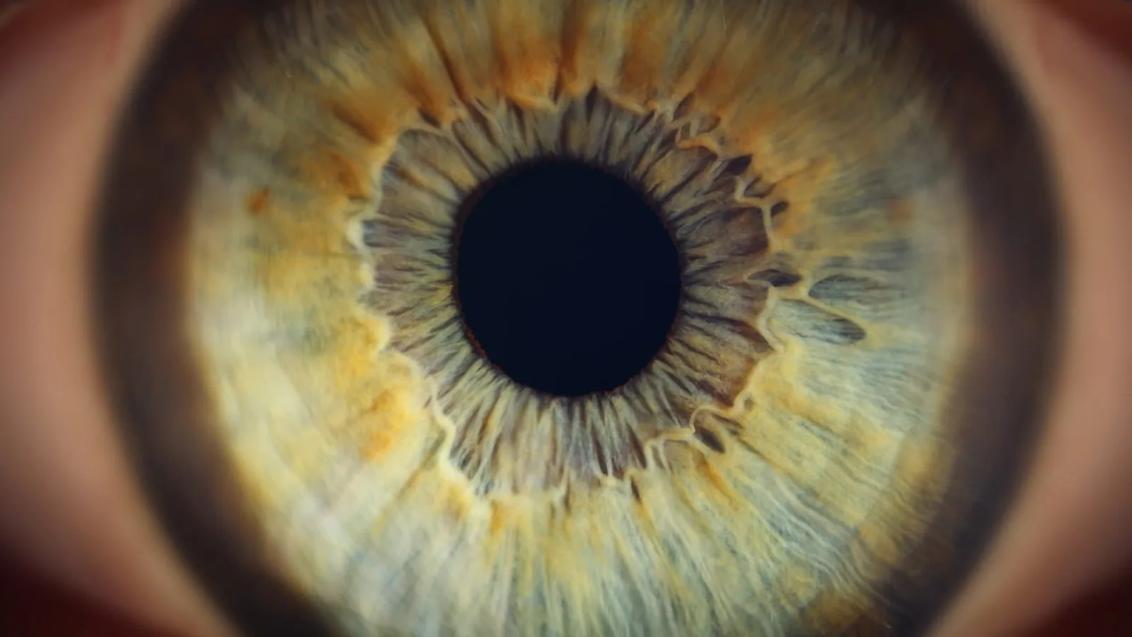
The palpebral aperture is the external opening between the upper (superior) and lower (inferior) eyelids, bounded medially and laterally by the canthi. It is the visible gateway through which the globe, conjunctiva, and cornea are accessed for examination, and its size, shape, and orientation vary substantially across domestic species.
The palpebral aperture matters because it directly governs how much of the ocular surface is exposed to the environment. A wide aperture increases corneal exposure and predisposes to drying and ulceration. A narrow or malformed aperture can trap hair against the globe, obstruct vision, or signal underlying neurologic or structural disease. In brachycephalic breeds, the aperture is measurably larger and the globe more protuberant than in nonbrachycephalic dogs, which has direct consequences for corneal health [1]. Understanding the normal aperture for each species is the first step in recognizing when something is wrong.
Anatomy of the Palpebral Aperture
Boundaries and Canthi
The palpebral aperture is a roughly elliptical space. Its dorsal border is the free margin of the upper eyelid, and its ventral border is the free margin of the lower eyelid. The medial canthus is the angle where the two eyelids meet closest to the midline of the face. The lateral canthus is the angle where they meet farthest from the midline.
In most domestic species, the lateral canthus sits lower (more ventral) than the medial canthus. This slight downward tilt at the lateral angle directs the tear film toward the medial canthus and the lacrimal puncta, supporting normal tear drainage. The medial canthus houses the caruncle, a small fleshy mound of modified conjunctiva, and the openings of the lacrimal canaliculi.
The eyelids themselves are thin, flexible folds of skin, muscle, and tarsal plate lined internally by palpebral conjunctiva. The orbicularis oculi muscle encircles the aperture and closes the lids. The levator palpebrae superioris and the superior tarsal muscle (in species that have one) open the upper lid. The lower lid is retracted primarily by gravity and by the inferior tarsal muscle.
The Palpebral Fissure Length
The palpebral fissure length (PFL) is the horizontal distance between the medial and lateral canthi. It is a standard morphometric measurement used in both clinical and research settings. In a study of broad-snouted caimans (Caiman latirostris), mean PFL was 28.9 ± 3.0 mm [2]. In dogs, PFL is measured as part of craniofacial assessment, and relative palpebral fissure length (RPFL) normalizes this value to skull size [3].
PFL is not simply a cosmetic measurement. It correlates with ocular protrusion and cephalic index. In a CT-based study of 32 dogs, horizontal palpebral fissure showed a moderate positive correlation with cephalic index (r = 0.372, P = .0024), meaning that dogs with wider, shorter skulls tend to have wider palpebral apertures [1]. This relationship helps explain why brachycephalic breeds are overrepresented in corneal exposure cases.
Eyelid Margin Anatomy
The free margin of each eyelid is a specialized structure. It bears the meibomian gland orifices, which secrete the lipid layer of the tear film. In species with cilia (eyelashes), the cilia emerge from the anterior aspect of the margin. The posterior aspect of the margin is smooth and apposes the globe directly.
The mucocutaneous junction (the line where skin transitions to conjunctiva) runs along the margin. This junction is clinically important because it is the landmark used to assess entropion and ectropion. When the margin rolls inward, the haired skin contacts the cornea. When it rolls outward, the conjunctiva is exposed and the punctum may be displaced.
Comparative Anatomy Across Species
Dogs
Dogs have a round to slightly oval palpebral aperture. The lateral canthus is positioned slightly lower than the medial canthus. The upper eyelid bears a row of cilia, though these are less prominent than in humans. The lower eyelid has no true cilia in most breeds, though some breeds (such as the Cocker Spaniel) may have scattered hairs along the lower margin.
Brachycephalic dogs (Pugs, French Bulldogs, Shih Tzus, Boston Terriers) have significantly larger palpebral fissure dimensions and greater ocular protrusion than nonbrachycephalic dogs [1]. A 2026 study of 75 brachycephalic dogs found that the French Bulldog had the widest skulls and largest palpebral width among the three breeds examined, while Pugs had the shortest muzzles and lowest craniofacial ratio [3]. These breed-specific differences mean that aperture size cannot be predicted from the brachycephalic label alone.
Cats
Cats have a round, wide palpebral aperture similar to dogs. The lateral canthus is lower than the medial canthus. Cats have prominent upper eyelid cilia and a well-developed third eyelid (nictitating membrane) that can glide across the globe. The palpebral aperture in cats is relatively larger in proportion to globe size than in dogs, which contributes to their characteristic wide-eyed expression.
Horses
Horses have an almond-shaped palpebral aperture. The long axis is oriented obliquely, with the lateral canthus positioned lower than the medial canthus. The upper eyelid bears long, stiff cilia that project outward and downward. The lower eyelid has shorter, finer cilia. The horse has a well-developed levator palpebrae superioris, giving it voluntary control over upper lid position. This is why horses can partially close the aperture voluntarily, unlike dogs and cats.
Ruminants
Cattle, sheep, and goats have an oval to almond-shaped aperture. The lateral canthus is lower than the medial canthus. The upper eyelid has cilia, and the lower eyelid has fine hairs. The palpebral aperture in ruminants is relatively smaller than in dogs and cats, and the globe is more deeply set within the orbit.
Rodents
Rodents (rats, mice, hamsters, guinea pigs) have a slit-like palpebral aperture. The opening is narrow and horizontally oriented. The lateral canthus is lower than the medial canthus. Rodents have very fine, short cilia or no true cilia. The narrow aperture reduces corneal exposure, which is consistent with their crepuscular or nocturnal habits. In the musk shrew (Suncus murinus), the Harderian gland duct opens into the anterior corner of the conjunctival sac, and the lacrimal duct opens at the posterior corner, a pattern that reflects the tight spatial constraints of the small aperture [4].
Birds
Birds have a round to oval palpebral aperture with a prominent upper eyelid and a well-developed lower eyelid. The lateral canthus is lower than the medial canthus. Most birds have no cilia. The third eyelid is highly developed and can sweep across the globe rapidly.
Reptiles
Reptiles show wide variation. In the broad-snouted caiman, the palpebral fissure is a horizontal slit with a mean length of 28.9 mm [2]. The aperture is bounded by thick, scaly eyelids. Some geckos and snakes have a transparent spectacle (brille) that permanently covers the globe, and the palpebral aperture is reduced or absent.
Summary Table: Palpebral Aperture by Species
| Species | Aperture Shape | Lateral Canthus Position | Cilia (Eyelashes) | Notable Features |
|---|---|---|---|---|
| Dog | Round to oval | Lower than medial | Upper lid cilia present, lower lid absent in most breeds | Brachycephalic breeds have larger apertures and more protuberant globes [1][3] |
| Cat | Round, wide | Lower than medial | Upper lid cilia present | Prominent third eyelid |
| Horse | Almond-shaped | Lower than medial | Upper and lower cilia present | Voluntary upper lid control |
| Cattle/Sheep/Goat | Oval to almond | Lower than medial | Upper lid cilia present | Globe deeply set |
| Rodent | Slit-like | Lower than medial | Fine or absent | Narrow aperture reduces exposure |
| Bird | Round to oval | Lower than medial | Absent | Highly developed third eyelid |
| Reptile (caiman) | Horizontal slit | Lower than medial | Absent | Thick scaly lids, PFL ~28.9 mm [2] |
How the Palpebral Aperture Is Assessed
Visual Inspection
The first step is direct observation. The examiner notes the shape, symmetry, and size of the aperture. Asymmetry between the two eyes suggests unilateral disease such as Horner syndrome, facial nerve paralysis, or a retrobulbar mass. The position of the canthi is recorded. The presence of discharge, crusting, or trichiasis (hair contacting the globe) is noted.
Palpebral Fissure Length Measurement
PFL is measured with calipers or a ruler placed at the medial and lateral canthi. In research settings, PFL is often normalized to body weight or skull length to produce relative palpebral fissure length (RPFL) [3]. This normalization allows comparison across breeds of different sizes.
Cephalic Index and Craniofacial Ratio
The cephalic index (CI) is the ratio of skull width to skull length. The craniofacial ratio (CFR) is the ratio of muzzle length to skull length. Both are used to quantify brachycephalic conformation. In a study of 75 brachycephalic dogs, all were classified as extreme brachycephalic (CFR < 0.2, CI > 0.81) [3]. Higher CI values correlate with larger palpebral fissures and greater ocular protrusion [1].
Ocular Protrusion Assessment
Ocular protrusion is the degree to which the globe extends beyond the orbital rim. It is measured on CT or by exophthalmometry. Brachycephalic dogs have significantly greater ocular protrusion than nonbrachycephalic dogs, even when normalized to body weight [1]. This protrusion, combined with a wide aperture, leaves the cornea exposed.
Schirmer Tear Test and Tear Film Assessment
The Schirmer tear test (STT) measures tear production. In the CT study of 32 dogs, there was no significant difference in STT I results between brachycephalic and nonbrachycephalic dogs [1]. This means that a wide aperture does not necessarily reflect increased tear production. The cornea may still dry out if the aperture is too large for the tear film to cover adequately.
Fluorescein Staining
Fluorescein dye is applied to the ocular surface to detect corneal ulcers. A wide palpebral aperture with poor tear film coverage predisposes to exposure keratitis, which appears as punctate or geographic uptake of fluorescein in the inferior or central cornea.
Clinical Relevance, Limitations and Common Mistakes
Entropion
Entropion is the inward rolling of the eyelid margin. The haired skin of the eyelid contacts the cornea, causing irritation, epiphora, blepharospasm, and potentially corneal ulceration. Entropion narrows the palpebral aperture and changes its shape. It is common in breeds with excess facial skin, such as the Shar Pei, Chow Chow, and English Bulldog. The lower eyelid is most often affected.
Entropion can be spastic (caused by pain and blepharospasm) or anatomic (caused by conformational factors). Spastic entropion resolves when the underlying painful condition is treated. Anatomic entropion requires surgical correction, though the specific technique is beyond the scope of this article.
Ectropion
Ectropion is the outward rolling of the eyelid margin. The conjunctiva is exposed, and the palpebral aperture appears wider and more triangular. The lower eyelid is most commonly affected. Ectropion predisposes to conjunctivitis, epiphora, and exposure keratitis. It is common in breeds with loose facial skin, such as the Bloodhound, Saint Bernard, and Cocker Spaniel.
Lagophthalmos
Lagophthalmos is the inability to fully close the palpebral aperture. It can be caused by facial nerve paralysis, exophthalmos, or conformational factors in brachycephalic breeds. When the aperture cannot close, the cornea remains exposed during blinking and sleep. This leads to exposure keratitis, which is inflammation of the cornea caused by drying.
In brachycephalic dogs, the combination of a wide aperture, shallow orbit, and large globe makes lagophthalmos common. The cornea may remain exposed even during sleep. Owners often report that the dog sleeps with its eyes partially open.
Exposure Keratitis
Exposure keratitis is corneal inflammation caused by inadequate eyelid coverage. The palpebral aperture is too large or the eyelids cannot close completely. The inferior and central cornea are most affected because they are farthest from the tear film reservoir. Clinical signs include conjunctival hyperemia, corneal edema, and fluorescein uptake. If untreated, exposure keratitis can progress to corneal ulceration, infection, and perforation.
Brachycephalic Ocular Syndrome
Brachycephalic ocular syndrome is a collection of ocular abnormalities associated with brachycephalic conformation. It includes a wide palpebral aperture, shallow orbit, ocular protrusion, lagophthalmos, exposure keratitis, and medial canthal entropion. The syndrome is most severe in breeds with extreme brachycephaly, such as the Pug and French Bulldog. A 2024 study confirmed that brachycephalic dogs have significantly greater ocular protrusion and larger palpebral fissure dimensions than nonbrachycephalic dogs [1].
Mycoplasmosis and Palpebral Changes
In a wild Agassiz's desert tortoise, bilateral eyelid reduction and increased palpebral fissure opening were associated with Mycoplasma agassizii infection. The tortoise developed dermal excoriation of periocular scales and increased bulbar exposure [5]. This case demonstrates that infectious disease can alter the palpebral aperture, not just conformational factors.
Common Mistakes
Mistake 1: Assuming all brachycephalic dogs have the same aperture anatomy. The brachycephalic phenotype is heterogeneous. French Bulldogs, Pugs, and Shih Tzus have different craniofacial profiles and different palpebral fissure dimensions [3]. Clinical assessment must be breed-specific and individual.
Mistake 2: Confusing entropion with blepharospasm. Blepharospasm is involuntary squinting caused by pain. It narrows the aperture but does not represent true entropion. The distinction matters because spastic entropion resolves with pain control, while anatomic entropion requires surgery.
Mistake 3: Overlooking lagophthalmos in brachycephalic breeds. Owners may not notice that the dog sleeps with its eyes open. A thorough examination includes observing the patient during sleep or asking the owner to check at home.
Mistake 4: Measuring PFL without normalizing. A 30 mm PFL in a Great Dane is normal. A 30 mm PFL in a Pug is abnormal. Relative measurements are more informative than absolute values.
Mistake 5: Assuming a wide aperture always means good vision. A wide aperture increases light entry but also increases exposure. Vision depends on the cornea, lens, retina, and optic nerve, not just the aperture.
Quick Review
- The palpebral aperture is the opening between the upper and lower eyelids, bounded by the medial and lateral canthi.
- The lateral canthus is lower than the medial canthus in most domestic species.
- Dogs and cats have round, wide apertures. Horses have almond-shaped apertures. Rodents have slit-like apertures.
- Brachycephalic dogs have significantly larger palpebral fissures and greater ocular protrusion than nonbrachycephalic dogs [1].
- Entropion narrows the aperture. Ectropion widens it. Lagophthalmos prevents closure.
- Exposure keratitis results from inadequate eyelid coverage and is common in brachycephalic breeds.
- PFL is the horizontal distance between canthi and should be normalized to body size or skull dimensions [3].
This article is educational and is not a substitute for veterinary diagnosis or treatment.
Frequently Asked Questions
What is the palpebral aperture?
The palpebral aperture is the opening between the upper and lower eyelids. It is bounded by the medial and lateral canthi and serves as the visible gateway to the ocular surface.
Why is the lateral canthus lower than the medial canthus?
The lower lateral canthus directs the tear film toward the medial canthus and the lacrimal puncta. This orientation supports normal tear drainage and helps prevent tear pooling at the lateral angle.
Do all dogs have the same palpebral aperture shape?
No. Brachycephalic breeds have larger, rounder apertures and more protuberant globes than nonbrachycephalic breeds. Even within brachycephalic breeds, French Bulldogs, Pugs, and Shih Tzus differ in palpebral fissure dimensions and craniofacial ratios [3].
What is the difference between entropion and ectropion?
Entropion is inward rolling of the eyelid margin, which narrows the aperture and causes hair to contact the cornea. Ectropion is outward rolling, which widens the aperture and exposes the conjunctiva.
What is lagophthalmos?
Lagophthalmos is the inability to fully close the palpebral aperture. It can result from facial nerve paralysis, exophthalmos, or conformational factors and leads to exposure keratitis if untreated.
How is palpebral fissure length measured?
Palpebral fissure length is measured as the horizontal distance between the medial and lateral canthi. It is often normalized to body weight or skull length to produce relative palpebral fissure length, which allows comparison across breeds of different sizes [3].
Related Articles
- Telomere Testing: Methods, Meaning, and Clinical Relevance
- Bovine Hoof Anatomy and Lameness Examination: A Clinical Approach
- Bovine Respiratory System: Anatomy and Clinical Examination
- Feline Renal Anatomy and Physiology: A Clinical Correlation
- Equine Forelimb Tendon and Ligament Anatomy: Clinical Relevance
- Ruminant Digestive Anatomy and Physiology: A Clinical Reference
- How to Find Mode: Mean, Median, Mode Guide
Further Reading
Sources
- Comparative analysis of ocular biometry, ocular protrusion, and palpebral fissure dimensions in brachycephalic and nonbrachycephalic dog breeds.
- Ophthalmic diagnostic tests, orbital anatomy, and adnexal histology of the broad-snouted caiman (Caiman latirostris).
- Comparative applied craniofacial morphometry in popular brachycephalic breeds: A tool for preventive medicine and risk stratification.
- Major ocular glands (harderian gland and lacrimal gland) of the musk shrew (Suncus murinus) with a review on the comparative anatomy and histology of the mammalian lacrimal glands.
- Bilateral palpebral reduction and concurrent mycoplasmosis in a wild Agassiz's desert tortoise (Gopherus agassizii).