Occipital Condyle: Anatomy and Articulation
By Dr. Zubair Khalid, DVM, MS, PhD ·
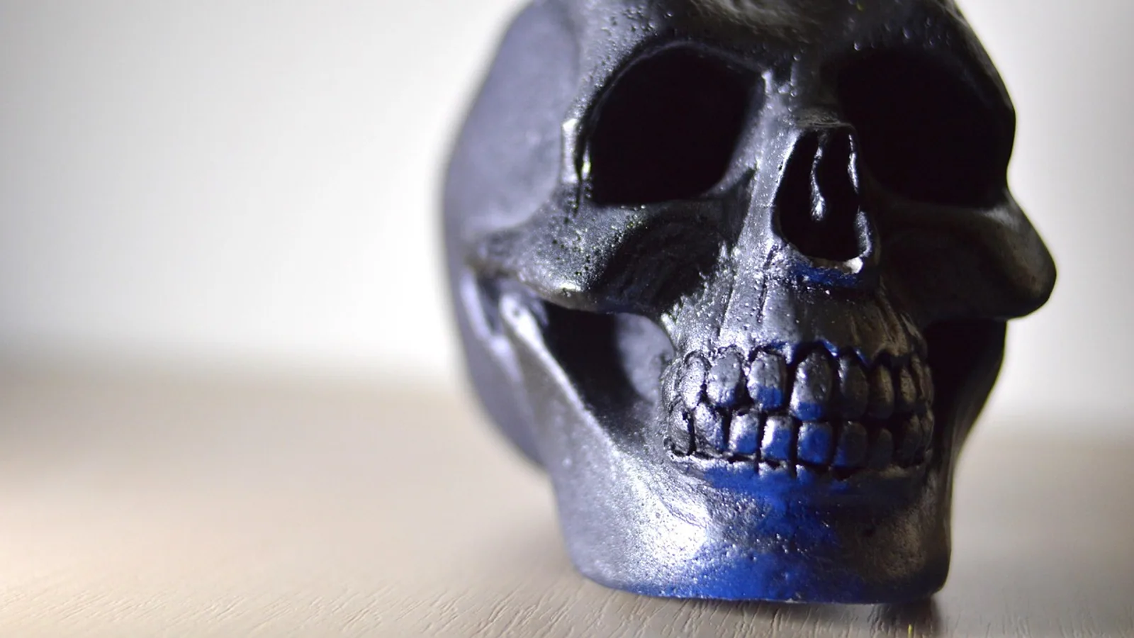
The occipital condyle is one of a pair of smooth, kidney-shaped articular surfaces on the ventral and lateral margins of the foramen magnum, the large opening in the occipital bone through which the brainstem becomes the spinal cord. Each occipital condyle articulates with the corresponding cranial articular fovea of the first cervical vertebra (the atlas) to form the atlanto-occipital joint, a synovial joint that permits mainly flexion and extension of the head.
This small joint carries a disproportionate clinical weight. It is the mechanical pivot between skull and spine, it sits within millimeters of the hypoglossal canal and jugular foramen, and it is the structure that must be stabilized when congenital or traumatic instability threatens the spinal cord. For veterinary students, the occipital condyle is also a comparative anatomy landmark: its shape, the strength of its ligamentous support, and even its number vary across species, from the paired convex condyles of the dog to the single midline condyle of birds.
Why the Occipital Condyle Matters
The atlanto-occipital joint is the most cranial of the synovial joints of the vertebral column, and it is the one that allows a horse to nod, a dog to track a thrown ball, and a bird to hold its head steady while the body moves. When this joint fails, the consequences are neurological rather than orthopedic. Occipital condylar fractures in horses can produce delayed paralysis of the vagus and glossopharyngeal nerves, causing dysphagia days after a minor head injury [1]. In dogs, congenital atlanto-occipital dislocation can coexist with atlantoaxial instability and compress the spinal cord, requiring surgical stabilization from the occipital bone to the second cervical vertebra [2]. In humans, condylar hypoplasia and atlas anomalies are studied as contributors to craniovertebral instability in Chiari malformation [3]. The same principles of bony geometry and ligamentous restraint apply across all of these species.
Anatomy of the Occipital Bone and Condyles
Position and Orientation
The occipital bone forms the caudal wall and much of the floor of the cranial cavity. Its most conspicuous feature is the foramen magnum, which in the dog is roughly oval and in the horse is more circular. The occipital condyles sit on either side of this opening, projecting ventrally and laterally from the exoccipital portion of the bone.
In lateral view, each condyle is oriented obliquely, with its long axis running craniomedial to caudolateral. This obliquity is what converts the joint into a hinge with a slight rotational component. The condyles converge rostrally toward the basioccipital and diverge caudally, so the pair forms a shallow V when viewed from below. The ventral surface of each condyle is smooth and covered by hyaline cartilage in the living animal.
Shape and Surface Features
The classic description of the occipital condyle is kidney-shaped or reniform, with a concave medial border and a convex lateral border. This shape is not universal. In dogs and cats, the condyles are more convex and rounded, giving the joint a ball-like quality that allows a small amount of lateral rotation. In horses and ruminants, the condyles are more distinctly elongated and flattened, with a pronounced articular ridge that interlocks with the atlas. Morphometric studies of the occipital condyles in other species have documented oval, irregular, kidney, and figure-of-eight shapes, with bilateral constrictions or partial separation of the articular surface occurring as normal variants [4]. The condylar canal, when present, transmits a small emissary vein and is recorded as an anatomical variant in morphometric surveys [5].
Relations to Nerves and Vessels
Two foramina are clinically tied to the occipital condyle. The hypoglossal canal lies just dorsal and lateral to the condyle and transmits the hypoglossal nerve (cranial nerve XII), which supplies the tongue. The jugular foramen lies further laterally and transmits the glossopharyngeal (IX), vagus (X), and accessory (XI) nerves along with the internal jugular vein. Because of this proximity, a fracture or mass at the condyle can produce lower cranial nerve deficits without directly injuring the brainstem. A synovial cyst arising from the atlanto-occipital joint has been reported to cause isolated unilateral hypoglossal nerve paralysis, illustrating how a joint lesion can compress a nerve that runs nearby [6]. The hypoglossal and first cervical nerves also send a small branch to the atlanto-occipital joint capsule itself, which is relevant to the pain generated by joint disease [7].
The Atlanto-Occipital Joint
Joint Type and Articular Surfaces
The atlanto-occipital joint is a synovial joint of the ellipsoid (condylar) type. The articular surfaces are the paired occipital condyles and the paired cranial articular foveae of the atlas. In the dog and cat, the foveae are concave and cup the convex condyles. In the horse, the atlas foveae are shallower and more elongated, matching the flatter condyles. The joint has a fibrous capsule lined by synovial membrane, and the capsule is reinforced by dorsal and ventral atlanto-occipital membranes.
Ligamentous Support
Ligamentous support varies with species and with the mechanical demands of the head. The dorsal atlanto-occipital membrane is a broad sheet that closes the dorsal gap between the occiput and the atlas. The ventral atlanto-occipital membrane lies between the basioccipital and the ventral arch of the atlas. A superficial anterior atlanto-occipital ligament has been described as a constant structure in the anterior joint, a narrow band of central thick fibers lying in front of the anterior atlanto-occipital membrane, with a mean length of 19.8 mm, width of 6.2 mm, and thickness of 0.6 mm in one cadaveric series [8]. Its force to failure was recorded at 38.8 N, indicating that it contributes to but does not dominate craniocervical stability [8]. In horses and ruminants, the nuchal ligament and the heavy dorsal musculature add substantial passive support, which is one reason these species tolerate the large head loads of grazing and locomotion.
Range of Motion
The atlanto-occipital joint permits mainly flexion and extension, the nodding motion of the head. Rotation around the longitudinal axis is limited by the interlocking shape of the condyles and foveae, and lateral bending is restricted by the joint capsule and membranes. In the horse, the dorsal and ventral outpouchings of the joint capsule are large enough to be identified on computed tomography and accessed arthroscopically, and the dorsal pouch provides access to roughly half of the dorsocranial occipital condyle and about 15 percent of the dorsocranial atlas articular surface [9]. This capsular volume is why septic arthritis of the atlanto-occipital joint can be debrided arthroscopically in a foal [9].
Comparative Anatomy Across Species
Dog and Cat
In dogs and cats, the occipital condyles are convex and the atlas foveae are correspondingly concave, producing a joint that is closer to a ball-and-socket in shape than in the horse. This allows a slightly greater range of lateral and rotational movement, which is useful for a predator that must track prey with its head while the body turns. The condyles are separated by the foramen magnum and are readily identified on transverse ultrasound images of the atlanto-occipital region, where they appear as two bony landmarks flanking the cisterna magna [10]. This ultrasound anatomy is the basis for indirect ultrasound-guided cisternal puncture in the dog and cat [10].
Horse
The equine occipital condyles are elongated and more flattened than those of the dog, and the joint is reinforced by strong membranes and the nuchal ligament. The dorsal and ventral joint pouches are well developed, and the joint can be approached arthroscopically from dorsal or ventral to the longissimus capitis tendon [9]. Dura perforation occurred with a blind dorsal approach in two of five cadaver joints but did not occur when the approach was guided by ultrasonography, which is a practical caution for equine surgeons [9]. Occipital condylar fracture in the horse is a recognized cause of delayed lower cranial nerve paralysis, with dysphagia appearing several days after a minor head injury [1].
Cow and Other Ruminants
Ruminants share the paired condyle pattern with the horse, but the condyles are set on a wider basioccipital and the joint is supported by the heavy nuchal ligament and the dorsal musculature of the poll. The articular surfaces are flatter than in the dog, and the joint is correspondingly more restricted in rotation. The same general anatomy applies to sheep and goats, with size being the main difference.
Bird
Birds depart from the mammalian plan. Instead of paired occipital condyles, birds have a single midline occipital condyle that articulates with the atlas in a manner that permits a wide range of head rotation. This single condyle is one of the clearest osteological differences between birds and mammals at the craniocervical junction. The avian atlas is correspondingly modified, with a single articular surface rather than paired foveae.
Comparison Table
| Species | Condyle shape and number | Joint type | Main motion | Ligamentous support |
|---|---|---|---|---|
| Dog | Paired, convex, kidney-shaped | Synovial ellipsoid | Flexion and extension, limited rotation | Capsule, dorsal and ventral membranes |
| Cat | Paired, convex, kidney-shaped | Synovial ellipsoid | Flexion and extension, limited rotation | Capsule, dorsal and ventral membranes |
| Horse | Paired, elongated and flattened, with articular ridge | Synovial ellipsoid | Flexion and extension, minimal rotation | Heavy capsule, membranes, nuchal ligament |
| Cow | Paired, flattened, wide-set | Synovial ellipsoid | Flexion and extension, minimal rotation | Heavy capsule, membranes, nuchal ligament |
| Bird | Single midline condyle | Synovial, modified | Wide range of head rotation | Capsule and short ligaments |
How the Joint Is Examined in Practice
Physical and Neurological Examination
A veterinarian assessing the atlanto-occipital region begins with observation of head posture and neck movement. Reluctance to lower the head, resistance to neck flexion, or a head tilt that does not fit a vestibular pattern can point to joint pain. Cranial nerve examination is essential because of the proximity of the hypoglossal canal and jugular foramen. Tongue weakness suggests hypoglossal involvement, while dysphagia, laryngeal hemiplegia, or soft palate dysfunction suggests vagus or glossopharyngeal involvement [1].
Imaging
Radiography of the craniocervical junction is challenging because of superimposed skull structures, but it can reveal rotation of the atlas or a bone fragment dorsal to the guttural pouches in horses with condylar fracture [1]. Computed tomography is the preferred modality for defining condylar shape, joint congruity, and fracture lines. In the horse, CT arthrography has been used to map the dorsal and ventral pouches of the atlanto-occipital joint before arthroscopy [9]. Magnetic resonance imaging is useful for assessing spinal cord compression in dogs with congenital atlanto-occipital dislocation [2]. Ultrasound is a practical bedside tool in small animals and rabbits, where the occipital condyles serve as transverse landmarks for cisternal puncture [11][10].
Arthroscopy and Surgery
Arthroscopy of the atlanto-occipital joint has been described in the horse, with dorsal and ventral approaches that provide access to the occipital condyle and atlas articular surfaces [9]. In dogs, surgical treatment of concurrent atlanto-occipital dislocation and atlantoaxial instability has been reported using ventral facetectomy of C1 followed by stabilization from the occipital bone to C2 with screws, wire, and polymethyl methacrylate [2]. These are specialized procedures performed by experienced surgeons, and they carry substantial risk to the spinal cord and cranial nerves.
Clinical Relevance, Limitations and Common Mistakes
Occipital condyle pathology is uncommon but consequential. The main clinical categories are trauma, congenital malformation, and inflammatory or degenerative joint disease.
Traumatic occipital condylar fracture is best documented in the horse. A seven-year-old Friesian developed delayed dysphagia caused by vagus nerve paralysis and suspected glossopharyngeal paralysis several days after a minor head injury, with endoscopic findings of right laryngeal hemiplegia and intermittent dorsal displacement of the soft palate [1]. Radiography showed rotation of the atlas and a large bone fragment dorsal to the guttural pouches, and the diagnosis was occipital condyle fracture with delayed cranial nerve paralysis [1]. The clinical lesson is that delayed dysphagia after head injury should prompt evaluation of the atlanto-occipital articulation and skull base [1].
Congenital malformation of the condyle or atlas can destabilize the craniocervical junction. In humans, condylar hypoplasia defined by an atlanto-occipital joint axis angle of 130 degrees or greater has been studied as a risk factor for occipitocervical fusion in children with Chiari malformation and syringomyelia [3]. A morphometric study of atlanto-occipital joint geometry classified joints into a typical ball-and-socket type, a shallower type, and an abnormal flat-tilt type, with instability observed in all flat-tilt joints and some shallow joints but not in typical joints [12]. In dogs, congenital atlanto-occipital dislocation has been reported in a Toy Poodle with progressive tetraparesis and respiratory difficulty, treated by ventral C1 facetectomy and occiput-to-C2 stabilization [2]. Congenital unilateral atlanto-occipital rotatory subluxation has also been described as a cause of C1 neuralgia in a human case report [13].
Inflammatory disease of the joint is rare but can cause nerve compression. A synovial cyst of the atlanto-occipital joint caused isolated unilateral hypoglossal nerve paralysis in one reported case, and the anatomy of the hypoglossal nerve and its relationship to the joint were reviewed in that report [6].
Common mistakes in studying this region include assuming that the occipital condyle is a single midline structure in all species (it is paired in mammals and single in birds), confusing the atlanto-occipital joint with the atlantoaxial joint (the latter is responsible for most head rotation), and underestimating the clinical significance of the hypoglossal canal and jugular foramen when evaluating a patient with lower cranial nerve signs. Another frequent error is to treat condylar shape as uniform across mammals. The convex condyles of the dog and the flattened condyles of the horse produce different joint mechanics and different imaging appearances.
This article is educational and is not a substitute for veterinary diagnosis or treatment.
Quick Review
- The occipital condyle is a paired, kidney-shaped articular surface flanking the foramen magnum.
- It articulates with the cranial articular fovea of the atlas to form the atlanto-occipital joint, a synovial ellipsoid joint.
- The joint permits mainly flexion and extension, with limited rotation.
- Dogs and cats have convex condyles, horses and ruminants have flatter, more elongated condyles with stronger ligamentous support, and birds have a single midline condyle.
- The hypoglossal canal and jugular foramen lie close to the condyle, so fractures or masses can cause lower cranial nerve deficits.
- Occipital condylar fracture in the horse can cause delayed dysphagia from vagus and glossopharyngeal paralysis.
- Congenital condylar hypoplasia or malformation can destabilize the craniocervical junction and may require surgical stabilization.
Frequently Asked Questions
What is the occipital condyle?
The occipital condyle is one of a pair of smooth articular surfaces on the occipital bone that flank the foramen magnum and join the skull to the first cervical vertebra.
Which bone articulates with the occipital condyle?
The atlas, or first cervical vertebra, articulates with the occipital condyle through its cranial articular fovea.
What type of joint is the atlanto-occipital joint?
It is a synovial ellipsoid joint that permits mainly flexion and extension of the head.
Do all animals have two occipital condyles?
No. Mammals including dogs, cats, horses, and cows have paired occipital condyles, but birds have a single midline occipital condyle.
What nerves are at risk with an occipital condyle injury?
The hypoglossal nerve and the glossopharyngeal, vagus, and accessory nerves are at risk because the hypoglossal canal and jugular foramen lie close to the condyle.
Can a condylar fracture cause delayed neurological signs?
Yes. In horses, occipital condylar fracture has caused delayed vagus and glossopharyngeal paralysis with dysphagia appearing several days after a minor head injury.
Related Articles
- Comparative Anatomy of the Mammalian Kidney
- Comparative Anatomy of the Mammalian Heart
- Gastrointestinal Adaptations in Exotic Herbivores
- Canine Respiratory System: Anatomy and Physiology
- Feline Cardiovascular Anatomy and Physiology
- Bovine Female Reproductive Anatomy and Physiology
- Shoulder Joint Anatomy: Articulation and Movement
- Knee Joint and Muscles: Anatomy, Movement, and Species Notes
Sources
- Delayed onset vagus nerve paralysis after occipital condyle fracture in a horse.
- C1 Facetectomy and Ventral Fixation of Occipitoatlantoaxial Complex for Concurrent Congenital Atlanto-Occipital Dislocation and Atlantoaxial Instability in a Toy Poodle
- The role of occipital condyle and atlas anomalies on occipital cervical fusion outcomes in Chiari malformation type I with syringomyelia: a study from the Park-Reeves Syringomyelia Research Consortium.
- Morphometric analysis of superior articular facets of atlas vertebra and its clinical applications in ergonomics of atlanto-occipital joints.
- Morphological and Morphometric Analysis of the Occipital Condyles and Their Clinical Significance
- Isolated unilateral hypoglossal nerve paralysis caused by an atlanto-occipital joint synovial cyst.
- Microsurgical Anatomy of the Hypoglossal and C1 Nerves: Description of a Previously Undescribed Branch to the Atlanto-Occipital Joint.
- Superficial anterior atlanto-occipital ligament: Anatomy of a forgotten structure with relevance to craniocervical stability
- Arthroscopic approach and intra-articular anatomy of the equine atlanto-occipital joint
- Ultrasonographic percutaneous anatomy of the atlanto-occipital region and indirect ultrasound-guided cisternal puncture in the dog and the cat.
- Ultrasonographic anatomy of the atlanto-occipital region and ultrasound-guided cerebrospinal fluid collection in rabbits (Oryctolagus cuniculus).
- Exploring the Pathogenesis of Atlanto-Occipital Instability in Chiari Malformation With Type II Basilar Invagination: A Systematic Morphological Study.
- Congenital Unilateral Atlanto-Occipital Rotatory Subluxation: Rare Cause of C1 Neuralgia