Nervous System Diagram: Labeled Overview
By Dr. Zubair Khalid, DVM, MS, PhD ·
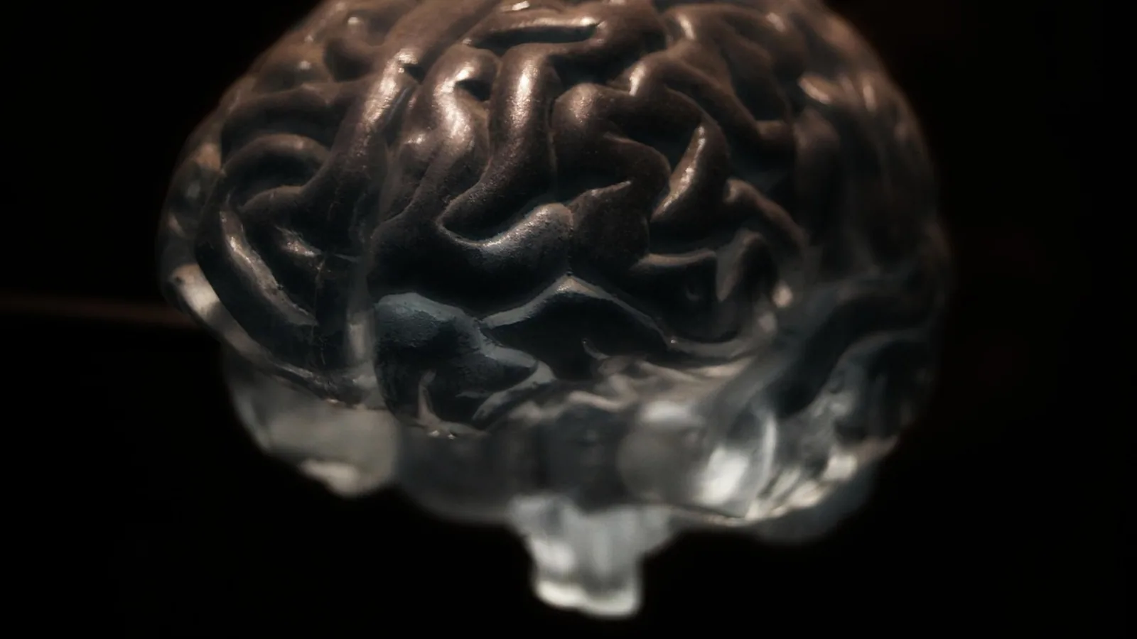
A labeled nervous system diagram divides the animal body's control network into two great halves: the central nervous system (CNS), which is the brain and spinal cord, and the peripheral nervous system (PNS), which is every nerve and ganglion outside those two organs. The CNS is the processing center, and the PNS is the wiring that carries sensory information in and motor commands out, including the autonomic nerves that run the heart, gut, and glands without conscious input.
This overview walks through each label you would expect to find on a veterinary nervous system diagram, explains what that structure does, and flags the places where dogs, cats, horses, and cattle differ from one another and from people. The labeled figure itself is provided separately, so the descriptions here use words, tables, and numbered lists rather than an image.
This article is educational and is not a substitute for veterinary diagnosis or treatment.
The Two Great Divisions of the Nervous System
Every vertebrate nervous system is organized around a central axis. The brain sits inside the skull, the spinal cord runs down the vertebral canal, and together they form the CNS. Everything else, the cranial nerves emerging from the brainstem, the spinal nerves leaving the vertebral column, and the autonomic ganglia and plexuses scattered through the body, belongs to the PNS.
The division matters because the two halves behave differently when injured. CNS neurons in mammals regenerate poorly after damage, while peripheral axons can regrow over long distances if the nerve sheath is intact. That difference is one reason a spinal cord injury in a dog carries a very different prognosis from a crushed peripheral nerve in the same dog.
A useful way to read any nervous system diagram is to follow the flow of information. Sensory (afferent) fibers travel from receptors toward the CNS. Motor (efferent) fibers travel away from the CNS toward muscles and glands. The diagram's arrows, when present, almost always point along that sensory-in, motor-out pattern.
Central Nervous System: Brain and Spinal Cord
The Brain
The brain is the rostral expansion of the neural tube. On a labeled diagram it is usually broken into the cerebrum, cerebellum, brainstem (midbrain, pons, medulla oblongata), diencephalon (thalamus and hypothalamus), and the olfactory bulbs. Each region has a recognizable surface signature.
The cerebrum carries the cerebral cortex, the folded gray matter sheet responsible for conscious perception, voluntary movement, and learned behavior. The cerebellum sits behind and below the cerebrum and coordinates posture, balance, and fine motor timing. The brainstem is the stalk that connects the cerebrum to the spinal cord and houses the nuclei of most cranial nerves plus the centers that regulate breathing and heart rate.
Brain surface folding is one of the most useful comparative labels. Animals with smooth, unfolded cortices are lissencephalic, and animals with heavily folded cortices are gyrencephalic. Dogs and cats are gyrencephalic. So are horses, cattle, and pigs. Many rodents and rabbits are closer to the lissencephalic pattern. The folding increases surface area without increasing skull volume, which is one reason a large-brained mammal can pack more cortex into a smaller cranium.
Skull shape and brain shape are linked in dogs. In a study of 62 dogs from 33 breeds plus 17 mixed or unknown-breed dogs, researchers found that as the neurocranium became broader and shorter, gray matter volume decreased significantly in the right olfactory bulb, frontal cortex, marginal gyrus, and cerebellum [1]. As the neurocranium became narrower and longer, gray matter volume decreased across a wider set of regions, including the olfactory bulb, frontal cortex, temporal cortex, amygdala, hypothalamus, hippocampus, periaqueductal gray, cerebellum, and brainstem [1]. That is a direct example of how the labels on a nervous system diagram can shift in size and shape across breeds without changing their names.
The Spinal Cord
The spinal cord is the caudal continuation of the brainstem. It runs through the vertebral canal and gives off paired spinal nerves at each segment. A labeled diagram typically marks the cervical, thoracic, lumbar, sacral, and caudal (or coccygeal) regions, plus the dorsal and ventral horns of the gray matter in cross-section.
The dorsal horn receives sensory input from the dorsal root of each spinal nerve. The ventral horn contains the motor neuron cell bodies whose axons leave through the ventral root. This dorsal-sensory, ventral-motor arrangement is one of the oldest and most reliable rules in vertebrate neuroanatomy.
Spinal cord segment numbers vary by species because the vertebral formula varies. Dogs and cats have cervical, thoracic, lumbar, sacral, and caudal segments that roughly track their vertebral counts. Horses and cattle have longer thoracic and lumbar series than dogs. The cervical enlargement supplies the forelimbs, and the lumbosacral enlargement supplies the hindlimbs. In a dog with a spinal cord lesion, the neurologist uses the segment number to localize the problem to a specific vertebra, which is why the segment labels on a diagram are clinically meaningful rather than decorative.
Peripheral Nervous System: Cranial Nerves, Spinal Nerves, and Autonomic Divisions
Cranial Nerves
Mammals have 12 pairs of cranial nerves, numbered I through XII from rostral to caudal. They emerge directly from the brain or brainstem rather than from the spinal cord. The standard list is olfactory (I), optic (II), oculomotor (III), trochlear (IV), trigeminal (V), abducens (VI), facial (VII), vestibulocochlear (VIII), glossopharyngeal (IX), vagus (X), accessory (XI), and hypoglossal (XII).
The 12-pair count is conserved across domestic mammals, though the size and branching of individual nerves differ. The trigeminal nerve is the great sensory nerve of the face and the motor nerve of the muscles of mastication. The facial nerve drives the muscles of facial expression and carries taste from the rostral tongue. The vagus nerve is the longest cranial nerve and the main parasympathetic supply to the thorax and abdomen.
Comparative work on the African wild dog brain found that the cranial nerve motor nuclei and the rest of the motor system matched what is seen in the domestic dog, with one notable exception: a distinct fascicle of protoplasmic commissural dendrites at the rostral pole of the hypoglossal nucleus that has not been reported in other mammals [2]. That is a reminder that even a structure as conserved as the hypoglossal nucleus can carry species-specific features.
Spinal Nerves
Spinal nerves form where the dorsal and ventral roots join just outside the vertebral canal. Each spinal nerve is therefore a mixed nerve carrying both sensory and motor fibers. After the join, the nerve typically splits into a dorsal ramus for the epaxial muscles and skin of the back, and a ventral ramus for the limbs and body wall.
The ventral rami of adjacent spinal nerves interlace to form plexuses. The cervical plexus supplies the neck, the brachial plexus supplies the forelimb, the intercostal nerves run between the ribs, and the lumbosacral plexus supplies the hindlimb and pelvis. A full dissection and plastination of a dog's nervous system preserved the brain, spinal cord with its meninges, cervical plexus, brachial plexus, intercostal and abdominal nerves, lumbosacral plexus, sympathetic trunk, vagus nerve, and phrenic nerves as a single teaching specimen [3]. That specimen list is essentially a checklist of the labels a complete veterinary nervous system diagram should carry.
Autonomic Divisions
The autonomic nervous system is the involuntary half of the PNS. It has two main divisions, sympathetic and parasympathetic, plus the enteric nervous system embedded in the gut wall.
The sympathetic division prepares the body for activity. Its preganglionic neurons sit in the thoracolumbar spinal cord, and its ganglia form a chain along the vertebral column called the sympathetic trunk. The parasympathetic division supports rest, digestion, and repair. Its preganglionic neurons sit in the brainstem and sacral spinal cord, and its ganglia are usually near or inside the target organ. The vagus nerve is the largest single parasympathetic outflow.
Autonomic wiring is not just a background system. A 2026 study in mice showed that glutamatergic neurons in the medial prefrontal cortex modulate the peripheral immune response during sepsis through autonomic pathways and adrenergic signaling, with the left cervical vagus and beta-2 adrenergic receptors both involved [4]. The takeaway for a veterinary reader is that the autonomic labels on a diagram connect the brain to immune and inflammatory function, not only to heart rate and gut motility.
Table: Central vs Peripheral Components at a Glance
| Component | Location | Main function | Comparative note |
|---|---|---|---|
| Cerebrum | Inside the skull, rostral | Conscious perception, voluntary movement, learning | Gyrencephalic in dogs, cats, horses, cattle. Lissencephalic in many rodents. |
| Cerebellum | Caudal to cerebrum, dorsal to brainstem | Posture, balance, motor coordination | Gray matter volume decreases with broader, shorter skulls in dogs [1] |
| Brainstem | Between cerebrum and spinal cord | Cranial nerve nuclei, respiration, cardiovascular control | Motor nuclei similar across carnivores, with a species-specific hypoglossal feature in the African wild dog [2] |
| Spinal cord | Vertebral canal | Relay and reflex center, ascending and descending tracts | Segment numbers track the vertebral formula, so they vary by species |
| Meninges | Around brain and spinal cord | Protection, CSF containment, vascular support | Preserved intact in a full dog dissection and plastination specimen [3] |
| Cranial nerves | Emerge from brain and brainstem | Sensory and motor supply to head, neck, and viscera | 12 pairs in mammals, with species differences in size and branching |
| Spinal nerves | Emerge from spinal cord at each segment | Mixed sensory and motor supply to body wall and limbs | Form cervical, brachial, and lumbosacral plexuses [3] |
| Sympathetic trunk | Along the vertebral column | Fight-or-flight outflow to viscera and vessels | Preserved as a labeled structure in the dog plastination specimen [3] |
| Vagus nerve | From brainstem through neck to thorax and abdomen | Main parasympathetic supply to thorax and abdomen | Modulates immune response via autonomic pathways in experimental models [4] |
Meninges, Cerebrospinal Fluid, and the Blood-Brain Barrier
Three connective tissue layers, the meninges, wrap the CNS. From outside in they are the dura mater, arachnoid mater, and pia mater. The dura is tough and fibrous. The arachnoid is a delicate middle layer. The pia is a thin membrane that clings to the brain and spinal cord surface and follows every fold.
Cerebrospinal fluid (CSF) fills the space between the arachnoid and pia, called the subarachnoid space, and the internal ventricles of the brain. CSF cushions the CNS, clears metabolic waste, and helps regulate intracranial pressure. A 2025 review of CSF circulation described how the anatomy of CSF flow contributes to waste clearance in the brain and spinal cord and how dysregulation can lead to conditions such as syringomyelia and hydrocephalus [5]. The same review noted that certain brachycephalic dog breeds have a high prevalence of CSF-related conditions because of artificial selection for neotenous traits, making them useful models for studying analogous human conditions such as Chiari-like malformation [5].
The blood-brain barrier is a selective interface formed by tight junctions between endothelial cells of CNS capillaries, supported by astrocytes and pericytes. It keeps large molecules, many pathogens, and most circulating drugs out of the brain and spinal cord. The barrier is why some infections and some cancers are hard to treat once they reach the CNS. A 2026 review of circulating tumor DNA and CNS recurrence in lung cancer noted that the blood-brain barrier limits the release of tumor DNA into peripheral blood, which can produce false-negative results when clinicians try to detect CNS metastasis from a blood sample [6]. That mechanism is not unique to cancer. Any molecule that needs to reach the CNS has to cross or bypass the same barrier.
Myelin: The Insulation That Makes the Diagram Work


Myelin is the fatty sheath that wraps axons and speeds up action potential conduction. In the CNS it is produced by oligodendrocytes. In the PNS it is produced by Schwann cells. The two cell types make chemically similar but not identical myelin, and that difference is one reason CNS and PNS injuries behave differently.
Sulfatide is a major glycolipid component of myelin in both divisions. A 2026 imaging mass spectrometry study in mice identified 13 sulfatide species in adult dorsal root ganglia and found that very-long-chain non-hydroxylated sulfatides were enriched in compact myelin-rich regions, while long-chain and hydroxylated species had broader distributions [7]. The study also showed that sulfatide diversification was established before mature myelin formation, with structurally diverse classes detectable by embryonic day 14.5 and all species present by postnatal day 2 [7].
Cholesterol is another major myelin component. In the CNS, more than 99 percent of cholesterol is unesterified, and roughly 70 percent of total CNS cholesterol sits in the myelin sheath. A 2026 study in a mouse model measured free cholesterol and cholesterol esters in brain and spinal cord during myelination, demyelination, and remyelination. Brain cholesterol increased steadily up to 38 weeks of age, while spinal cord cholesterol increased until postnatal day 42 and then stayed steady out to 38 weeks [8]. During demyelination, cholesterol dropped significantly in both tissues and did not return to normal even during remyelination, and cholesterol esters made up 19 percent of the measured cholesterol pool in the brain and 66 percent in the spinal cord at peak demyelination [8].
Myelin staining is a standard teaching and diagnostic tool. A 2026 methods paper validated an Eriochrome Cyanine R protocol for thick sections of central and peripheral nervous system tissue across seven vertebrate species and multiple demyelinating conditions, producing homogeneous myelin labeling with sharp white matter and gray matter contrast in whole brains, cortical slabs, spinal cord, and peripheral nerves [9]. The same protocol resolved single myelinated axons and intracortical bands and could be combined with Nissl-like counterstains and immunohistochemistry [9]. For a student reading a nervous system diagram, that is the practical link between the labeled structure and the histology slide underneath it.
How the Labels Connect: A Worked Example
Take a dog that steps on a sharp stone with its left forelimb. The flow of information through the labeled structures runs like this.
- Sensory receptors in the foot generate a signal.
- The signal travels up the sciatic nerve and into the lumbosacral plexus.
- It enters the spinal cord through the dorsal root of a spinal nerve in the lumbosacral enlargement.
- Ascending tracts carry the signal toward the brainstem and thalamus.
- The cerebrum registers the sensation as pain.
- Motor commands descend through the corticospinal and extrapyramidal pathways.
- Motor neurons in the ventral horn send axons back out through the ventral root.
- The signal returns to the forelimb muscles through the brachial plexus and peripheral nerves.
- The dog lifts its foot.
Every step in that sequence corresponds to a label on a complete veterinary nervous system diagram. When a clinician localizes a neurologic lesion, they are essentially asking which of those steps is broken.
Clinical Relevance, Limitations and Common Mistakes
The single most common mistake when reading a nervous system diagram is confusing the CNS and PNS labels. A structure inside the skull or vertebral canal is CNS. A structure outside those boundaries is PNS, even if it is physically close to the CNS. The optic nerve is a classic trap because it is a cranial nerve but is embryologically an outpouching of the brain and is wrapped in meninges.
A second mistake is assuming that the 12 cranial nerve pairs are identical in size and function across species. The numbering is conserved, but the relative development of each nerve varies. The olfactory nerve is proportionally larger in dogs than in humans. The facial nerve carries a different mix of motor and sensory fibers in different species.
A third mistake is treating the autonomic nervous system as a single unit. The sympathetic and parasympathetic divisions have separate anatomy, separate neurotransmitters, and often opposing effects on the same organ. A diagram that shows only one division is incomplete.
A fourth mistake is reading a normal anatomy diagram as if it predicts disease. It does not. Anatomy tells you where a lesion could be. It does not tell you what the lesion is. Individual cases need a veterinarian for diagnosis and treatment.
Species Differences Worth Knowing
Domestic mammals share the same basic nervous system plan, but the details shift in ways that matter for anatomy study and clinical practice.
Dogs show the widest within-species variation because of breed selection. Skull shape correlates with brain region volumes, as the 62-dog MRI study showed [1]. A 2023 MRI study of the DE50-MD dog model of Duchenne muscular dystrophy found reduced regional gray matter in the piriform lobe, hippocampus, and cingulate gyrus compared with wild-type littermates, plus larger lateral ventricle volume, with no progression over the 14 to 33 month study window [10]. That is a disease model, but it illustrates how a single gene defect can reshape the labeled brain regions on a diagram.
Horses and cattle have longer spinal cords relative to body length and correspondingly more thoracic and lumbar segments than dogs. Their cranial nerve anatomy is broadly similar to the dog, but the size and course of the vagus and trigeminal nerves reflect their different feeding and head anatomy.
Cats sit between dogs and horses in most comparative measures. Their brain is gyrencephalic, their cranial nerve count is 12 pairs, and their autonomic anatomy follows the standard mammalian pattern.
Frequently Asked Questions
What are the two main divisions of the nervous system?
The central nervous system is the brain and spinal cord. The peripheral nervous system is everything else, including the 12 pairs of cranial nerves, the spinal nerves, and the autonomic ganglia and plexuses.
How many cranial nerve pairs do mammals have?
Mammals have 12 pairs of cranial nerves, numbered I through XII from front to back. The count is conserved across dogs, cats, horses, and cattle, though the size and branching of individual nerves differ by species.
What is the difference between the CNS and the PNS?
The CNS is the brain and spinal cord, which sit inside the skull and vertebral canal. The PNS is all neural tissue outside those boundaries, including cranial nerves, spinal nerves, and autonomic ganglia.
What do the meninges do?
The meninges are three protective layers (dura mater, arachnoid mater, and pia mater) that wrap the brain and spinal cord. They contain the cerebrospinal fluid, support blood vessels, and cushion the CNS against impact.
What is cerebrospinal fluid and where does it come from?
Cerebrospinal fluid is a clear fluid produced mainly by the choroid plexus inside the brain ventricles. It circulates through the ventricles and the subarachnoid space, cushions the CNS, clears metabolic waste, and helps regulate intracranial pressure [5].
What does the blood-brain barrier do?
The blood-brain barrier is a selective filter formed by tight junctions between CNS capillary endothelial cells. It keeps many molecules, pathogens, and drugs out of the brain and spinal cord, which is why some CNS infections and tumors are hard to treat.
Do spinal cord segment numbers differ between species?
Yes. Spinal cord segments track the vertebral formula of each species, so dogs, cats, horses, and cattle have different numbers of thoracic, lumbar, sacral, and caudal segments. The cervical enlargement supplies the forelimbs and the lumbosacral enlargement supplies the hindlimbs in all of them.
What is the difference between lissencephalic and gyrencephalic brains?
A lissencephalic brain has a smooth cortex with few or no folds. A gyrencephalic brain has a heavily folded cortex. Dogs, cats, horses, and cattle are gyrencephalic. Many rodents and rabbits are closer to the lissencephalic pattern.
Related Articles
- Protein Synthesis Diagram Labeled
- Ribosome Diagram: Structure, Function, and Labeled Parts
- Microscope Parts and Functions: A Visual Guide to Labeled Diagrams
- DNA Drawing & Structure Diagram: Labeled Double Helix Step-by-Step
- Veterinary Computer Communication Systems: An Overview
- Canine Skeletal System: Axial and Appendicular Overview
- Endomembrane System: Parts, Function, and Diagram
Sources
- Covariation of Skull and Brain Morphology in Domestic Dogs.
- The Brain of the African Wild Dog. VI. The Motor System.
- Dissection and Plastination of the Nervous System of a Dog: A Teaching Tool.
- Medial Prefrontal Cortex Modulation of the Peripheral Immune Response in Sepsis Via the Autonomic Nervous System.
- A review of cerebrospinal fluid circulation with respect to Chiari-like malformation and syringomyelia in brachycephalic dogs.
- [[Research Progress on Circulating Tumor DNA Status and Postoperative Central Nervous System Recurrence Risk and Overall Survival in Non-small Cell Lung Cancer].](https://pubmed.ncbi.nlm.nih.gov/42705855/)
- Spatiotemporal Heterogeneity of Sulfatide Molecular Species During Schwann Cell Development in the Peripheral Nervous System.
- Cholesterol Quantification in Brain and Spinal Cord During Development, Demyelination, and Remyelination.
- Eriochrome Cyanine R revisited: a standardized myelin stain protocol for thick sections of the central & peripheral nervous system across species, pathologies and disease models.
- Brain magnetic resonance imaging in the DE50-MD dog model of Duchenne muscular dystrophy reveals regional reductions in cerebral gray matter.