Membrana Nictitans: Anatomy and Clinical Notes
By Dr. Zubair Khalid, DVM, MS, PhD ·
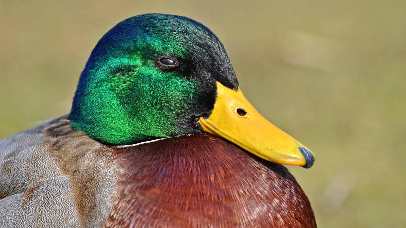
The membrana nictitans, also called the third eyelid or nictitating membrane, is a fold of conjunctiva that sweeps across the eye from the medial canthus to protect and lubricate the cornea. It is supported internally by a T-shaped piece of cartilage, moved actively by the retractor bulbi muscle and passively by pressure changes within the orbit, and it houses a tear-producing gland that supplies a large share of the aqueous tear film.
This article covers the gross and functional anatomy of the third eyelid, the role of the superficial gland of the third eyelid (the nictitans gland), the mechanics of prolapse, and how prolapse differs from conjunctivitis, neoplasia, and other causes of a pink mass at the medial corner of the eye. It closes with a species comparison table and a set of practical clinical notes.
This article is educational and is not a substitute for veterinary diagnosis or treatment.
What the Membrana Nictitans Is and Where It Sits
The membrana nictitans is a crescent-shaped fold of conjunctiva, the thin mucous membrane that lines the eyelids and covers the front of the eyeball. In the dog and cat it occupies the ventromedial corner of the orbit, tucked beneath the lower eyelid. Its free margin is usually visible as a thin, pale rim at the medial canthus. The rest of the membrane is hidden, folded back on itself like a pocket.
The membrane has two epithelial surfaces. The palpebral (outer) surface faces the inside of the lower eyelid. The bulbar (inner) surface faces the globe. Between them sits a layer of connective tissue, the substantia propria, which contains the cartilage and the gland. Because the membrane folds over the cartilage, the same piece of tissue presents two surfaces that meet at the leading edge.
The third eyelid is not a spare part. It contributes to the tear film, spreads tears across the cornea during blinking, and provides a physical barrier that can sweep across the eye in a fraction of a second. In species that dive or burrow, it also acts as a protective shutter.
The Conjunctival Fold
The conjunctiva of the third eyelid is continuous with the conjunctiva of the lower eyelid and the bulbar conjunctiva that covers the sclera. This continuity matters clinically. Inflammation that starts in the lower conjunctiva can spread into the third eyelid, and a mass that appears to arise from the third eyelid may actually be an extension of a lesion elsewhere on the conjunctival surface.
The fold creates a space called the conjunctival sac or fornix, which is deepest at the medial canthus. When the third eyelid is prolapsed or everted, this sac can become a trap for debris, hair, and discharge.
The T-Shaped Cartilage
The core of the third eyelid is a flexible plate of hyaline cartilage shaped like a capital T or an anchor. The vertical bar of the T anchors the cartilage near the medial orbital rim. The horizontal bar, sometimes called the crossbar or the scrolled portion, sits within the free margin of the membrane and curves slightly to follow the contour of the globe.
The cartilage gives the membrane its shape and spring. When the membrane is pushed across the eye, the cartilage keeps it flat against the cornea. When the membrane retracts, the cartilage helps it fold back into its resting position. The crossbar is the part that can scroll or curl abnormally, producing a condition called cartilage eversion.
Cartilage eversion is distinct from gland prolapse, though the two can occur together. In a cat case report, an apparent prolapsed gland turned out to be an everted cartilage of the nictitating membrane, and the scrolled portion was removed through an incision on the bulbar surface [1]. A separate feline study described third eyelid rotation associated with both nictitans gland prolapse and cartilage eversion, a combination described as rarely encountered [2]. Cartilage eversion has also been reported in an adult American Quarter Horse mare, where the abnormal scrolled cartilage was excised and histopathology showed normal cartilage without neoplasia or inflammation [3].
Why the Cartilage Shape Matters Clinically
A normal crossbar lies flat within the membrane. When it curls forward, the leading edge of the third eyelid rolls outward and exposes the bulbar conjunctiva. Owners often describe this as a pink or red rim that will not sit flat. The everted cartilage itself is not a tumor and is not infected, but it can cause ocular irritation and epiphora. Thermal cautery has been used to remodel the cartilage in dogs by applying low-energy heat to the bulbar conjunctival surface over the convexity of the cartilage, causing gradual contraction and softening that returns the membrane to a more normal position [4]. In the horse case, the scrolled cartilage was excised with a handheld cautery unit [3].
Musculature and Movement
The third eyelid moves by two mechanisms. The first is active and muscular. The retractor bulbi muscle is a cone-shaped muscle that surrounds the optic nerve and inserts on the globe. When it contracts, it pulls the globe backward into the orbit. That retrobulbar movement pushes the orbital fat and connective tissue forward, and the third eyelid slides across the cornea. This is why the membrane flashes across the eye when an animal is startled or when the globe is retracted.
The second mechanism is passive. Any increase in orbital pressure, whether from retraction of the globe, swelling, or a space-occupying lesion, can push the third eyelid forward. This is why the membrane protrudes in conditions as different as orbital cellulitis, retrobulbar masses, and severe conjunctivitis.
A useful way to think about it is that the third eyelid has no dedicated muscle of its own in the domestic species discussed here. It is a passive sail that is deployed by the machinery that moves the globe. This has two clinical consequences. First, a protruding third eyelid is often a sign of orbital disease rather than primary third eyelid disease. Second, surgical procedures that anchor the gland must preserve the membrane's ability to slide, or they will defeat its protective function.
The intranictitans tacking technique was designed with this in mind. It anchors the prolapsed gland to the cartilage of the nictitans with a nonabsorbable suture, so the third eyelid retains its normal mobility and protective functions [5]. By contrast, older techniques that tacked the gland to the orbital rim could restrict movement.
The Superficial Gland of the Third Eyelid (Nictitans Gland)
The superficial gland of the third eyelid, also called the nictitans gland or the third eyelid gland, sits at the base of the cartilage within the bulbar aspect of the membrane. It is a lobulated, pinkish gland that produces the aqueous component of the tear film. In dogs, the lacrimal gland and the third eyelid gland are the two intraorbital glands that secrete the aqueous portion of tears [6].
In dogs and cats, the nictitans gland contributes roughly 30 to 50 percent of tear production. That figure is a standard teaching value and explains why removing the gland is a bad idea. A dog that loses its nictitans gland may develop keratoconjunctivitis sicca (KCS, or dry eye) later in life, because the remaining lacrimal tissue cannot always compensate.
Gross anatomy studies in dogs have measured the third eyelid gland at approximately 10.5 mm long, 11.0 mm wide, and 3.3 mm thick, with a mean weight of about 263 mg [6]. The lacrimal gland was slightly larger and heavier in the same study. These measurements come from dissections of dog heads and show that the third eyelid gland is a substantial structure, not a minor accessory.
The Deep Gland and Comparative Notes
Some species have a second accessory gland associated with the third eyelid, called the deep gland of the third eyelid. In pigs, both the superficial and deep glands of the third eyelid are classified as accessory lacrimal glands, and immunohistochemical study has mapped their innervation by vasoactive intestinal peptide, dopamine beta-hydroxylase, substance P, neuropeptide Y, and galanin [7]. This work is comparative and does not change canine or feline clinical practice, but it shows that the third eyelid gland is under autonomic control and is not simply a passive reservoir.
Rabbits have a prominent deep gland of the nictitating membrane, and a pink mass on the dorsomedial aspect of a rabbit's eye may represent prolapse of this deep gland rather than the classic cherry eye of dogs [8]. This is a useful reminder that the same clinical sign can have a different anatomical basis in different species.
Prolapse of the Nictitans Gland (Cherry Eye)
Prolapse of the nictitans gland is the most common disorder of the third eyelid in dogs [9]. It appears as a pink, fleshy, round mass at the medial canthus, which is why it is called cherry eye. The mass is the gland itself, displaced from its normal position behind the membrane and exposed to the air.
The cause is thought to be a defect in the connective tissue that secures the gland in place [10]. The gland is not primarily inflamed or neoplastic in the typical case. It prolapses because its anchoring tissue is weak or abnormally formed.
Cherry eye is most often seen in dogs under two years of age [10]. A large UK study of primary care records found an annual prevalence of 0.20 percent, with a median age at first diagnosis of 0.63 years [9]. Dogs under one year had 10.82 times the odds compared with dogs aged two to under four years [9]. Neutered animals had higher odds than entire animals within both sexes, and purebred dogs had 1.43 times the odds of crossbreeds [9].
Breed predispositions are strong. Compared with crossbred dogs, the breeds with the highest odds in that study included the Neapolitan Mastiff (odds ratio 34.26), English Bulldog (24.08), Cane Corso (14.66), Lhasa Apso (12.37), and American Cocker Spaniel (11.57) [9]. Breeds with brachycephalic skull conformation had 6.71 times the odds compared with mesocephalic breeds [9]. A genetic study identified a single peak of genome-wide significance on canine chromosome 18, at the location of an FGF4 insertion previously linked to chondrodysplasia in several breeds [10]. This suggests that at least part of the risk is inherited and may relate to cartilage and connective tissue development.
In cats, prolapse of the nictitans gland is less common and is often accompanied by cartilage eversion and rotation of the third eyelid. In one feline case series, the most common breeds were Persian and British Shorthair, each accounting for 38.4 percent of cases, with most cats affected unilaterally and males overrepresented [2].
What Prolapse Is Not
A pink mass at the medial canthus is not automatically cherry eye. Several other conditions can look similar, and the distinction matters because the treatment is different.
Conjunctivitis is inflammation of the conjunctiva. It produces redness, swelling, and discharge, but the tissue is diffuse rather than a discrete mass. The third eyelid may be elevated, but it is not displaced by a gland.
Neoplasia of the third eyelid is rare but real. Nonpigmented malignant spindle cell tumors of the membrana nictitans are uncommon in dogs, with only three cases diagnosed in Scandinavia over twenty-three years [11]. Those cases included one hemangiosarcoma and two amelanotic melanomas, all presenting as firm or multilobulated hyperemic masses on the right nictitating membrane [11]. A basal cell adenocarcinoma has also been reported on the bulbar conjunctiva of the third eyelid in a dog, presenting as an exophytic mass with swelling and proptosis of the third eyelid [12]. A similar tumor has been reported in the gland of the third eyelid of a brown bear [13]. The lesson is that a mass that is firm, irregular, ulcerated, or progressive should be evaluated as a possible tumor rather than assumed to be a prolapsed gland.
Prolapse of the lacrimal gland is a separate entity. The lacrimal gland sits dorsolaterally under the orbital ligament [6], so its prolapse would appear in a different location. In practice, the main confusion is between the nictitans gland and the deep gland in species such as rabbits [8].
Cartilage eversion can also mimic gland prolapse. In the cat case report, the presumed diagnosis was prolapsed nictitans gland, but surgery revealed an everted cartilage instead [1]. When the cartilage is scrolled, the leading edge of the membrane rolls outward and the exposed tissue can look like a prolapsed gland.
Species Comparison
The table below summarizes key anatomical and clinical differences in the species most often seen in general practice. Cartilage shape is described qualitatively because detailed morphometric data are not available for every species in the same format.
| Species | Cartilage shape | Gland size and type | Prolapse common? |
|---|---|---|---|
| Dog | T-shaped with a scrolled crossbar, hyaline cartilage | Large superficial gland, about 263 mg mean weight, contributes roughly 30 to 50 percent of tears | Yes, the most common third eyelid disorder in dogs, with strong breed predispositions [9] |
| Cat | T-shaped with a scrolled crossbar, similar to dog | Superficial gland present, smaller than in dogs | Less common than in dogs, often associated with cartilage eversion and third eyelid rotation [2] |
| Horse | T-shaped cartilage, can scroll abnormally | Superficial gland present | Rare, cartilage eversion reported in an adult mare [3] |
| Cattle | T-shaped cartilage, prominent membrane | Superficial gland present, contributes to tear film | Uncommon, prolapse not a major clinical entity in this species |
| Bird | Cartilage present but often thinner and more flexible | Glandular tissue present, varies by species | Rare, third eyelid is highly mobile and normally sweeps across the eye |
Bird third eyelids deserve a note. Histological study of woodpeckers showed regional variation in the marginal plait, leading edge, epithelial surfaces, and stromal pigmentation of the nictitating membrane [14]. Birds rely heavily on the third eyelid for corneal protection during flight and foraging, and its structure reflects that demand.
Hedgehogs have an oval-shaped cartilage that is elastic rather than hyaline, and their third eyelid surfaces are covered by non-keratinized stratified squamous epithelium without glands [15]. This is a reminder that the "typical" mammalian third eyelid is not universal.
Clinical Relevance, Limitations and Common Mistakes
The most common mistake is to treat cherry eye as a cosmetic problem and remove the gland. Excision of the nictitans gland reduces tear production and increases the risk of KCS. Surgical replacement is the standard approach. In a survey of veterinarians, 96.2 percent suggested surgical replacement of cherry eye, with a pocketing technique being the most frequently nominated procedure [16]. General practitioners were more likely to suggest gland excision after a failed surgery, while veterinarians with postgraduate ophthalmology training more often nominated techniques incorporating a periosteal anchor for salvage repair [16].
A second mistake is to assume every medial canthal mass is a prolapsed gland. Firm, irregular, ulcerated, or rapidly growing masses should be evaluated for neoplasia [11][12]. Cartilage eversion can also mimic prolapse, and the two can occur together [2][1].
A third mistake is to judge success only by appearance in the first few days. After replacement, the gland may remain swollen for a period. In the intranictitans tacking technique, glands reduced in size and took on a normal appearance over several weeks [5].
Surgical success rates vary by technique and population. A study of 132 dogs found a 91.6 percent success rate with the Morgan pocket technique and chondrectomy alone, and 100 percent when combined with a wedge conjunctivectomy, with recurrence and complication rates significantly lower in giant breed dogs for the combined procedure [17]. Another study of 353 dogs reported 95 percent successful repositioning overall, with recurrence in 5 percent, and lower recurrence in large breeds such as the English Bulldog and Boxer when a combined technique was used [18]. A modified pocket technique with temporary ventral scleral gland fixation achieved successful repositioning in 99.2 percent of 126 eyes after one surgery [19]. A suture anchor technique around the insertion of the ventral rectus muscle reported no recurrences in 122 eyes [20].
These numbers are not directly comparable because the study populations, follow-up periods, and definitions of success differ. They do show that replacement techniques generally work well and that recurrence is the main failure mode.
Complications include lacrimal cysts and corneal erosion. In one series, postoperative complications occurred in 6.9 percent of eyes treated with the pocket technique alone and 4 percent with the combined technique, including lacrimal cysts in eight eyes and corneal erosion in three [17]. Another series reported lacrimal cysts in 4 percent of eyes and corneal ulceration in 1.6 percent [19]. These are manageable but worth discussing with an owner before surgery.
What remains uncertain is the exact mechanism that weakens the gland's anchoring tissue in the first place. The genetic association with an FGF4 insertion on chromosome 18 is a strong clue [10], but the pathway from gene to connective tissue failure is not fully mapped. It is also unclear why some breeds respond better to one surgical technique than another, and whether early intervention changes the long-term risk of KCS.
Individual cases need a veterinarian. A general practice examination can distinguish many of these conditions, but a mass that is not typical of cherry eye, a recurrent prolapse, or a dog with reduced tear production should be referred for ophthalmic evaluation.
Frequently Asked Questions
What is the membrana nictitans?
The membrana nictitans is the third eyelid, a fold of conjunctiva supported by T-shaped cartilage that sits at the medial corner of the eye and helps protect and lubricate the cornea.
What causes cherry eye in dogs?
Cherry eye is caused by a defect in the connective tissue that holds the nictitans gland in place, allowing the gland to prolapse. Genetics play a role, and certain breeds are at much higher risk [9][10].
Is cherry eye painful for my dog?
Cherry eye is usually not severely painful, but it can cause irritation, discharge, and dry eye over time if the gland is not replaced. Some dogs rub at the eye.
Can cherry eye go away on its own?
Cherry eye rarely resolves without treatment. In mild cases, anti-inflammatory medication may reduce swelling, but surgical replacement is usually needed [10].
Why should the gland not be removed?
The nictitans gland produces a large share of the aqueous tear film in dogs and cats. Removing it increases the risk of keratoconjunctivitis sicca, or dry eye, later in life.
How do I tell cherry eye from conjunctivitis?
Cherry eye appears as a discrete pink fleshy mass at the medial canthus. Conjunctivitis causes diffuse redness, swelling, and discharge without a distinct mass.
Can a tumor look like cherry eye?
Yes. Firm, irregular, ulcerated, or progressive masses on the third eyelid should be evaluated for neoplasia rather than assumed to be a prolapsed gland [11][12].
Do cats get cherry eye?
Cats can develop prolapse of the nictitans gland, but it is less common than in dogs and is often accompanied by cartilage eversion or rotation of the third eyelid [2].
Related Articles
- Clinical Research Notes Templates
- Veterinary Anatomy Notes: How to Create and Use Them for NAVLE
- The Best Note-Taking Tools for Clinical Research
- Project-Based Note Organization for Clinical Research
- Bovine Hoof Anatomy and Lameness Examination: A Clinical Approach
- Bovine Respiratory System: Anatomy and Clinical Examination
- Epiphyseal Plate: Growth, Anatomy and Clinical Notes
- Pleural Cavities: Anatomy, Boundaries, and Clinical Notes
Sources
- Everted third eyelid cartilage in a cat: a case report and literature review.
- Surgical treatment of nictitans gland prolapse and cartilage eversion accompanying the nictitating membrane (third eyelid) rotation in cats.
- Third eyelid cartilage eversion in an adult mare.
- Thermal cautery of the canine third eyelid for treatment of cartilage eversion.
- Intranictitans tacking for replacement of prolapsed gland of the third eyelid in dogs.
- Gross anatomy and morphometric evaluation of the canine lacrimal and third eyelid glands.
- Immunohistochemical study on the expressions of neuropeptides in the superficial and deep gland of the third eyelid of pigs.
- Pink mass on the dorsomedial aspect of a rabbit's eye: cherry eye or prolapse of the deep gland of the nictating membrane.
- Breed and conformational predispositions for prolapsed nictitating membrane gland (PNMG) in dogs in the UK: A VetCompass study.
- Association of FGF4L1 Retrogene Insertion with Prolapsed Gland of the Nictitans (Cherry Eye) in Dogs.
- Challenges in diagnosing canine spindle cell tumours using immunohistochemistry, illustrated by three nonpigmented malignant cases from the nictitating membrane.
- Basal cell adenocarcinoma on bulbar conjunctiva of third eyelid in a dog.
- Basal cell adenocarcinoma in the gland of the third eyelid of a brown bear (Ursus arctos).
- Histological Diversity of Eyelids and the Nictitating Membrane in Six Woodpecker Species (Picidae).
- Morphological and histological study of the third eyelid in hedgehogs.
- Variation in the Reported Management of Canine Prolapsed Nictitans Gland and Feline Herpetic Keratitis.
- Treatment of prolapsed gland and cartilage deformity of the nictitating membrane with pocket technique and chondrectomy alone, or combined with a wedge conjunctivectomy: 132 dogs (1998-2018).
- Pocket technique or pocket technique combined with modified orbital rim anchorage for the replacement of a prolapsed gland of the third eyelid in dogs: 353 dogs.
- Surgical Correction of Prolapse of Nictitating Membrane Gland Using a Variant of the Pocket Technique: A Retrospective Study on 101 Dogs and 126 Eyes.
- Suture anchor placement technique around the insertion of the ventral rectus muscle for the replacement of the prolapsed gland of the third eyelid in dogs: 100 dogs.