Mandibular Fossa: Anatomy, Borders, and Clinical Notes
By Dr. Zubair Khalid, DVM, MS, PhD ·
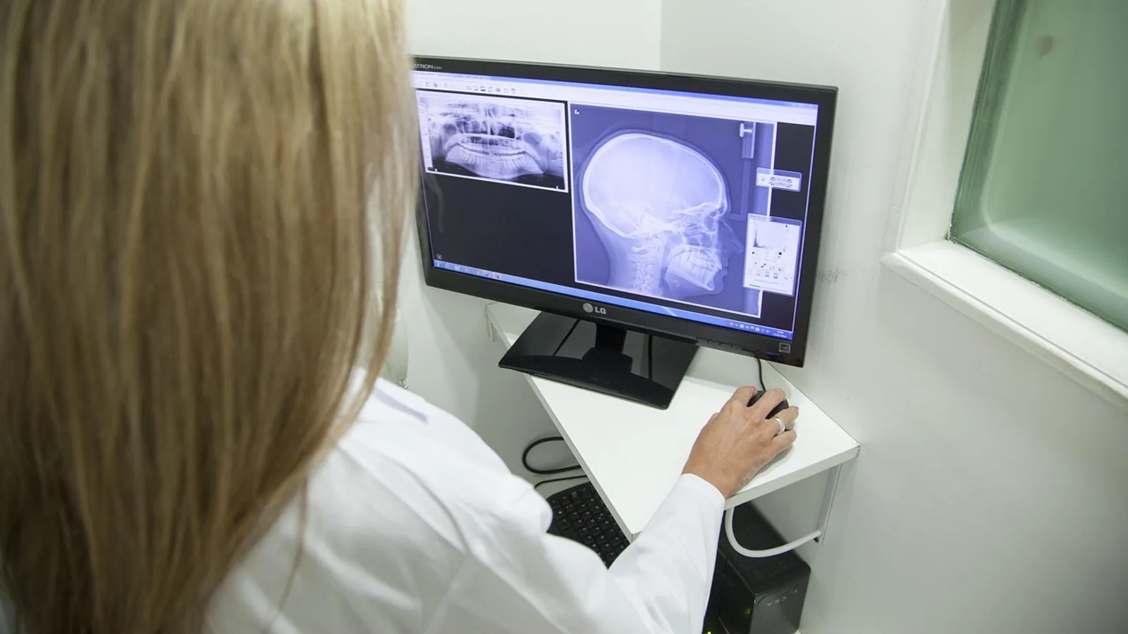
The mandibular fossa is the shallow depression on the ventral surface of the squamous part of the temporal bone that receives the head of the mandible (the mandibular condyle) to form the temporomandibular joint. It is bounded anteriorly by the articular tubercle, posteriorly by the tympanic plate, and separated from the tympanic part of the temporal bone by the squamotympanic fissure.
This article walks through the bony anatomy of the mandibular fossa, its borders, the soft tissues that line it, and how the temporomandibular joint (TMJ) is built across dogs, cats, horses, and humans. The TMJ is a synovial joint. In humans and some other species it contains a fibrocartilaginous articular disc that divides the joint into two compartments, while dogs and cats have a more hinge-like joint with limited translation. That structural difference shapes how each species chews, how disease presents, and how the joint is imaged.
This article is educational and is not a substitute for veterinary diagnosis or treatment.
What the Mandibular Fossa Is
The mandibular fossa (also called the glenoid fossa in human anatomy texts, and the fossa mandibular in Latin-based nomenclature) is a depression in the squamous part of the temporal bone. The squamous part is the flat, scale-like portion of the temporal bone that forms much of the lateral wall of the skull. The fossa sits on the ventral aspect of this squamous plate, just rostral (in animals) or anterior (in humans) to the ear canal.
The fossa receives the mandibular condyle, which is the rounded articular process at the caudal end of the mandible. Together, the fossa and the condyle form the osseous framework of the temporomandibular joint. The fossa is not a deep socket like the acetabulum of the hip. It is a shallow, gently concave surface that allows the condyle to rotate and, in some species, to translate forward.
The word "fossa" comes from the Latin for ditch or trench. In anatomy, a fossa is any depression or hollow, usually on a bone. The mandibular fossa is one of several fossae in the skull, and its name tells you what it holds: the mandible.
The Squamous Part of the Temporal Bone
The temporal bone has several parts. The squamous part is the flat, fan-shaped plate on the lateral side of the skull. The petrous part is the dense, pyramid-shaped portion that houses the inner ear. The tympanic part forms the bony ear canal. The mastoid part is the rear projection behind the ear.
The mandibular fossa belongs to the squamous part, but its posterior border is formed by the tympanic part. This is why the squamotympanic fissure matters: it is the seam where the squamous and tympanic parts meet, and it marks the back edge of the fossa.
Borders of the Mandibular Fossa
The fossa has three named borders. Knowing them helps you orient on a skull, on a radiograph, or on a CT scan.
Anterior Border: The Articular Tubercle
The articular tubercle is a rounded, transverse ridge at the front of the fossa. It is the anterior limit of the articular surface. In humans, the articular tubercle is prominent and the condyle slides forward onto it during opening of the mouth. In dogs and cats, the tubercle is lower and the condyle does not translate as far.
The articular tubercle is sometimes called the articular eminence. The two terms refer to the same structure. The tubercle is the bony ridge. The eminence is the raised surface. In veterinary anatomy, "articular tubercle" is the more common term.
Posterior Border: The Tympanic Plate
The tympanic plate (the tympanic part of the temporal bone) forms the posterior wall of the fossa. It is a thin, curved plate of bone that also forms the floor and anterior wall of the external acoustic meatus (the ear canal). The posterior border of the fossa is therefore not a sharp edge but a transition into the tympanic region.
In dogs and cats, the retroarticular process is a bony projection that sits just behind the mandibular fossa. It helps prevent the condyle from sliding too far caudally. The retroarticular process is a useful landmark on oblique radiographs of the feline and canine TMJ [1][2].
The Squamotympanic Fissure
The squamotympanic fissure (also called the petrotympanic fissure in humans, though the two are not identical) is the gap between the squamous and tympanic parts of the temporal bone. It runs transversely and marks the boundary between the mandibular fossa and the tympanic plate.
The fissure is not just a seam. It transmits small vessels and the chorda tympani nerve in humans. In animals, it is a landmark for surgical and imaging orientation. On a lateral radiograph of the skull, the fissure can sometimes be seen as a thin radiolucent line just behind the condyle.
Medial and Lateral Borders
The fossa is bounded medially by the spine of the sphenoid bone and the petrous part of the temporal bone. Laterally, it is bounded by the root of the zygomatic process of the temporal bone. The zygomatic process is the bony bar that forms the cheekbone arch. Its root is the part that attaches to the squamous temporal bone, and it forms the lateral wall of the fossa.
These medial and lateral borders are less commonly named in clinical descriptions, but they matter for imaging. A condyle that sits too far medially or laterally is a malposition, and the borders define what "normal" position looks like.
The Temporomandibular Joint
The temporomandibular joint is the synovial joint between the mandibular fossa and the mandibular condyle. It is one of the few synovial joints in the body with a fibrocartilaginous articular surface rather than hyaline cartilage. This is a key point. Most synovial joints, such as the knee, are lined with hyaline cartilage. The TMJ is lined with fibrocartilage, which is more resistant to shear forces and better suited to the sliding and grinding movements of chewing [3].
The joint is enclosed by a fibrous joint capsule. The capsule is lined by a synovial membrane that produces synovial fluid. The fluid lubricates the joint and nourishes the fibrocartilage, which has no blood supply of its own.
The Articular Disc
In humans, the TMJ contains a complete fibrocartilaginous articular disc. The disc divides the joint into two compartments: an upper (superior) compartment between the disc and the mandibular fossa, and a lower (inferior) compartment between the disc and the condyle. The two compartments do not communicate. The upper compartment allows translation (sliding forward), and the lower compartment allows rotation (hinging).
The disc is not present in all species. Dogs and cats have a thin, incomplete disc or no true disc at all, depending on how strictly you define it. The canine TMJ is often described as having a thin fibrocartilaginous disc that is visible on MRI in about 70% of joints on T1-weighted images and 65% on T2-weighted images [4]. This means the disc is present but not always clearly identifiable on imaging, and it does not divide the joint into two fully separate compartments the way the human disc does.
Horses have a well-developed articular disc. Histological studies of healthy equine TMJs show the disc is composed of an inner core of fibrocartilage and hyaline-like cartilage meshwork, covered by cell-rich dense connective tissue and fibrocartilage on its dorsal and ventral aspects [5]. The equine disc is a real structure, and it separates the joint into two compartments.
How the Joint Moves
The movement of the TMJ depends on the shape of the fossa and the presence of a disc.
In humans, the joint is a true ginglymoarthrodial joint. That means it both hinges (ginglymus) and slides (arthrodia). When you open your mouth, the condyle first rotates in the lower compartment, then translates forward onto the articular tubercle in the upper compartment. This translation is why the human jaw can open wide.
In dogs and cats, the joint is more hinge-like. The condyle rotates but does not translate far. The fossa is shallower, the articular tubercle is lower, and the retroarticular process limits caudal movement. The result is a joint that opens and closes with limited sliding. This is why dogs and cats cannot open their mouths as wide as humans, and why their jaw movement is more of a simple hinge than a complex glide.
In horses, the joint is adapted for the complex movements of the chewing cycle. The equine TMJ has a well-developed disc, and the collagen fiber architecture of the articular surfaces reflects the different forces applied during chewing. Split-line analysis of the equine TMJ shows that collagen fibers in the central two-thirds of the articular surfaces run in a rostrocaudal direction, while the lateral and medial aspects have curved or punctate patterns [6]. This suggests the joint is built to handle forces from multiple directions, not just simple opening and closing.
Species Variation in the Mandibular Fossa and TMJ
The table below summarizes the key differences in fossa depth, condyle shape, and disc presence across dogs, cats, horses, and humans.
| Feature | Dog | Cat | Horse | Human |
|---|---|---|---|---|
| Fossa depth | Shallow | Shallow | Moderately deep | Deep |
| Condyle shape | Transversely elongated, flattened | Transversely elongated, flattened | Transversely elongated, slightly convex | Ellipsoid, wider mediolaterally |
| Articular disc | Thin, often incomplete or not consistently visible on MRI [4] | Thin, often incomplete | Well-developed, complete, fibrocartilaginous [5] | Complete, fibrocartilaginous |
| Joint movement | Hinge-like, limited translation | Hinge-like, limited translation | Complex, adapted for chewing cycle [6] | Hinge and glide, wide translation |
| Retroarticular process | Present | Prominent | Present | Absent or rudimentary |
| Articular surface tissue | Fibrocartilage | Fibrocartilage | Fibrocartilage, with dense connective tissue over the fossa [5] | Fibrocartilage |
| Clinical imaging | Oblique radiography, MRI [2][4] | Oblique radiography, CT [1] | Radiography, CT, MRI | MRI, CBCT |
Dogs
The canine mandibular fossa is shallow and the condyle is flattened and transversely elongated. The joint is hinge-like. The articular disc is thin and not always visible on MRI. In a study of 10 dogs, the disc was visible in 14 of 20 TMJs on T1-weighted images and 13 of 20 on T2-weighted images [4]. The lateral collateral ligament was not identified in any joint in that study, which suggests the canine TMJ relies more on the joint capsule and surrounding muscles for stability than on a discrete ligament.
The canine TMJ is best imaged with oblique radiography. A study comparing five oblique views found that "nose-up" laterorostral-laterocaudal oblique projections at 10° and 20° rotation provided the clearest visualization of the mandibular fossa, condylar process, joint space, and retroarticular process [2]. A 10° rotation in either axis is needed to project the two joints independently of each other [7].
Cats
The feline mandibular fossa is similar to the dog's but the retroarticular process is more prominent. The joint is hinge-like with limited translation. Radiographic assessment of the feline TMJ is challenging because of superimposition of overlying structures. A study of five feline skulls found that the dependent TMJ anatomy was best seen on latero-10°-ventral-laterodorsal, latero-15°-ventral-laterodorsal, and latero-20°-ventral-laterodorsal oblique views [1]. Opposite lateral oblique views at these angulations can help characterize the anatomy in clinical patients.
Horses
The equine mandibular fossa is deeper than the dog's or cat's, and the joint has a well-developed disc. The articular surfaces of the equine TMJ have a distinct histological zoning pattern. Apart from the mandibular fossa itself, the osseous aspects of the joint are covered by three tissue layers: a superficial cell-rich dense connective tissue layer, a middle fibrocartilage layer, and a deep hyaline-like cartilage layer [5]. The mandibular fossa is different. It is covered only by dense connective tissue, often supplemented by synovial membrane. This suggests the fossa experiences low biomechanical stress compared to the condyle and disc [5].
The collagen fiber texture of the equine TMJ also reflects its function. Split-lines in the central two-thirds of the articular surfaces of the articular tubercle, disc, and mandibular head run in a rostrocaudal direction. The lateral and medial aspects have curved arrangements in the disc and punctate split-lines in the bony components. Mediolateral orientated split-lines are found in the rostral and caudal borders of the disc and in the mandibular fossa [6]. This pattern is consistent with a joint that handles complex, multi-directional forces during chewing.
Humans
The human mandibular fossa is deep and the condyle is ellipsoid. The articular tubercle is prominent. The joint has a complete fibrocartilaginous disc that divides it into two compartments. The human TMJ allows both rotation and translation, which is why the jaw can open wide. The retroarticular process is absent or rudimentary in humans.
The human TMJ is commonly imaged with MRI. A study comparing 0.55 T and 1.5 T MRI for temporomandibular disorders found that image quality for disc morphology and osseous joint morphology was inferior at 0.55 T compared to 1.5 T, but disc position was comparable between the two field strengths [8]. A sufficient diagnostic image quality was maintained in 92% of cases at 0.55 T and 100% at 1.5 T [8]. Another study comparing a dental-dedicated 0.55 T system with a 1.5 T system found that images from the two systems were rated similarly, though inter-rater agreement was low [9].
Clinical Relevance, Limitations and Common Mistakes
The mandibular fossa and TMJ are clinically important in veterinary medicine because disorders of this joint can cause pain, difficulty eating, and malocclusion. However, the incidence of published TMJ disorders in horses is low, which has led researchers to question whether the equine TMJ is well adapted to its biomechanical requirements or able to remodel its articular surfaces in response to modified loading conditions [5].
In dogs and cats, TMJ disease is often suspected when an animal shows signs of jaw pain, reluctance to chew, or malocclusion. The joint is difficult to assess on plain radiography because of superimposition. Oblique views are needed, and the angle of obliquity matters. For dogs, 10° to 30° lateral rotation is useful in mesaticephalic and dolichocephalic breeds, and 20° to 30° in brachycephalic breeds [7]. For cats, latero-10° to latero-20° ventral-laterodorsal oblique views are best [1].
MRI is the modality of choice for evaluating the soft tissues of the TMJ, including the disc. In dogs, MRI allows evaluation of the osseous and certain soft tissue structures of the TMJ [4]. The condylar process and mandibular fossa are hyperintense to muscle and isointense to hypointense to fat on T1-weighted images, and mildly hyperintense to muscle on T2-weighted images [4]. The disc is isointense to hyperintense to muscle on T1-weighted images and varies from hypointense to hyperintense to muscle on T2-weighted images [4].
A common mistake is to assume that the TMJ is the same across species. The human TMJ has a complete disc and allows wide translation. The dog and cat TMJ have a thin or incomplete disc and limited translation. The horse TMJ has a complete disc but is adapted for the complex movements of the chewing cycle. These differences affect how disease presents and how the joint should be imaged.
Another common mistake is to rely on a single radiographic view. The TMJ is a three-dimensional structure, and a single lateral view will superimpose the two joints. At least 10° of rotation in either axis is needed to project the joints independently [7].
A third mistake is to assume that a visible disc on MRI means the joint is normal. The disc can be visible but displaced. In humans, disc displacement with or without reduction is a common finding in temporomandibular disorders [10]. In dogs, the disc is visible in only about 70% of joints on T1-weighted images, so a non-visible disc is not necessarily abnormal [4].
Degenerative joint disease of the TMJ has been described in carnivores, including a lioness. In that case, spontaneous degenerative joint disease was observed unilaterally, with features comparable to other carnivores. The diseased disc had altered direction-dependent mechanical properties, and tensile strength and stiffness differed substantially between the diseased and healthy disc [11]. This suggests that TMJ disease in carnivores can affect the mechanical function of the disc, not just the bone.
The limitations of our current knowledge are significant. The role of degenerative joint disease of the TMJ in pain or disability is not well understood in carnivores [11]. The incidence of TMJ disorders in horses is low, and it is not clear whether this reflects a genuinely well-adapted joint or a lack of diagnosis [5]. Imaging of the TMJ in dogs and cats is challenging, and the best protocols are still being refined. Individual cases need a veterinarian for diagnosis and treatment.
Frequently Asked Questions
What is the mandibular fossa?
The mandibular fossa is the depression in the squamous part of the temporal bone that receives the mandibular condyle to form the temporomandibular joint. It is bounded anteriorly by the articular tubercle, posteriorly by the tympanic plate, and separated from the tympanic part by the squamotympanic fissure.
What are the borders of the mandibular fossa?
The anterior border is the articular tubercle, the posterior border is the tympanic plate, and the squamotympanic fissure separates the fossa from the tympanic part of the temporal bone. Medially, the fossa is bounded by the sphenoid and petrous temporal bone. Laterally, it is bounded by the root of the zygomatic process.
Do dogs and cats have an articular disc in the TMJ?
Dogs and cats have a thin, often incomplete articular disc that is not always visible on imaging. In dogs, the disc is visible on MRI in about 70% of joints on T1-weighted images and 65% on T2-weighted images.
How is the TMJ different in horses?
The equine TMJ has a well-developed, complete articular disc and is adapted for the complex movements of the chewing cycle. The mandibular fossa is covered only by dense connective tissue, suggesting it experiences low biomechanical stress compared to the condyle and disc.
Why is the TMJ called a synovial joint?
The TMJ is a synovial joint because it has a joint capsule lined by synovial membrane, produces synovial fluid, and has articular surfaces lined with cartilage. The cartilage is fibrocartilage rather than hyaline cartilage, which is unusual for a synovial joint.
What is the squamotympanic fissure?
The squamotympanic fissure is the gap between the squamous and tympanic parts of the temporal bone. It marks the posterior border of the mandibular fossa and is a landmark for imaging and surgical orientation.
How is the TMJ imaged in dogs and cats?
Oblique radiography is commonly used. For dogs, "nose-up" laterorostral-laterocaudal oblique views at 10° and 20° rotation provide the clearest images. For cats, latero-10° to latero-20° ventral-laterodorsal oblique views are best. MRI is used for soft tissue evaluation.
Can TMJ disease cause pain in animals?
TMJ disease can cause pain and difficulty eating in animals, but the role of degenerative joint disease in pain or disability is not well understood in carnivores. Signs such as jaw pain, reluctance to chew, or malocclusion should be evaluated by a veterinarian.
Related Articles
- Clinical Research Notes Templates
- Veterinary Anatomy Notes: How to Create and Use Them for NAVLE
- The Best Note-Taking Tools for Clinical Research
- Project-Based Note Organization for Clinical Research
- Cross-Border Regulatory Compliance
- Bovine Hoof Anatomy and Lameness Examination: A Clinical Approach
- Epiphyseal Plate: Growth, Anatomy and Clinical Notes
Sources
- Radiographic Assessment of the Feline Temporomandibular Joint with Oblique Projections: A Preliminary Ex Vivo Study.
- Assessment of five oblique radiographic projections of the canine temporomandibular joint.
- Is the temporomandibular joint affected by rheumatoid arthritis? A comparative investigation with knee arthritis in an experimental rat model.
- Magnetic resonance imaging features of the temporomandibular joint in normal dogs.
- Functional anatomy of the equine temporomandibular joint: Histological characteristics of the articular surfaces and underlining tissues.
- Functional anatomy of the equine temporomandibular joint: Collagen fiber texture of the articular surfaces.
- The effect of obliquity on the radiographic appearance of the temporomandibular joint in dogs.
- MRI of Temporomandibular Joint Disorders: A Comparative Study of 0.55 T and 1.5 T MRI.
- Comparison of a 0.55 T dental-dedicated magnetic resonance imaging system with a 1.5 T system in evaluation of the temporomandibular joint regarding subjective image quality assessment and rater agreement.
- Temporomandibular joint osseous characteristics in skeletal short and long facial types with and without temporomandibular disorders: A three-dimensional comparative study.
- Degenerative temporomandibular joint disease in a lioness (Panthera leo).