Mammillary Body: Location, Function, and Anatomy
By Dr. Zubair Khalid, DVM, MS, PhD ·
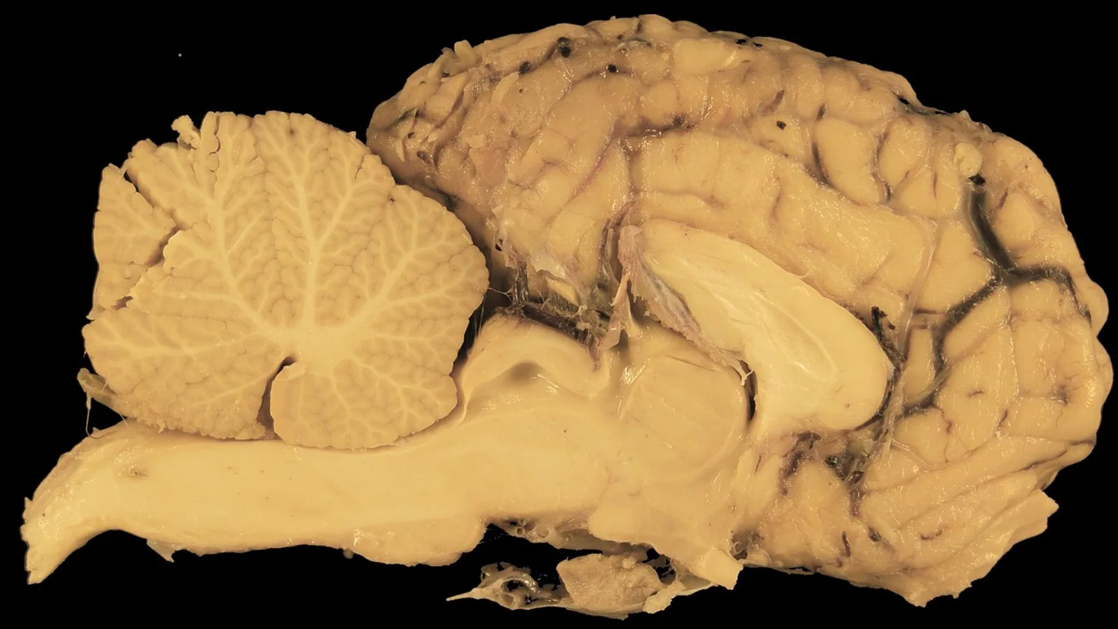
The mammillary body is a paired, spherical nuclear mass on the caudal (posterior) surface of the hypothalamus, one on each side of the midline, that receives input through the fornix and projects to the anterior thalamic nuclei as a relay station in the Papez circuit. Each mammillary body is divided into a medial and a lateral mammillary nucleus, and together these nuclei support recollective memory and spatial navigation rather than serving as a passive pass-through.
The mammillary bodies matter in veterinary practice for two reasons. First, they are a reliable anatomical landmark and an imaging target in the diencephalon, so knowing their position prevents them from being mistaken for a mass or a lesion. Second, they are selectively vulnerable to thiamine (vitamin B1) deficiency, hypoxia-ischemia, and direct extension of disease along the Papez circuit, which is why a patient with sudden memory loss, abnormal mentation, or a brain MRI showing signal change in this region deserves a structured workup. This article covers their location, nuclei, connections, comparative anatomy across species, and the clinical patterns that point to them.
This article is educational and is not a substitute for veterinary diagnosis or treatment.
Where the Mammillary Bodies Sit in the Diencephalon
The diencephalon is the division of the forebrain that sits between the brainstem and the cerebral hemispheres. Its major parts are the thalamus, hypothalamus, epithalamus, and subthalamus. The mammillary bodies belong to the hypothalamus, specifically to its caudal or posterior region, and they form the most conspicuous paired surface feature of the ventral diencephalon.
On the ventral surface of the brain, the mammillary bodies appear as two rounded eminences immediately caudal to the tuber cinereum and the infundibulum, and immediately rostral to the interpeduncular fossa of the midbrain. Each body sits just medial to the origin of the oculomotor nerve (cranial nerve III). That relationship is useful in dissection and in imaging: a structure sitting between the infundibulum rostrally and the interpeduncular fossa caudally, flanking the midline, is the mammillary body until proven otherwise.
Internally, each mammillary body is a nuclear complex rather than a single uniform nucleus. Two divisions are conventionally described:
- The medial mammillary nucleus is the larger component and forms the bulk of the visible surface. It is the principal target of the postcommissural fornix and the principal source of the mammillothalamic tract.
- The lateral mammillary nucleus is a thinner component that lies lateral and slightly dorsal to the medial nucleus. In many species it forms only a shell over the medial nucleus rather than a distinct bulge.
This medial-lateral division is not a trivial anatomical detail. The two nuclei have different inputs, different outputs, and different physiological signatures. In mice, the mammillary body contains at least four neuronal subtypes occupying different spatial subregions, and two of these subtypes are tagged by parvalbumin (PV) and dopamine receptor-D2 (Drd2) markers. PV neurons are spontaneously active, whereas Drd2 neurons are inactive at rest and generate rebound bursts, and the two populations receive input from two discrete classes of subicular neurons and support different memory tasks [1]. The medial and lateral nuclei therefore represent separable processing streams inside a structure that looks like a single sphere.
A morphological variant worth knowing is the accessory mammillary body. Postmortem examination of the hypothalamus of a 79-year-old woman who died of cardiac arrest without recorded neurologic symptoms revealed well-defined spherical protrusions located rostro-laterally to otherwise normal-appearing mammillary bodies. Cytoarchitectonically these accessory bodies were formed by an enlarged lateral mammillary nucleus, which is normally a thin shell over the medial nucleus [2]. The clinical importance is practical: an enlarged lateral mammillary body can be mistaken on imaging for a neoplastic or other pathologic process of the basal diencephalon, so this variant should be recognized rather than over-interpreted [2].
The Papez Circuit and the Mammillary Body as a Relay
James Papez described a closed circuit linking the hippocampal formation, mammillary bodies, anterior thalamus, and cingulate cortex as an anatomical basis for emotion. Later work consolidated the limbic functions of this circuit but showed a more important role in memory [3]. Modern dissection and ultrahigh-field imaging studies describe the network as connecting the anterior and posterior cingulate cortex, entorhinal cortex, hippocampus, fimbria, dentate gyrus, fornix, mammillary bodies, and anterior thalamus [3].
The mammillary body is the diencephalic hub of that loop. The canonical flow is:
- Hippocampus to mammillary body. Subicular axons leave the hippocampal formation, travel in the fornix (the major white matter tract of the hippocampal output), and terminate mainly in the medial mammillary nucleus. The portion of the fornix that runs behind the anterior commissure is called the postcommissural fornix, and it is the dominant hippocampal input to the mammillary bodies.
- Mammillary body to anterior thalamus. The medial mammillary nucleus projects through the mammillothalamic tract to the anterior thalamic nuclei, chiefly the anteromedial and anteroventral nuclei.
- Anterior thalamus to cingulate cortex. Anterior thalamic neurons project to the cingulate gyrus, including the anterior cingulate cortex.
- Cingulate cortex back to hippocampus. Cingulate and retrosplenial cortex project through the cingulum bundle to the entorhinal cortex and parahippocampal region, closing the loop back to the hippocampus.
Tractography in living human subjects has refined this picture. Diffusion spectrum imaging in eight healthy volunteers and analysis of Human Connectome Project data showed thalamic fibers connecting with both the posterior cingulate cortex and the retrosplenial cortex, with the retrosplenial cortex mainly responsible for direct hippocampal connections and the posterior cingulate cortex not [4]. That finding supports treating the retrosplenial and posterior cingulate cortices as separate functional hubs within the circuit rather than as one undifferentiated cortical target [4].
Afferent and efferent connections at a glance
| Direction | Pathway or source | Main target within the mammillary body | Notes |
|---|---|---|---|
| Afferent | Postcommissural fornix from the subiculum | Medial mammillary nucleus | Classic hippocampal input, topographically organized |
| Afferent | Gudden's ventral tegmental nucleus | Mammillary body (medial) | Nonhippocampal input critical for function |
| Afferent | Other brainstem tegmental nuclei | Mammillary body | Contributes to the ascending relay |
| Efferent | Mammillothalamic tract | Anterior thalamic nuclei (anteromedial, anteroventral) | Principal output, damaged in Korsakoff's syndrome |
| Efferent | Descending projections to tegmental nuclei | Midbrain tegmentum | Reciprocal limb of the circuit |
| Efferent | Projections to medial dorsal thalamus | Mediodorsal thalamic nucleus | Part of the wider limbic network |
The table captures the key point that the mammillary body has both hippocampal and nonhippocampal inputs. For decades the dominant model treated the mammillary bodies as a hippocampal relay, passing information to the anterior thalamic nuclei and from there to the cingulate cortex, with no independent role in memory [5]. That model also neglected nonhippocampal inputs [5]. Contemporary work has changed the picture.
Why the Mammillary Body Is More Than a Hippocampal Relay
The strongest evidence against the simple relay model comes from lesion experiments in rats. When researchers removed the two principal inputs to the mammillary bodies, the postcommissural fornix from the hippocampal formation and Gudden's ventral tegmental nucleus, only the tegmental lesion impaired behavioral tests of spatial working memory and, in particular, disrupted the use of extramaze spatial landmarks. Disconnecting the principal mammillary output, the mammillothalamic tract, produced the same impairment, while postcommissural fornix lesions did not. The same effective lesions also produced widespread reductions in immediate-early gene (c-fos) expression across a network of memory-related regions, which was not seen after fornix lesions [6]. In other words, projections from the tegmental nuclei of Gudden, and not the hippocampal formation, are critical for sustaining mammillary body function, and the mammillary bodies have a role independent of their subicular inputs [5].
Anatomical tracing studies add a second layer. Retrograde tracer experiments comparing rats and macaque monkeys showed that subicular projections to the mammillary bodies and to the anterior thalamic nuclei arise from different populations of cells with laminar separation, consistent with parallel information streams whose segregation appears more marked in the rat brain [7]. In rats, the mammillary and anterior thalamic projections had complementary topographies in the proximal-distal plane, consistent with differential involvement in object-based (proximal subiculum) and context-based (distal subiculum) information. Medial mammillary inputs arose along the anterior-posterior extent of the subiculum and favored the central subiculum and the more proximal subiculum, whereas anterior thalamic inputs were largely confined to the dorsal subiculum [7]. The circuit is therefore not a single wire but a set of parallel channels.
Physiological recordings in mice align with this. Large-scale silicon probe recordings of mammillary body activity across brain states found that mammillary body cells are highly diverse in their relationship to theta, ripple, and slow oscillations, and several of these physiological features are inherited by the topographically organized inputs to the mammillary cells [8]. The structure is a processing node whose output reflects the specific input channel that drives it.
Comparative Anatomy: Species Differences in Size and Prominence
The mammillary bodies are present in all mammals, but their relative size, surface prominence, and internal organization vary across species. The general pattern is that the mammillary complex is proportionally larger and more clearly demarcated in rodents and carnivores than in primates, including humans.
In rats and mice the mammillary bodies are large, paired, and easily identified on the ventral diencephalon, and the medial and lateral nuclei are well separated. The lateral mammillary nucleus is a distinct structure rather than a thin shell, which is why rodent preparations are used so heavily in circuit-level experiments. The tracing studies cited above were performed in rats and macaque monkeys precisely because the subicular projection patterns can be compared across a rodent and a primate [7].
In carnivores such as dogs and cats, the mammillary bodies are prominent paired eminences and are routinely identified on ventral brain dissection. Their size relative to the rest of the hypothalamus is greater than in humans, and the mammillothalamic tract is a robust, dissectible bundle.
In primates, including humans, the mammillary bodies are smaller in absolute and relative terms, and the lateral mammillary nucleus is reduced to a thin shell over the medial nucleus. This is the arrangement that makes the accessory mammillary body variant described above, in which an enlarged lateral nucleus forms a rostro-lateral protrusion, anatomically possible and radiologically confusing [2].
For veterinary students, the practical takeaway is that species differences in prominence affect how easily the structure is seen on gross dissection and on cross-sectional imaging, not whether it exists or what it does. The Papez circuit is conserved, and the mammillary body occupies the same nodal position in dogs, cats, rodents, ruminants, and primates.
How the Mammillary Body Is Observed and Tested in Practice
In a live animal, the mammillary bodies are not directly accessible to physical examination. They are evaluated by imaging and by inference from clinical signs.
Imaging. On MRI, the mammillary bodies are assessed for size, signal intensity, and symmetry. In humans, mammillary body and fornix volume loss has been demonstrated in chronic traumatic encephalopathy and is measurable on MRI, which has driven interest in these structures as in vivo imaging biomarkers [9]. In a study of 177 individuals (61 controls, 46 fighters who were positive for traumatic encephalopathy syndrome, and 70 who were negative), automated measurements of mammillary body and fornix size were compared with manual measurements to confirm the automated results and to demonstrate clinical relevance [9]. The same principle applies in veterinary imaging: mammillary body volume and signal are measurable, and change in them is meaningful.
Signal change as a lesion marker. In a retrospective review of 235 full-term human neonates with clinically confirmed hypoxic-ischemic encephalopathy, MRI performed within 10 days of birth showed abnormal high mammillary body signal on T2-weighted sequences in 31 neonates (13.2%), including 4 with mild, 25 with moderate, and 2 with severe hypoxic-ischemic encephalopathy. Restricted diffusion was seen in 6 neonates imaged between days 5 and 7. For those 31 neonates, the most common MRI pattern (41.9%) was abnormal signal restricted to the mammillary bodies with the rest of the brain appearing normal [10]. Follow-up imaging showed mammillary body and hippocampal atrophy in some of these patients [10]. This is the pattern that makes the mammillary body a useful early marker: it can be abnormal when the rest of the brain looks unremarkable.
Experimental models. In a neonatal mouse model of hypoxia-ischemia induced in 9-day-old pups, immunohistochemistry at days 1 to 3 after injury showed cell death, microglia activation, and early gliosis in the ipsilateral mammillary body. Glial scarring was still present at day 28 with concomitant ipsilateral mammillary body atrophy, and the injury also induced mammillothalamic tract atrophy and reduced expression of calbindin-positive but not parvalbumin-positive cells. Therapeutic hypothermia and intranasal mesenchymal stem cell therapy both reduced cortical and hippocampal lesions but did not confer equivalent protection to the mammillary bodies [11]. This is a species-relevant model because it demonstrates that the mammillary bodies can be injured in a pattern distinct from the hippocampus, and that standard neuroprotective strategies may not cover them.
Functional assessment. In animals, memory and spatial navigation are assessed behaviorally rather than by direct mammillary body testing. The rodent lesion literature uses spatial working memory tasks and tests of extramaze landmark use, which are the behavioral readouts most tightly linked to mammillary body and mammillothalamic tract integrity [6].
Clinical Relevance, Limitations and Common Mistakes
The classic clinical link for the mammillary bodies is Wernicke-Korsakoff syndrome, a neurologic disorder caused by thiamine (vitamin B1) deficiency. Wernicke's encephalopathy is the acute, potentially reversible stage, and Korsakoff's syndrome is the chronic amnestic stage. Volumetric MRI studies comparing Korsakoff's syndrome with Alzheimer's disease found that mammillary body and mediodorsal thalamic volumes were reduced in all patient groups compared with controls but were more severely damaged in Korsakoff's syndrome than in Alzheimer's disease, and that mammillothalamic tract volumes were damaged in Korsakoff's syndrome only [12]. That pattern is the reason the mammillary bodies and the mammillothalamic tract are treated as signature sites of thiamine-related injury.
A detailed case illustrates the functional consequence. A 43-year-old woman developed dense anterograde and retrograde amnesia suddenly as a result of Wernicke-Korsakoff's syndrome. Volumetric MRI showed severely reduced fornix and mammillary body volumes with intact hippocampi [13]. The dissociation between mammillary body and fornix damage on one hand and hippocampal preservation on the other is a recurring theme: memory impairment can arise from damage at the mammillary body node alone.
In animals, thiamine deficiency produces comparable lesions. Ruminants (especially cattle and sheep) and cats are the species most often affected, and the clinical picture includes abnormal mentation, ataxia, and in some cases seizures or blindness. The neuropathology in these species centers on selective vulnerability of brainstem and diencephalic nuclei, and the mammillary bodies are among the structures that can show signal change and atrophy on MRI. The pediatric literature is explicit that the main causes of mammillary body pathology on MRI are thiamine deficiency, hypoxia-ischemia, direct damage from masses or hydrocephalus, and deafferentation resulting from pathology elsewhere in the Papez circuit [14]. Every one of those mechanisms has a veterinary counterpart.
Other conditions that involve the mammillary bodies in humans, and which have direct comparative value, include:
- Direct tumor extension. A limbic system glioblastoma centered in the piriform cortex infiltrated the amygdala, hippocampus, bilateral fornices, mammillary bodies, anterior cingulate gyrus, and anterior commissure. Despite this extensive involvement, the patient had a remarkable paucity of symptoms, and diffusion tensor imaging tractography showed that the tumor displaced rather than destroyed the adjacent white matter tracts [15]. Displacement without destruction can preserve function, which is a useful reminder when interpreting imaging in animals with mass lesions.
- Chemotherapy toxicity. A young patient treated with methotrexate and cytarabine via an Ommaya reservoir developed acute short-term memory deterioration, with MRI showing signal alteration in the forniceal columns and mammillary bodies [16]. This is a novel mechanism of Papez circuit injury and further clinical evidence of the circuit's function.
- Metabolic disease. In type 2 diabetes, patients showed reduced effective connectivity from the hippocampus to the mammillary body, along with structural atrophy in the left hippocampus and left thalamus and enlargement of the choroid plexus. The hippocampus-to-mammillary-body connectivity change was partially mediated by choroid plexus volume [17]. This links metabolic disease to circuit-level dysfunction, a relevant concept as veterinary patients live longer with endocrine and metabolic disease.
- Neurodegenerative disease. In behavioral variant frontotemporal dementia, bilateral atrophy of the entorhinal cortex and mammillary bodies distinguished patients from those with amyotrophic lateral sclerosis, and the frontotemporal group showed marked atrophy in Papez circuit gray matter with lower delayed recall performance [18]. By contrast, in amyotrophic lateral sclerosis the thalamus, mammillary bodies, and fornix were preserved even though other circuit components showed change [19].
Common mistakes students make
- Treating the mammillary body as a passive hippocampal relay. The lesion evidence shows that Gudden's ventral tegmental nucleus input, not the fornix, is the critical input for spatial working memory, and that mammillary body function is partly independent of subicular input [5][6].
- Confusing the mammillary bodies with the mamillary or mammary glands. The terms sound similar and are unrelated. The mammillary body is a hypothalamic nucleus. The mammary gland is the milk-producing organ.
- Assuming a normal-appearing hippocampus rules out memory circuit injury. In the Wernicke-Korsakoff case, the hippocampi were intact while the fornix and mammillary bodies were severely reduced [13].
- Assuming a large lesion always causes severe signs. The glioblastoma case showed extensive Papez circuit involvement with minimal symptoms because the tumor displaced rather than destroyed white matter tracts [15].
- Forgetting that the mammillary bodies can be abnormal in isolation. In the neonatal hypoxia-ischemia series, the most common MRI pattern was abnormal signal restricted to the mammillary bodies with the rest of the brain appearing normal [10].
- Over-reading an accessory mammillary body as pathology. An enlarged lateral mammillary nucleus can form a rostro-lateral protrusion that mimics a basal diencephalic mass [2].
Quick Review
- The mammillary bodies are paired nuclei on the caudal hypothalamus, one on each side of the midline, between the infundibulum and the interpeduncular fossa.
- Each body has a medial mammillary nucleus (larger, main fornix target, main source of the mammillothalamic tract) and a lateral mammillary nucleus (thinner, shell-like in primates).
- They are the diencephalic hub of the Papez circuit: hippocampus to fornix to mammillary body to mammillothalamic tract to anterior thalamus to cingulate cortex and back.
- They receive both hippocampal (postcommissural fornix) and nonhippocampal (Gudden's ventral tegmental nucleus) input, and the tegmental input is critical for spatial working memory.
- They are proportionally larger and more clearly demarcated in rodents and carnivores than in primates.
- Thiamine deficiency causes Wernicke-Korsakoff syndrome in humans and comparable lesions in cats and ruminants.
- The mammillary bodies can show signal change or atrophy from thiamine deficiency, hypoxia-ischemia, masses, hydrocephalus, or deafferentation elsewhere in the Papez circuit.
Frequently Asked Questions
What is the mammillary body?
The mammillary body is a paired nuclear mass on the posterior hypothalamus that relays information between the hippocampus and the anterior thalamus as part of the Papez circuit.
What are the two nuclei of the mammillary body?
Each mammillary body contains a medial mammillary nucleus, which is the larger component and the main target of the fornix, and a lateral mammillary nucleus, which is thinner and forms only a shell in primates.
What does the mammillary body do?
It supports recollective memory and spatial navigation by processing hippocampal and tegmental inputs and forwarding them through the mammillothalamic tract to the anterior thalamus.
What happens if the mammillary bodies are damaged?
Damage produces anterograde and retrograde amnesia, and in animals it impairs spatial working memory and the use of spatial landmarks.
Are the mammillary bodies affected by thiamine deficiency in animals?
Yes. Thiamine deficiency in cats and ruminants can produce lesions in the mammillary bodies and other diencephalic and brainstem nuclei, similar to the Wernicke-Korsakoff pattern in humans.
How are the mammillary bodies seen on imaging?
They are assessed on MRI for size, signal intensity, and symmetry, and abnormal T2 signal or restricted diffusion in them can be an early marker of injury even when the rest of the brain looks normal.
Related Articles
- RNA Location: Types, Functions, and Cellular Distribution
- Z-DNA Found in Biology: Location, Function, and Detection
- Prokaryotic DNA: Location, Structure, and Function
- Equine Larynx Anatomy and Function in Respiration
- Canine Lymphatic System: Anatomy and Function
- Equine Hoof Anatomy: Structures and Function
Sources
- Cell-Type Specific Circuits in the Mammillary Body for Place and Object Recognition Memory.
- Accessory mammillary bodies formed by the enlarged lateral mammillary nuclei: cytoarchitecture.
- Postmortem Dissections of the Papez Circuit and Nonmotor Targets for Functional Neurosurgery.
- In vivo visualization of connections among revised Papez circuit hubs using full q-space diffusion spectrum imaging tractography.
- The mammillary bodies and memory: more than a hippocampal relay.
- Dismantling the Papez circuit for memory in rats.
- Complementary subicular pathways to the anterior thalamic nuclei and mammillary bodies in the rat and macaque monkey brain.
- Physiological characteristics of neurons in the mammillary bodies align with topographical organization of subicular inputs.
- Volume Loss in the Mammillary Bodies, Fornix, and Other Papez Circuit Structures in Fighters with Traumatic Encephalopathy Syndrome.
- Signal Change in the Mammillary Bodies after Perinatal Asphyxia.
- Hypothermia and Stem Cells Confer Limited Protection to Mammillary Bodies in a Neonatal Mouse Model of Hypoxia-Ischemia.
- Korsakoff's Syndrome and Alzheimer's Disease-Commonalities and Specificities of Volumetric Brain Alterations within Papez Circuit.
- Into the future with little past: exploring mental time travel in a patient with damage to the mammillary bodies/fornix.
- The Mammillary Bodies: A Review of Causes of Injury in Infants and Children.
- Limbic System Glioblastoma Extending to the Papez Circuit: A Case Report.
- A novel mechanism of toxic injury to the Papez circuit from chemotherapy.
- Papez circuit remodeling in type 2 diabetes: Enlarged choroid plexus volume partially mediates functional impairments linked to insulin resistance.
- Papez Circuit Gray Matter and Episodic Memory in Amyotrophic Lateral Sclerosis and Behavioural Variant Frontotemporal Dementia.
- Structural and functional papez circuit integrity in amyotrophic lateral sclerosis.