Lumbar Plexus: Roots, Branches, and What It Supplies
By Dr. Zubair Khalid, DVM, MS, PhD ·
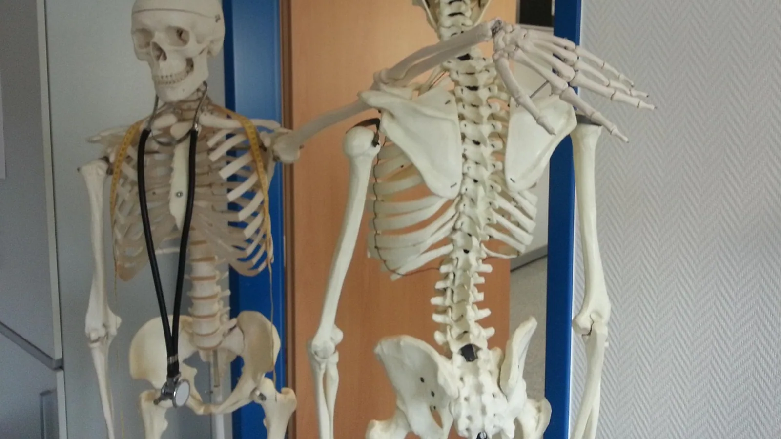
The lumbar plexus is a somatic nerve network formed by the ventral rami of spinal nerves L1 through L4, with occasional contributions from T12 or L5 depending on species. It sits within or deep to the psoas major muscle and gives off the iliohypogastric, ilioinguinal, genitofemoral, lateral femoral cutaneous, femoral, and obturator nerves, which together supply the caudal abdominal wall, the external genitalia, and much of the thigh.
This article walks through the root-to-branch organization of the plexus, the muscles and skin each branch reaches, how the lumbosacral trunk links the lumbar and sacral plexuses, and where domestic species differ from one another. It is written for veterinary and biomedical students who need a working map, not just a list of names.
What a Ventral Rami Plexus Actually Is
Every spinal nerve splits into a dorsal ramus and a ventral ramus just after it exits the intervertebral foramen. The dorsal ramus turns caudally and supplies the epaxial muscles and the skin over the back. The ventral ramus supplies everything else: the hypaxial muscles, the body wall, and the limbs.
A plexus is what happens when adjacent ventral rami exchange fibers before distributing them. Instead of each spinal nerve running alone to a single target, the rami intermingle and re-sort, so a muscle can receive axons that entered the cord at two or three different segmental levels. This re-sorting is why a single nerve like the femoral nerve can carry fibers from L4, L5, and L6 in a dog, and why a lesion at one spinal level rarely silences an entire muscle group.
The lumbar plexus is a ventral rami plexus. That distinction matters in the clinic. Dorsal rami lesions produce back pain and paraspinal muscle wasting. Ventral rami lesions produce limb weakness, altered gait, and sensory loss on the limb or ventral body wall. The two problems look nothing alike on examination.
The plexus lies in the retroperitoneal space, posterior to the psoas major in humans and within or deep to the iliopsoas in domestic species [1]. Nerves can be grouped by where they run relative to that muscle: intrapsoas, retroperitoneal, and distal to the inguinal ligament [1]. Each of those zones is a potential site of nerve injury during surgery, which is why surgeons who work in the flank or inguinal region need to know the plexus in detail [1].
Roots: Which Spinal Nerves Form the Lumbar Plexus
The classic textbook root range is L1 to L4 [2]. In humans, the plexus is formed by the anterior rami of L1 through L4 nerve roots and lies deep within the psoas muscle [2]. A contribution from T12 is common enough to be described as a normal variant, and a contribution from L5 is the bridge to the sacral plexus.
In domestic mammals the same general plan holds, but the segmental numbers shift with the number of vertebrae and the length of the lumbar column. Dogs and cats have seven lumbar vertebrae, horses and ruminants have six or seven depending on species, and the plexus roots are named by the spinal nerves that exit at those levels. The functional grouping is what matters: a cranial group that supplies the caudal abdominal wall and the skin of the groin, a middle group that supplies the cranial thigh, and a caudal group that supplies the adductor compartment and contributes to the lumbosacral trunk.
The puma dissection reported by Londoño-Osorio and colleagues illustrates how much the root numbers can move even within one family. In that animal the lumbosacral plexus was formed by L4 through S3, and the femoral nerve arose from the union of L4 and L5, whereas in other felids the femoral nerve is formed by L5 and L6 [3]. The takeaway for students is that root numbers are a guide, not a law. Learn the pattern, then check the species.
Branches: The Named Nerves and Their Targets
The named branches of the lumbar plexus fall into two functional sets. The cranial set is the iliohypogastric, ilioinguinal, and genitofemoral nerves, which supply the caudal abdominal wall and the inguinal region. The caudal set is the lateral femoral cutaneous, femoral, and obturator nerves, which supply the thigh.
Iliohypogastric and Ilioinguinal Nerves
These two nerves arise from the cranial roots, typically L1 and L2. They run caudally across the caudal abdominal wall, giving off muscular branches to the internal oblique and transverse abdominis muscles and cutaneous branches to the skin of the caudal flank and inguinal region. In the dog and cat they are small and easily missed in a routine dissection, but they matter in two situations: flank incisions that cut across them, and herniorrhaphy, where the surgeon relies on the inguinal region being sensate and the abdominal wall being innervated.
Genitofemoral Nerve
The genitofemoral nerve arises from L1 and L2 and divides into a genital branch and a femoral branch. The genital branch supplies the cremaster muscle and the skin of the scrotum or labia. The femoral branch supplies a small patch of skin over the femoral triangle.
This nerve is the most variable branch of the plexus in humans. Stoyanov and Pavlov described a complex variation in which the lateral femoral cutaneous nerve, the femoral branch of the genitofemoral nerve, and the anterior femoral nerve were all involved in an unusual pattern found during routine dissection [1]. The genitofemoral nerve is also one of the named somatic nerves that traverse the pelvis and must be recognized during minimally invasive pelvic surgery [4]. For veterinary students, the practical point is that the genitofemoral nerve is small, variable, and easy to damage in the inguinal canal.
Lateral Femoral Cutaneous Nerve
This is a pure sensory nerve. It arises from L2 and L3 and supplies the skin over the lateral and cranial thigh. It has no motor component, so a lesion produces numbness or altered sensation rather than weakness. In humans it is the nerve involved in meralgia paresthetica, a compression syndrome at the inguinal ligament. In dogs and cats the nerve is small and its sensory territory overlaps with branches of the femoral nerve, which is why isolated injury is rarely diagnosed in practice.
Femoral Nerve
The femoral nerve is the largest branch of the lumbar plexus and the dominant motor nerve of the cranial thigh. It arises from the middle roots, typically L4 through L6 in the dog, and runs caudally between the psoas major and iliacus muscles before emerging in the femoral triangle.
Its motor branches supply the quadriceps femoris group (rectus femoris, vastus lateralis, vastus medialis, vastus intermedius) and the iliopsoas. In the puma, the sartorius muscle received branches from both the saphenous and femoral nerves, a pattern that differs from the cat [3]. The femoral nerve also gives off the saphenous nerve, which is purely sensory and supplies the skin of the medial stifle and medial tarsus.
The femoral nerve is the nerve you test when you assess a dog with acute non-weight-bearing lameness and a loss of the patellar reflex. A femoral nerve lesion produces a weak or absent patellar reflex, an inability to extend the stifle, and sensory loss over the medial aspect of the limb.
Obturator Nerve
The obturator nerve arises from the caudal roots, typically L5 through L7 in the dog, and runs caudally along the medial wall of the pelvis to exit through the obturator foramen. It supplies the adductor muscles of the medial thigh: the pectineus, adductor, gracilis, and the external obturator.
The obturator nerve is a pure motor nerve in most domestic species, with a small or absent cutaneous component. It is the nerve that is at risk during difficult obstetrical deliveries, pelvic fractures, and surgery on the medial thigh. A lesion produces weakness in adduction, a widened stance, and a tendency for the limb to slip outward on smooth flooring.
The obturator nerve is one of the named somatic nerves that traverse the pelvis and must be identified during pelvic surgery [4]. In the barn owl, the obturator nerve arises from the lumbar plexus, while the ischiadic nerve arises from the sacral plexus, showing that the division of labor between the two plexuses is conserved across birds and mammals even though the root numbers differ [5].
The Lumbosacral Trunk
The lumbosacral trunk is the bridge between the two plexuses. It is formed by the caudal roots of the lumbar plexus (typically L4 and L5 in humans, L5 and L6 in the dog) and descends into the pelvis to join the sacral plexus. The lumbosacral trunk is one of the named somatic nerves that traverse the pelvis and must be recognized during minimally invasive pelvic surgery [4].
This trunk carries fibers that will become the sciatic nerve. A lesion of the lumbosacral trunk therefore produces a partial sciatic deficit, with weakness in the caudal thigh and crus but sparing of the femoral and obturator territories. The trunk is also the reason a lumbar plexus lesion and a sacral plexus lesion can look similar on a cursory examination.
Table: Nerves, Roots, Motor Targets, and Sensory Territory
| Nerve | Spinal roots (dog) | Motor targets | Sensory territory |
|---|---|---|---|
| Iliohypogastric | L1, L2 | Internal oblique, transverse abdominis | Skin of caudal flank |
| Ilioinguinal | L1, L2 | Internal oblique, transverse abdominis | Skin of inguinal region, cranial medial thigh |
| Genitofemoral | L1, L2 | Cremaster | Scrotum or labia, small patch over femoral triangle |
| Lateral femoral cutaneous | L2, L3 | None | Skin of lateral and cranial thigh |
| Femoral | L4, L5, L6 | Quadriceps femoris, iliopsoas, sartorius | Skin of medial stifle and medial tarsus via saphenous nerve |
| Obturator | L5, L6, L7 | Pectineus, adductor, gracilis, external obturator | Minimal or none |
| Lumbosacral trunk | L5, L6 | Contributes to sacral plexus and sciatic nerve | None directly |
Root numbers are typical values for the dog and vary by species. In the puma, the femoral nerve arises from L4 and L5 rather than the L5 and L6 seen in other felids [3]. In humans, the plexus is formed by the anterior rami of L1 through L4 [2].
How the Plexus Forms: A Root-to-Branch Flow
The flow from spinal root to named nerve follows a consistent pattern. Ventral rami emerge from the intervertebral foramina, pass through or deep to the iliopsoas, exchange fibers, and then split into the named branches. The diagram below traces that path.
flowchart TD
    A[Ventral rami L1 to L4] --> B[Intrapsoas exchange]
    B --> C[Cranial branches]
    B --> D[Middle branches]
    B --> E[Caudal branches]
    C --> F[Iliohypogastric nerve]
    C --> G[Ilioinguinal nerve]
    C --> H[Genitofemoral nerve]
    D --> I[Lateral femoral cutaneous nerve]
    D --> J[Femoral nerve]
    E --> K[Obturator nerve]
    E --> L[Lumbosacral trunk]
    L --> M[Sacral plexus]
The cranial branches supply the caudal abdominal wall and the inguinal region. The middle branches supply the cranial thigh. The caudal branches supply the adductor compartment and hand off to the sacral plexus through the lumbosacral trunk.
Species Differences in the Lumbar Plexus
The lumbar plexus is conserved in its overall plan across mammals, but the root contributions and the relative size of the branches vary.
Dogs and Cats
In dogs and cats the femoral and obturator nerves are the dominant branches of the lumbar plexus. The femoral nerve is large and carries fibers from L4 through L6. The obturator nerve carries fibers from L5 through L7. The cranial branches (iliohypogastric, ilioinguinal, genitofemoral) are small and supply a relatively limited territory.
The cat and the puma differ in the details. In the puma, the lumbosacral plexus is formed by L4 through S3, and the femoral nerve arises from L4 and L5, whereas in other felids it is formed by L5 and L6 [3]. The sartorius muscle in the puma receives branches from both the saphenous and femoral nerves, a pattern not seen in the cat [3]. These differences are small but they matter for anyone doing comparative dissection or interpreting a neurological examination in a wild felid.
Horses
In the horse the lumbar plexus is formed by the ventral rami of L2 through L5, with some variation. The femoral nerve is large and supplies the quadriceps group. The obturator nerve supplies the adductor muscles. The cranial branches are present but small. The lumbosacral trunk is formed by L5 and L6 and joins the sacral plexus to form the sciatic nerve.
The horse has a long lumbar column and a heavy pelvic limb, so the femoral and obturator nerves are correspondingly robust. A horse with a femoral nerve lesion will show a characteristic stance with the stifle flexed and the limb unable to bear full weight.
Ruminants
In cattle and sheep the lumbar plexus is formed by L2 through L5, with the femoral nerve arising from L4 and L5 and the obturator nerve from L5 and L6. The cranial branches are small. The lumbosacral trunk is formed by L5 and L6.
Ruminants have a slightly different arrangement of the psoas muscles, and the plexus lies deep to the psoas major and minor. This makes the plexus less accessible for clinical examination but also protects it from many traumatic injuries that would affect a dog.
Birds
The barn owl shows how far the plan can be stretched. The lumbosacral plexus of the barn owl is formed by the ventral rami of ten synsacral spinal nerves [5]. The lateral femoral cutaneous nerve, obturator nerve, cranial coxal nerve, medial femoral cutaneous nerve, cranial femoral cutaneous nerve, and femoral nerve arise from the lumbar plexus, while the ischiofemoral nerve, caudal femoral cutaneous nerve, caudal coxal nerve, and ischiadic nerve arise from the sacral plexus [5]. The division of labor between the two plexuses is recognizable, but the root numbers are completely different from mammals.
Clinical Relevance, Limitations and Common Mistakes
The lumbar plexus is clinically relevant because its branches are the nerves that control the quadriceps and the adductor compartment, and because its sensory branches supply the skin of the medial thigh and the inguinal region. A lesion anywhere along the plexus produces a predictable pattern of weakness and sensory loss.
Lumbar plexus disorders can present as low back pain and can mimic lumbar radiculopathy, which is a common reason for delayed diagnosis or inappropriate management [2]. The plexus lies deep within the psoas muscle and can be affected by traumatic, compressive, inflammatory, infectious, and neoplastic processes [2]. Retroperitoneal tumors, psoas hematomas, inflammatory plexopathies, and iatrogenic injuries are among the most common categories [2]. Magnetic resonance imaging and magnetic resonance neurography have improved the ability to visualize these lesions [2].
The most common mistakes students and clinicians make with the lumbar plexus are these:
- Confusing the lumbar plexus with the lumbosacral plexus. The lumbar plexus is L1 to L4. The lumbosacral plexus is the lumbar plexus plus the sacral plexus, joined by the lumbosacral trunk.
- Forgetting that the femoral nerve is a mixed nerve. It has a large motor component to the quadriceps and a sensory component (the saphenous nerve) to the medial limb. A lesion produces both weakness and sensory loss.
- Assuming the obturator nerve has a large sensory component. In most domestic species it is predominantly motor.
- Missing the cranial branches. The iliohypogastric, ilioinguinal, and genitofemoral nerves are small, but they matter in flank surgery and herniorrhaphy.
- Forgetting the lumbosacral trunk. A lesion of the trunk produces a partial sciatic deficit that can be mistaken for a sacral plexus lesion.
- Assuming root numbers are fixed. They vary by species and by individual. The puma is a good example [3].
The lumbar plexus is also relevant in surgery. Nerves of the plexus can be injured during surgical interventions in the retroperitoneal space, and the intrapsoas, retroperitoneal, and distal-to-inguinal-ligament sections are all liable to injury [1]. The obturator nerve, genitofemoral nerve, lateral femoral cutaneous nerve, lumbosacral trunk, and femoral nerve are the named somatic nerves that traverse the pelvis and must be recognized during minimally invasive pelvic surgery [4].
This article is educational and is not a substitute for veterinary diagnosis or treatment.
What Is Still Uncertain
The root contributions to the lumbar plexus vary more than most textbooks admit. The human literature describes the plexus as one of the most variable nerve plexuses in the body, and variations involving the lateral femoral cutaneous nerve, the genitofemoral nerve, and the femoral nerve are well documented [1]. Comparable variation studies in dogs, cats, horses, and ruminants are less complete.
The functional consequences of small variations are also uncertain. A puma with a femoral nerve from L4 and L5 rather than L5 and L6 may have a slightly different pattern of quadriceps innervation, but the clinical significance of that difference is not known [3]. The same is true for the sartorius receiving branches from both the saphenous and femoral nerves [3].
Imaging of the plexus has improved, but the ability to visualize small branches and to distinguish them from surrounding structures is still limited [2]. This is an area where more work is needed.
Frequently Asked Questions
What spinal roots form the lumbar plexus?
The lumbar plexus is formed by the ventral rami of L1 through L4 in humans [2]. In dogs the roots are typically L1 through L4 with a contribution from L5 to the lumbosacral trunk, and in horses and ruminants the range is L2 through L5.
What is the difference between the lumbar plexus and the lumbosacral plexus?
The lumbar plexus is the L1 to L4 network. The lumbosacral plexus is the lumbar plexus plus the sacral plexus, connected by the lumbosacral trunk. The trunk carries fibers from the lumbar roots into the sacral plexus to help form the sciatic nerve.
Which nerve is the largest branch of the lumbar plexus?
The femoral nerve is the largest branch. It supplies the quadriceps femoris group and the iliopsoas, and it gives off the saphenous nerve to the medial limb.
Does the obturator nerve have a sensory component?
In most domestic species the obturator nerve is predominantly motor, supplying the adductor muscles of the medial thigh. Any cutaneous component is small or absent.
What does the genitofemoral nerve supply?
The genitofemoral nerve supplies the cremaster muscle and the skin of the scrotum or labia through its genital branch, and a small patch of skin over the femoral triangle through its femoral branch.
Why is the lumbar plexus called a ventral rami plexus?
It is called a ventral rami plexus because it is formed by the ventral rami of the spinal nerves, not the dorsal rami. Dorsal rami supply the epaxial muscles and the skin over the back. Ventral rami supply the limbs and the body wall.
Can lumbar plexus disorders cause back pain?
Yes. Lumbar plexus disorders can present as low back pain and can mimic lumbar radiculopathy, which often leads to delayed diagnosis [2]. Imaging with MRI or magnetic resonance neurography can help identify the cause.
Do all mammals have the same lumbar plexus roots?
No. Root numbers vary by species and by individual. The puma, for example, has a femoral nerve from L4 and L5, while other felids have a femoral nerve from L5 and L6 [3]. The overall plan is conserved, but the details are not.
Related Articles
- Branches of Biology
- Phylogenetic Root Choice
- Valley Vet Supply
- Biology Roots: Understanding the Foundational Principles of Life
- Canine Thoracic Limb Nerves: Brachial Plexus and Innervation Patterns
- Essential Dog Grooming Supplies
Further Reading
Sources
- A multilevel variation of the sensory branches of the lumbar plexus with an accessory genitofemoral nerve.
- Lumbar Plexus Disorders Presenting as Low Back Pain: A Narrative Review of a Practical Diagnostic Imaging Approach.
- Anatomical description of the origin and distribution of the lumbosacral plexus in one puma (Puma concolor).
- Neuropelviology in minimally invasive surgery: a surgical anatomy-based demonstration.
- The lumbosacral Plexus of the Barn Owl (tyto alba): Formation, branches and distribution.