Labeled Femur: Anatomy Diagram and Landmarks
By Dr. Zubair Khalid, DVM, MS, PhD ·
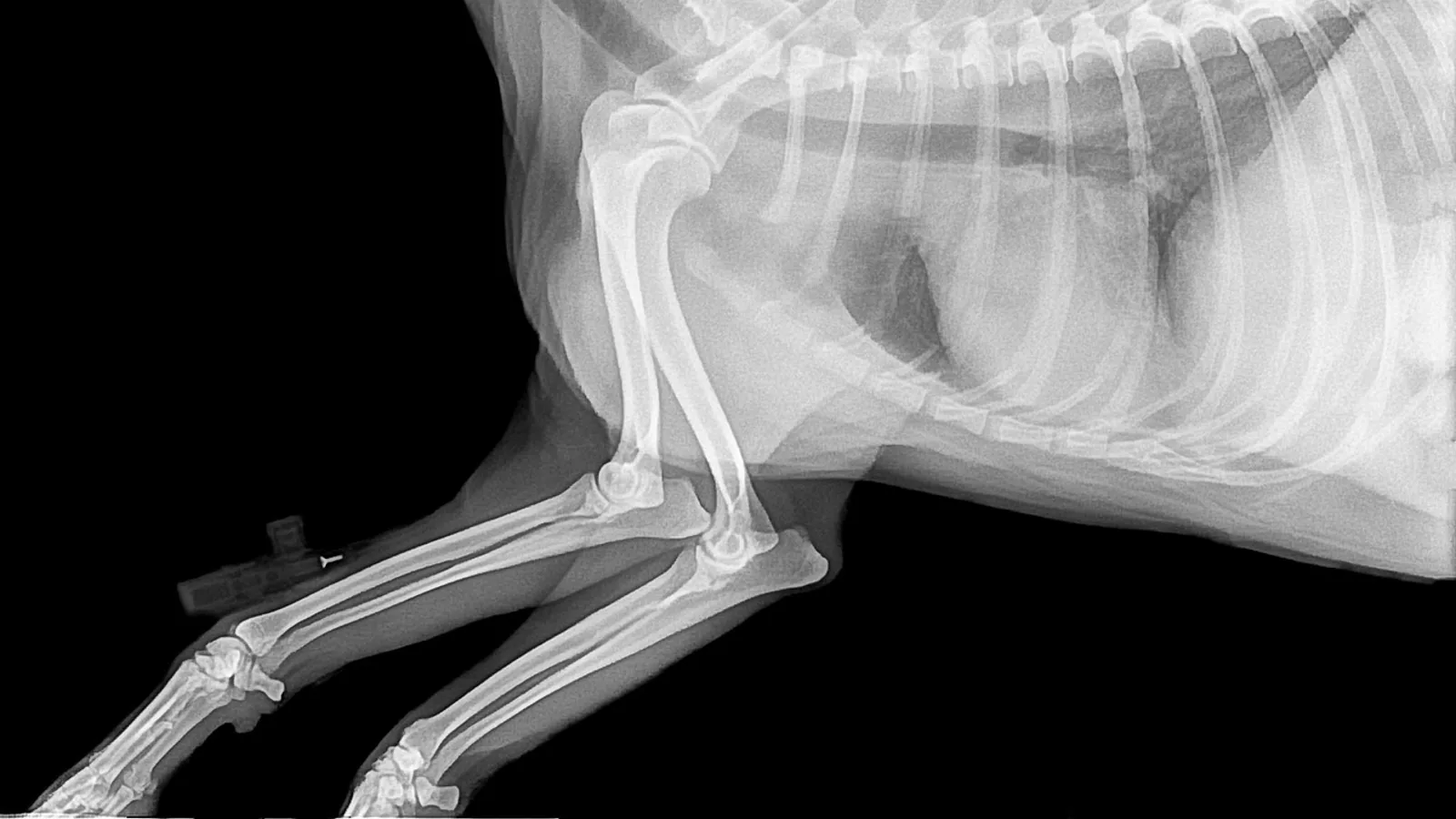
A labeled femur identifies the proximal landmarks (head, fovea capitis, neck, greater and lesser trochanters, third trochanter, trochanteric fossa) and the distal landmarks (medial and lateral condyles, patellar groove, and the condylar fossa between them). The femur is the strongest bone in the mammalian body and the primary load-bearing element of the pelvic limb, so its landmarks are the anchor points for the hip and stifle joints, the attachment sites for the major muscles of the thigh, and the reference structures used across veterinary anatomy, radiology, and orthopedic practice.
This article is educational and is not a substitute for veterinary diagnosis or treatment.
Orientation: How to Label the Femur Correctly
Before any landmark can be labeled, the bone must be oriented. The femur has a proximal end that articulates with the acetabulum of the pelvis and a distal end that articulates with the tibia and patella. The head always points medially and slightly cranially. The greater trochanter sits laterally and proximally. The patellar groove (trochlea) faces cranially at the distal end.
A reliable way to label the femur is to work from the two ends inward:
- Identify the femoral head, the smooth hemispherical ball that fits into the hip socket.
- Locate the fovea capitis, the small depression on the head.
- Follow the neck to the trochanteric region.
- Identify the greater trochanter laterally and the lesser trochanter medially.
- Note the trochanteric fossa between them.
- Move distally along the shaft (diaphysis) to the condyles.
- Finish with the patellar groove and the intercondylar fossa.
This sequence works for every domestic species. The differences lie in proportion and in which landmarks are prominent, not in the fundamental plan.
The proximal femur is a region of active growth and remodeling in young animals. In canids, the growth centers of the femoral head and greater trochanter meet dorsally, and this junction sits close to the attachment of the coxofemoral joint capsule and a segment of the proximocaudal femoral shaft physis [1]. That spatial relationship matters because it explains why the proximal femoral morphology of juvenile dogs changes so much during development and why a mineralized prominence sometimes forms at that meeting point, though not in every individual [1].
Proximal Landmarks
Femoral Head (Caput Femoris)
The femoral head is the spherical articular surface that sits in the acetabulum. It is covered by hyaline cartilage except at the fovea. The head transmits load from the pelvis to the femur, and its trabecular architecture is arranged to resist the compressive forces of weight bearing. In humans, finite element modeling of the femur under a 2000 N load shows the head displaces about 1.3 mm vertically and about 9.0 mm horizontally, while the neck experiences compressive strain inferiorly and tensile strain superiorly [2]. That strain pattern, compression below and tension above, is the mechanical signature of a bone built to bend under load.
Fovea Capitis
The fovea capitis is a small, non-articular depression on the medial aspect of the femoral head. It anchors the ligament of the head (ligamentum capitis femoris, also called the round ligament), which runs from the fovea to the acetabular notch. In the dog, this ligament carries a small artery, but the main blood supply to the head arrives through the joint capsule and the medullary vessels of the neck. The fovea is therefore both an anatomical landmark and a clinically meaningful one, because it marks the one spot on the head where cartilage is absent and where the intra-articular ligament attaches.
Femoral Neck
The neck connects the head to the shaft and is set at an angle to the long axis of the bone. The neck-shaft angle (also called the neck-body angle) describes that relationship. A morphometric study of 73 dry human femurs found a mean neck-body angle of 126.0 degrees with a standard deviation of 4.6 degrees, and the angle differed significantly between left and right sides [3]. In quadrupeds the neck is shorter and the angle is different, but the principle holds: the neck is a lever arm that converts muscle force into hip motion and that concentrates bending stress at the head-neck junction.
The neck is the weakest region of the proximal femur under bending and torsion. That is why femoral neck fractures are a persistent clinical problem. In young human adults, fracture union rates after modern internal fixation are high, with one fixation technique reporting a 96.7 percent union rate at one year [4]. The neck's blood supply and its mechanical vulnerability are the two reasons this region receives so much attention in both human and veterinary orthopedics.
Greater Trochanter
The greater trochanter is the large, laterally placed prominence at the proximal end. It is the insertion point for the middle gluteal muscle and the deep gluteal muscle, which are the principal hip extensors and abductors. In the horse, the greater trochanter is large and divided into a cranial part and a caudal part. In the dog and cat, it is a single robust process. In the cow, it is prominent but shaped differently from the horse.
The greater trochanter is also a growth site. In children with Legg-Calvé-Perthes disease, the greater trochanter can overgrow relative to a compromised femoral head and neck, a condition called relative overgrowth of the greater trochanter, which impairs hip function [5]. The same principle of balanced growth between the head, neck, and trochanter applies across species, even though the disease itself is human-specific.
Lesser Trochanter
The lesser trochanter is a smaller prominence on the medial and caudal aspect of the proximal femur. It is the insertion point for the iliopsoas muscle, a powerful hip flexor. In the dog and cat, the lesser trochanter is a distinct, rounded tubercle. In the horse, it is a low, rough ridge. In the cow, it is present but modest. The lesser trochanter is a useful landmark when orienting a femur because it identifies the medial side unambiguously.
Third Trochanter
The third trochanter is an additional prominence on the lateral aspect of the proximal shaft, distal to the greater trochanter. It is the insertion point for the superficial gluteal muscle. This landmark is the single most useful species discriminator on the femur:
- Horses: the third trochanter is large and prominent, a defining feature of the equine femur.
- Dogs: the third trochanter is present and well developed.
- Cats: present but smaller than in the dog.
- Ruminants (cattle, sheep, goats): the third trochanter is absent.
A student who is asked to label the femur of a cow and finds no third trochanter has not made a mistake. The bone simply does not have one.
Trochanteric Fossa
The trochanteric fossa is the deep depression on the medial side of the greater trochanter, at the junction of the trochanter and the neck. It is the insertion point for the obturator externus and obturator internus muscles, which are lateral rotators of the hip. In the dog, the trochanteric fossa is deep and distinct. In the horse, it is also well marked. In the cow, it is shallower. The fossa is a key landmark for identifying the proximal femur because it sits in a predictable position relative to the greater trochanter and the head.
Distal Landmarks
Medial and Lateral Condyles
The distal femur ends in two large articular condyles, the medial condyle and the lateral condyle. They articulate with the tibial plateau to form the stifle (knee) joint. The medial condyle is generally larger in the dog and cat. In the horse, the medial condyle is also larger and extends further distally. The condyles are covered in hyaline cartilage and are separated by the intercondylar fossa.
The condyles are the origin of the gastrocnemius muscle and the insertion point for the collateral ligaments of the stifle. The medial collateral ligament attaches to the medial condyle, and the lateral collateral ligament attaches to the lateral condyle. In the dog, the lateral collateral ligament is closely associated with the tendon of the popliteus muscle.
Patellar Groove (Trochlea Femoris)
The patellar groove is the smooth, saddle-shaped articular surface on the cranial aspect of the distal femur. It articulates with the patella and guides the patella during stifle flexion and extension. In the dog, the patellar groove is relatively shallow, which is one reason medial patellar luxation is common in small breeds. In the horse, the groove is deep and has a distinct medial and lateral ridge. In the cow, the groove is also well defined.
The patellar groove is a critical landmark for radiography. In dogs, the caudal margin of the intercondylar fossa on the femur is a repeatable radiographic landmark for measuring cranial tibial subluxation in the cruciate-deficient stifle [6]. That measurement uses the femur's distal landmarks as reference points, which is why accurate labeling of the condyles and fossa matters in clinical practice, not just in the dissection lab.
Intercondylar Fossa
The intercondylar fossa is the notch between the two condyles on the caudal aspect of the distal femur. It houses the origins of the cranial and caudal cruciate ligaments. The cranial cruciate ligament originates from the medial wall of the lateral condyle, and the caudal cruciate ligament originates from the lateral wall of the medial condyle. The fossa is a key landmark for identifying the distal femur and for understanding cruciate ligament anatomy.
Landmark Table
| Landmark | Muscle attachments or ligaments | Species differences |
|---|---|---|
| Femoral head | Ligament of the head (via fovea) | Similar across dog, cat, horse, cow. Head shape varies with body size. |
| Fovea capitis | Ligament of the head | Present in all domestic species. Position slightly varies. |
| Femoral neck | Joint capsule, retinacular vessels | Short in quadrupeds. Neck-shaft angle differs by species and side [3]. |
| Greater trochanter | Middle gluteal, deep gluteal muscles | Large and divided in horse. Single process in dog and cat. Prominent in cow. |
| Lesser trochanter | Iliopsoas muscle | Rounded tubercle in dog and cat. Low ridge in horse. Modest in cow. |
| Third trochanter | Superficial gluteal muscle | Prominent in horse and dog. Smaller in cat. Absent in ruminants. |
| Trochanteric fossa | Obturator externus, obturator internus | Deep in dog and horse. Shallower in cow. |
| Medial condyle | Medial collateral ligament, gastrocnemius | Larger than lateral in dog, cat, and horse. |
| Lateral condyle | Lateral collateral ligament, popliteus (dog) | Smaller than medial in most species. |
| Patellar groove | Patella, joint capsule | Shallow in dog (predisposes to luxation). Deep in horse and cow. |
| Intercondylar fossa | Cruciate ligaments | Present in all species. Shape varies with stifle conformation. |
Comparative Notes: Dog, Cat, Horse, and Cow
Dog
The canine femur is relatively straight with a distinct head, a short neck, and a prominent greater trochanter. The third trochanter is well developed. The lesser trochanter is a rounded tubercle. The patellar groove is shallow, which contributes to the high incidence of medial patellar luxation in small breeds. The distal condyles are roughly equal in size, with the medial condyle slightly larger. The intercondylar fossa is deep and houses the cruciate ligaments. The caudal margin of the intercondylar fossa is a standard radiographic landmark for measuring cranial tibial subluxation [6]. Canine proximal femoral development has been studied in detail across Canidae, showing that the growth centers of the head and greater trochanter meet dorsally near the joint capsule attachment, and that a mineralized prominence sometimes forms at that site [1].
Cat
The feline femur is similar to the dog's but more slender. The third trochanter is present but smaller. The lesser trochanter is a distinct tubercle. The patellar groove is shallow, and the condyles are similar in size. The cat's femur is adapted for a more crouched posture and greater flexibility, so the neck is shorter and the shaft is more curved than in the dog.
Horse
The equine femur is massive and adapted for weight bearing and propulsion. The greater trochanter is large and divided into cranial and caudal parts. The third trochanter is prominent and serves as the insertion for the superficial gluteal muscle. The lesser trochanter is a low ridge. The trochanteric fossa is deep. The distal condyles are large, with the medial condyle extending further distally. The patellar groove is deep and has well-defined ridges. The horse's femur is one of the bones commonly used in veterinary osteology teaching, and mixed augmented reality platforms have been developed using equine femur models to help students understand complex spatial relationships [7].
Cow
The bovine femur lacks a third trochanter, which is the single most important species difference for labeling. The greater trochanter is prominent but not divided. The lesser trochanter is modest. The trochanteric fossa is shallower than in the dog or horse. The distal condyles are large and the patellar groove is well defined. The cow's femur is adapted for a heavy body and a relatively upright stance, so the neck is short and the shaft is robust.
Clinical Relevance, Limitations and Common Mistakes
The most common labeling mistake is confusing the greater and lesser trochanters. The greater trochanter is lateral and larger. The lesser trochanter is medial and smaller. A second common mistake is missing the third trochanter in the dog or horse because it is mistaken for part of the greater trochanter. A third mistake is assuming the third trochanter is present in the cow. It is not.
Another frequent error is misidentifying the fovea capitis as a fracture or a defect. It is a normal anatomical feature that anchors the ligament of the head. In radiographs, the fovea appears as a small radiolucent area on the medial aspect of the head.
The patellar groove is sometimes confused with the intercondylar fossa. The patellar groove is cranial and articulates with the patella. The intercondylar fossa is caudal and houses the cruciate ligaments. They are on opposite sides of the distal femur.
In clinical practice, accurate landmark identification matters for radiographic measurement, implant placement, and surgical planning. In dogs, loss of conventional anatomic landmarks after femoral head and neck ostectomy complicates intraoperative orientation during total hip replacement, and one case report described eccentric reaming and a 5 mm malalignment of a custom acetabular prosthesis because the normal landmarks were gone [8]. That is a direct example of why knowing the labeled femur is not an academic exercise.
The limitations of this article are the limitations of any anatomical description. Individual animals vary in bone shape, size, and the prominence of specific landmarks. A veterinarian examining a specific patient needs to interpret anatomy in the context of that patient's breed, age, body condition, and clinical history.
Frequently Asked Questions
What is the fovea capitis and what does it do?
The fovea capitis is a small depression on the femoral head that anchors the ligament of the head. It is the only part of the head not covered by articular cartilage.
Which species has a third trochanter?
Horses and dogs have a prominent third trochanter. Cats have a smaller one. Ruminants such as cattle do not have a third trochanter at all.
What is the difference between the greater and lesser trochanters?
The greater trochanter is the large lateral prominence at the proximal femur. The lesser trochanter is the smaller medial prominence. They serve as attachment sites for different muscle groups.
Where is the trochanteric fossa located?
The trochanteric fossa is the depression on the medial side of the greater trochanter, near the junction of the trochanter and the neck. It is the insertion point for the obturator muscles.
What attaches to the greater trochanter?
The middle gluteal and deep gluteal muscles attach to the greater trochanter. These are the main hip extensors and abductors.
What is the patellar groove?
The patellar groove is the smooth articular surface on the cranial distal femur that articulates with the patella. It guides the patella during stifle movement.
Why is the femoral neck clinically important?
The femoral neck is the weakest part of the proximal femur under bending and torsion. It is a common fracture site and a focus of orthopedic research and treatment.
How do I tell a left femur from a right femur?
Orient the bone so the head points medially and the patellar groove faces cranially. The greater trochanter will be lateral, and the lesser trochanter will be medial. If the head points to your right, it is a left femur.
Related Articles
- Protein Synthesis Diagram Labeled
- Bovine Heart Anatomy and Auscultation Landmarks
- Ribosome Diagram: Structure, Function, and Labeled Parts
- Diagram of Yeast Cell: Structure, Function, and Labeling Guide
- Microscope Parts and Functions: A Visual Guide to Labeled Diagrams
- DNA Drawing & Structure Diagram: Labeled Double Helix Step-by-Step
- Brain Labeling: Labeled Diagrams of Brain Anatomy
Sources
- Developmental features of the canid proximocaudal femur.
- Subject-Specific Finite Element Analysis of the Human Femur Using Radiation-Free Three-Dimensional Zero-Echo-Time Magnetic Resonance Imaging (3D ZTE MRI): A Feasibility Study.
- Anatomical Variability of the Proximal Femur: A Morphometric Study Relevant to Orthopedic Implant Design.
- Functional and Radiological Outcome of Biplane Double-Supported Screw Fixation for Fracture Neck Femur in Young Adults to Preserve the Natural Head.
- Lateral Pillar Predicts Success of Greater Trochanter Apophyseodesis in Legg-Calvé-Perthes Disease.
- Radiographic landmarks for measurement of cranial tibial subluxation in the canine cruciate ligament deficient stifle.
- Application of mixed augmented reality with holographic platform for interactive teaching of veterinary osteology.
- Custom acetabular prosthesis for total hip replacement: A case report in a dog with acetabular bone loss after femoral head and neck ostectomy.