How Many Teeth Do Dogs Have? Dental Anatomy Guide
By Dr. Zubair Khalid, DVM, MS, PhD ·
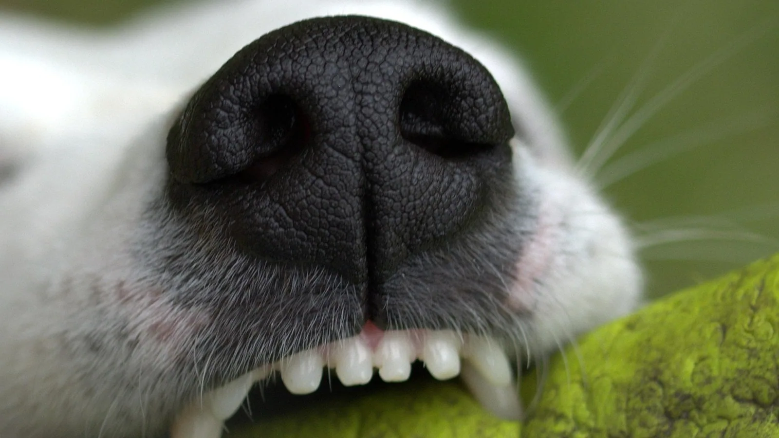
An adult dog has 42 permanent teeth. A puppy has 28 deciduous teeth, which are shed and replaced by the permanent set between roughly 4 and 7 months of age.
Those two numbers answer the search question, but they are only the entry point. The canine dentition is a precise, regionally specialized system. Tooth counts differ by arcade, eruption follows a predictable sequence, and skull shape changes the geometry of the mouth enough that brachycephalic dogs routinely carry fewer teeth in a more crowded arch. This guide covers the full dental formula, the eruption timeline, what each tooth type does, and the clinical problems that arise when deciduous teeth do not fall out on schedule.
This article is educational and is not a substitute for veterinary diagnosis or treatment.
The Short Answer: 42 Adult Teeth, 28 Puppy Teeth
The permanent dentition of the domestic dog (Canis lupus familiaris) contains 42 teeth. The deciduous dentition contains 28.
The difference of 14 teeth is explained almost entirely by the molars. Puppies have no molars at all. Every molar in the adult mouth is a new tooth that erupts into a jaw that was previously occupied only by incisors, canines, and premolars.
The count is fixed in the species-typical dog, but it is not invariant in the individual. Developmental anomalies do occur. A radiologically examined early medieval dog skull from Wolin, Poland showed an unerupted left maxillary canine together with an absent alveolus for the left maxillary first molar and missing incisors, a pattern most consistent with a congenital or very early developmental origin rather than ante-mortem tooth loss or post-depositional damage [1]. That case is archaeological, and the authors caution that a single specimen cannot support population-level conclusions [1]. The clinical point stands: a dog can be born with a dental formula that does not match the textbook.
The Canine Dental Formula, Arcade by Arcade
Veterinary dentists record the dentition as a formula written per side of each jaw. The upper number in each fraction is the maxillary (upper) count for one side. The lower number is the mandibular (lower) count for one side. Multiply by two to get the whole mouth.
Adult (permanent) dentition:
I3/3 C1/1 P4/4 M2/3
Deciduous (puppy) dentition:
I3/3 C1/1 P3/3
The letters stand for incisor (I), canine (C), premolar (P), and molar (M). Reading the adult formula in full:
- Incisors: 3 upper and 3 lower per side. Six upper and six lower across the mouth, 12 total. These are the small, chisel-edged teeth at the front used for nibbling, grooming, and gripping.
- Canines: 1 upper and 1 lower per side. Four total. These are the long, robust, slightly recurved teeth used for grasping and holding.
- Premolars: 4 upper and 4 lower per side. Sixteen total. These sit behind the canines and include the largest tooth in the dog's mouth, the maxillary fourth premolar.
- Molars: 2 upper and 3 lower per side. Ten total. These are the caudal grinding teeth, and the lower arcade carries one more molar per side than the upper.
Total: 12 + 4 + 16 + 10 = 42.
The puppy formula differs in two places. Premolars drop from 4/4 to 3/3 per side, and molars are absent entirely. Total: 12 + 4 + 12 + 0 = 28.
Why the Lower Jaw Carries an Extra Molar
The mandible is slightly shorter than the maxilla in most dogs, and the upper fourth premolar and lower first molar are positioned to shear past one another. This is the carnassial pair, the dominant cutting apparatus of a carnivore dentition. The extra mandibular molar sits caudal to that shear point and adds grinding surface without interfering with the occlusion.
Deciduous Teeth Are Not Small Copies of Permanent Teeth
Deciduous teeth are smaller, have thinner enamel, and have more slender, more divergent roots. The deciduous canine in particular has a narrow crown and a long, fine root that curves slightly. That root morphology matters clinically. When a deciduous tooth fractures and the crown is lost, the root can remain buried in the alveolus for years without visible evidence on the crown surface.
Table: Tooth Type, Adult Count, and Eruption Age
| Tooth type | Adult count (whole mouth) | Deciduous count (whole mouth) | Deciduous eruption | Permanent eruption |
|---|---|---|---|---|
| Incisors | 12 | 12 | 3 to 4 weeks | 4 months (central), then progressively caudal |
| Canines | 4 | 4 | 3 to 4 weeks | 5 to 6 months |
| Premolars | 16 | 12 | 4 to 6 weeks | 4 to 6 months |
| Molars | 10 | 0 | Not present | 5 to 7 months |
Eruption ages are approximate and follow the general sequence described below. Individual dogs vary, and small-breed dogs tend to run later than large-breed dogs.
Eruption Timeline: From 3 Weeks to 7 Months
Deciduous Eruption (3 to 6 Weeks)
Puppies are born edentulous. The deciduous teeth begin to break through the gingiva at about 3 weeks of age, and the full deciduous set is typically present by 6 weeks.
The sequence is rostral to caudal. Incisors come first, around 3 to 4 weeks. Canines follow at roughly 3 to 4 weeks. Premolars erupt at about 4 to 6 weeks and complete the deciduous arcade.
This timing coincides with the developmental window in which puppies transition from nursing to solid food. The deciduous teeth are sharp, and owners frequently notice them during play biting at 5 to 8 weeks of age.
Permanent Eruption (4 to 7 Months)
The permanent teeth begin erupting at about 4 months. The sequence again runs rostral to caudal, and the general rule is that incisors erupt first and molars erupt last.
- Incisors, about 4 months. The permanent central incisors appear first, followed by the intermediate and lateral incisors. The deciduous incisors are pushed out ahead of them.
- Canines, about 5 to 6 months. The permanent canines are substantially larger than their deciduous predecessors. Owners often notice the "double canine" stage, in which a permanent canine is erupting alongside a deciduous canine that has not yet loosened.
- Premolars, about 4 to 6 months. The permanent premolars erupt in the space vacated by the deciduous premolars, and the first maxillary premolar erupts as a new tooth because there is no deciduous counterpart at that position in the upper arcade.
- Molars, about 5 to 7 months. The molars erupt last because they have no deciduous predecessors and require the most caudal alveolar space. The mandibular first molar is typically the first molar to appear, and the maxillary molars follow.
By 7 months, most dogs have a complete permanent dentition. The teeth continue to mature after eruption. Apical closure, the completion of the root apex, lags well behind eruption. A retrospective radiographic and CT study of 788 teeth from 149 dogs found that 50 percent of studied apices were closed by 10 months and 95 percent by 17 months, with mandibular first molars and maxillary fourth premolars closing earlier than canines (95 percent closed by 7 months) and canine apices closing across a 7 to 18 month window [2]. The same study found that dogs over 25 kg reached closed apices at younger ages and over a narrower range than dogs under 25 kg [2]. This is why endodontic planning in a young dog depends on the individual tooth and the individual patient rather than on a single age cutoff.
What Each Tooth Type Does
Incisors
The incisors are the rostral-most teeth. They have a single root each, a small crown, and a straight, slightly spatulate edge. Dogs use them for grooming, for picking up small objects, and for the initial nip in a bite sequence. The incisor region is also a common site for periodontal disease. A visual oral examination study of 1,082 dogs in Türkiye found an association between sex and dental disease specifically in the incisor and premolar teeth, though not in the molars [3].
Canines
The canines are the longest teeth in the mouth and have the longest roots. They are single-rooted, deeply anchored, and positioned at the transition between the incisor and premolar regions. They function as grasping and holding teeth and as the primary weapon in a defensive bite.
Canine crown and root size shows measurable sexual dimorphism in carnivores. A morphometric study of 90 adult raccoon dog skulls, a related canid, found that canine measurements showed the most significant sex differences of all measurements taken [4]. The same study found that molars and tooth rows were not reliable for sex determination [4]. This is comparative anatomy rather than clinical canine dentistry, but it illustrates the general principle that canine tooth dimensions track body size and sex more closely than molar dimensions do.
The canine is also the tooth most often implicated in retained deciduous root remnants. A ten-year retrospective study of 5,432 medical records identified 237 retained deciduous root remnants in 143 dogs, and the remnants were most commonly associated with the left maxillary deciduous canine (39.2 percent) and the right maxillary deciduous canine (33.7 percent) [5].
Premolars
The premolars are the shearing teeth. The maxillary fourth premolar is the largest tooth in the dog's mouth and, together with the mandibular first molar, forms the carnassial pair. These two teeth do the heavy work of cutting food into pieces small enough to swallow.
The maxillary fourth premolar is also a tooth of clinical importance for reasons unrelated to chewing. Its root apices sit close to the orbit. A CT-based anatomical study of 193 dogs found that the shortest maxillary molar tooth root apex-to-globe distance was 2.1 mm in mesocephalic dogs under 5 kg [6]. The same study tabulated distances relevant to dental nerve blocks, caudal maxillary extractions, and caudal maxillectomy [6]. Anatomy in this region is tight, and skull conformation changes the margins.
Periodontal defects around the maxillary fourth premolar are a recognized diagnostic challenge. A study using an experimental model of induced bone loss in large-breed mesaticephalic and dolichocephalic dog cadavers compared a modified interproximal radiographic technique against the simplified bisecting angle technique and found that observers systematically underestimated the distance between the cemento-enamel junction and the bone margin with both methods [7]. The modified interproximal technique performed better overall in accuracy and sensitivity [7]. The practical takeaway is that radiographic staging of periodontal disease in this tooth is technically demanding and that a single standard view may understate the defect.
Molars
The molars are the caudal grinding teeth. The mandibular first molar is the largest tooth in the lower jaw and completes the carnassial shear with the maxillary fourth premolar. The upper arcade has two molars per side and the lower arcade has three.
Molars erupt last and are the teeth most likely to be affected by crowding in dogs with short jaws. They are also the teeth that most often develop periodontal disease in older dogs, in part because they are difficult for owners to brush.
Brachycephalic Versus Mesaticephalic Dogs
Skull conformation changes the dental equation. Mesaticephalic dogs have a skull of medium proportions, with a muzzle length roughly in the middle of the canine range. Dolichocephalic dogs have long, narrow skulls. Brachycephalic dogs have short, broad skulls with a compressed rostrum.
The tooth count in the species-typical formula is the same across these groups, but the space available to hold 42 teeth is not. In a brachycephalic dog, the same number of teeth is packed into a substantially shorter arcade. The practical consequences are:
- Crowding. Teeth rotate, overlap, or erupt out of line because there is not enough arch length.
- Reduced or absent teeth. Brachycephalic breeds are overrepresented among dogs with missing or unerupted teeth, and the crowded arch can leave no room for a tooth to erupt at all. The archaeological case described earlier, an unerupted maxillary canine with an absent alveolus for a molar, is a radiographic example of what a developmental failure of eruption looks like on imaging [1].
- Retained deciduous teeth. A permanent tooth that cannot find space may fail to resorb the root of its deciduous predecessor, leaving the deciduous tooth in place. Yorkshire Terriers were overrepresented in a ten-year retrospective study of retained deciduous root remnants, comprising 41.2 percent of affected patients [5].
- Anatomical margins. The distances between tooth roots and adjacent structures such as the globe vary with skull type, and the reference values generated from CT imaging are tabulated by skull conformation and body weight for exactly this reason [6].
Brachycephalic dogs also tend to have a more rostral and more crowded arrangement of the maxillary fourth premolar and molar region, which is relevant when a veterinarian plans an extraction or a nerve block [6].
Small breed size is a separate risk factor. A visual oral examination study of 1,082 dogs found that small breeds showed higher rates of dental disease than larger breeds [3]. Age also mattered, with lower rates in the 0 to 9 month and 10 to 18 month groups than in older dogs [3]. Feeding method was associated with disease prevalence as well, with home-cooked and mixed feeding linked to higher rates and dry food feeding linked to lower rates [3]. That last finding is an association and not proof of cause. Diet consistency is one of several risk factors examined in the periodontal disease literature, alongside age and breed size [8].
Retained Deciduous Teeth: A Common Clinical Problem
A retained deciduous tooth is a deciduous tooth that is still present after the permanent tooth at the same position has erupted. This is one of the most common dental abnormalities in dogs, and it is more than a cosmetic issue.
The mechanism is straightforward. Normally, the erupting permanent tooth resorbs the root of the deciduous tooth above it, and the deciduous crown loosens and falls out. When the permanent tooth erupts in an abnormal position or the deciduous root is not resorbed, the deciduous tooth stays.
A ten-year retrospective study at a referral clinic reviewed 5,432 medical records and identified 237 retained deciduous root remnants in 143 patients [5]. Direct problems associated with those remnants were found in 117 dogs, or 81.8 percent of the affected group [5]. The most common radiographic finding was periapical radiolucency, seen in 63 dogs (44.1 percent), followed by root resorption and ankylosis in 54 dogs (37.8 percent) [5]. Dental malocclusion affecting the corresponding permanent dentition was also recorded [5].
Two details from that study are worth carrying forward. First, retained deciduous root remnants do not resorb on their own [5]. A root fragment left in place is not a self-resolving problem. Second, the remnants are frequently invisible on a crown-level examination because the crown has fractured off and only the root remains. The study authors emphasize routine full-mouth radiography for identification of these abnormalities [5].
Retained deciduous teeth are most often discussed in the context of the canines, and the data support that emphasis. The maxillary deciduous canines accounted for the largest share of identified root remnants in the retrospective series [5]. A retained deciduous canine also displaces the permanent canine, which can drive the permanent tooth into an abnormal position and create a malocclusion that persists for life.
Clinical Relevance, Limitations and Common Mistakes
What Owners Commonly Get Wrong
Miscounting the puppy teeth. Owners who count 28 teeth in an 8-week-old puppy and then count 42 in the same dog at 8 months sometimes assume teeth were added. They were. The 14 molars are new teeth with no deciduous predecessors.
Assuming the double canine will resolve. A permanent canine erupting next to a retained deciduous canine does not always push the deciduous tooth out. When both are present at 6 to 7 months, the deciduous tooth is retained by definition and needs veterinary assessment.
Treating the tooth count as a diagnostic test. A dog with 40 teeth is not automatically unhealthy, and a dog with 42 teeth is not automatically healthy. Missing teeth can reflect congenital agenesis, failed eruption, extraction, or trauma, and these have different implications. Radiography distinguishes them. The Wolin case illustrates the differential: the authors used radiographic evidence of a fully formed but unerupted canine and a smooth bony surface at the molar region to argue against ante-mortem loss and taphonomic damage and in favor of a congenital or early developmental origin [1].
Ignoring the caudal mouth. The molars and the maxillary fourth premolar are the teeth owners see least often and brush least effectively. Periodontal disease is the most prevalent oral condition in companion animals, affecting over 80 percent of dogs and cats by three years of age, and it is frequently underdiagnosed [8].
What the Numbers Do Not Tell You
A tooth count is a starting point for an oral examination, not a substitute for one. A visual oral examination study of 1,082 dogs found that dental disease prevalence varied significantly with age, feeding method, sex, and breed group, and that small breeds carried higher rates [3]. None of those associations replaces an individual examination.
Awake oral examination also has real limits. A study of 100 dogs with histologically confirmed glossitis found that awake oral examination failed to detect 55 percent of the lesions [9]. That figure is about tongue lesions specifically, not teeth, but it makes the general point that a visual check in a conscious dog misses a substantial amount of oral pathology.
When to Involve a Veterinarian
Any dog with a persistent double tooth, a visibly rotated or crowded tooth, a fractured tooth, or a tooth that has not erupted by 7 months should be examined. Full-mouth radiography is the standard tool for identifying retained root remnants and for assessing the teeth that cannot be seen directly [5]. Individual cases require individual assessment, and this article cannot substitute for that examination.
Frequently Asked Questions
How many teeth does a dog have?
An adult dog has 42 permanent teeth. A puppy has 28 deciduous teeth.
How many teeth does a puppy have?
A puppy has 28 deciduous teeth, made up of 12 incisors, 4 canines, and 12 premolars. Puppies have no molars.
At what age do puppies get all their teeth?
Deciduous teeth begin erupting at about 3 weeks and the full set is usually present by 6 weeks. Permanent teeth begin erupting at about 4 months and the full permanent set is typically complete by 7 months.
Which teeth erupt first and which erupt last?
Incisors erupt first in both dentitions. Molars erupt last in the permanent dentition, at about 5 to 7 months, because they have no deciduous predecessors and need the most caudal alveolar space.
Do all dog breeds have 42 teeth?
The species-typical formula is 42, but individual dogs can have fewer because of congenital agenesis, failed eruption, or tooth loss. Brachycephalic breeds are more prone to missing and crowded teeth because the same number of teeth is packed into a shorter jaw.
What are retained deciduous teeth?
Retained deciduous teeth are puppy teeth that remain in the mouth after the permanent tooth at the same position has erupted. They are a common clinical problem. A ten-year retrospective study found direct problems in 81.8 percent of dogs with retained deciduous root remnants, most commonly periapical radiolucency [5].
Why do retained deciduous teeth matter?
Retained deciduous root remnants do not resorb, and they can cause periapical radiolucency, root resorption and ankylosis, and malocclusion of the permanent dentition [5]. They are often invisible without radiography because the crown may have fractured off [5].
Do brachycephalic dogs have fewer teeth?
Brachycephalic dogs often have fewer erupted teeth than the standard 42 because a shortened jaw leaves insufficient arch length for every tooth to erupt into position. Crowding, rotation, and retained deciduous teeth are all more common in these breeds.
Related Articles
- Snake Anatomy: Do Snakes Have Teeth, Spines, and Other Structures?
- Horse Dental Care and Floating Teeth
- Do Frogs Have Teeth? What It Means for Feeding and Handling
- Axolotl Teeth and Biting: What Owners Should Know
- Why Is My Dog grinding teeth? Bruxism Dental Pain and GI Nausea
- Dog Dental Cleaning Cost: What to Expect and How to Save
- How Many Teeth Do Cats Have? Feline Dental Guide
- Do Ducks Have Teeth? Duck Bill Anatomy Explained
- Large Centipedes: Leg Count and Danger Explained
Sources
- Multiple Dental Agenesis with an Impacted Maxillary Canine in an Early Medieval Dog (Canis lupus familiaris) from Wolin, Poland-A Case Study.
- Evaluation of Apical Closure Status of Canine, Maxillary Fourth Premolar, and Mandibular First Molar Teeth Using Retrospective Single-Time-Point Radiographic and Computed Tomography Data in Dogs.
- Assessment of Dental Pathology Using Visual Oral Examination in 1082 Dogs in Türkiye.
- Morphometric Analysis of the Common Raccoon Dog (Nyctereutes procyonoides) Teeth in Lithuania.
- Retained Deciduous Tooth Root Remnants in Dogs: A Ten-Year Retrospective Study.
- Retrospective study of clinically relevant maxillary and mandibular anatomy in all skull types of dogs and cats.
- Evaluation of a Modified Interproximal Technique as a Novel Radiographic View for the Diagnosis and Staging of Periodontal Defects of the Maxillary Fourth Premolar Using an Experimental Model of Induced Bone Loss in Dogs.
- Overview of Periodontal Disease in Dogs and Cats.
- Glossitis in dogs arises from diverse etiologies but shows predominantly chronic ulcerative pathology and low clinical detection rates.