Hip Muscles Anatomy: Origins, Actions, and Function
By Dr. Zubair Khalid, DVM, MS, PhD ·
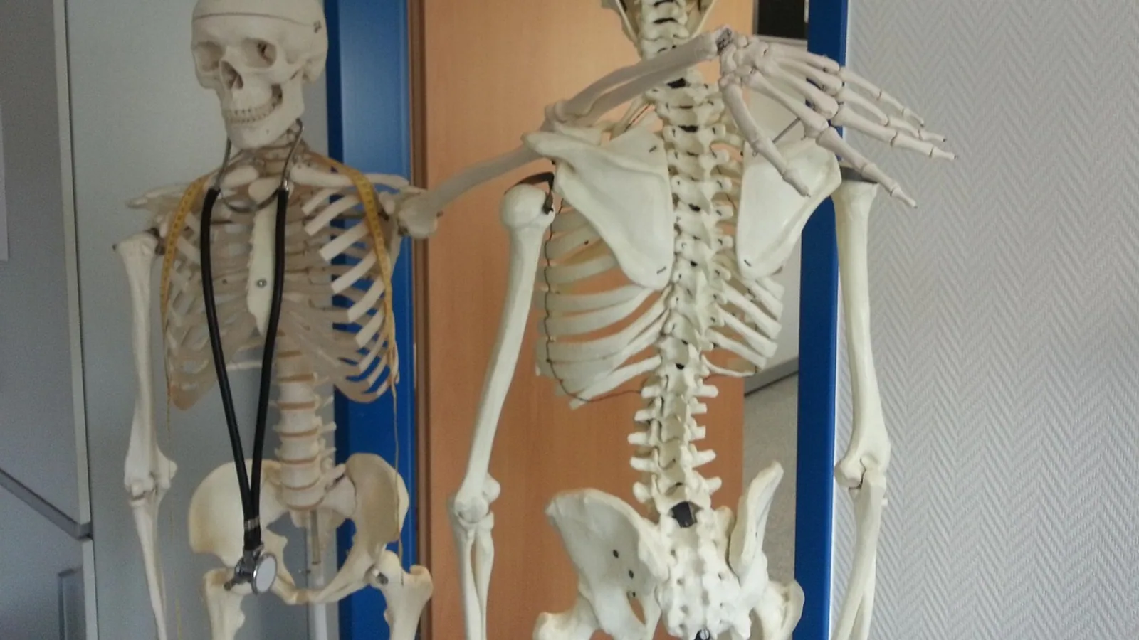
Hip muscles anatomy is the study of the skeletal muscles that attach to the pelvis, proximal femur, and adjacent fascia and cross the coxofemoral (hip) joint to produce extension, flexion, abduction, adduction, and rotation of the pelvic limb. The musculature of the hip is organized into functional groups (gluteals, iliopsoas complex, adductors, deep external rotators, and the hamstring group) whose actions depend on the position of the limb and the direction of the muscle's line of pull relative to the joint's center of rotation.
This matters clinically because the hip is a ball-and-socket joint that depends on muscle for both movement and stability. Weakness or surgical disruption of the gluteals produces a characteristic pelvic drop and a shortened stride, a pattern that shows up after total hip arthroplasty, in developmental dysplasia of the hip, and in neuromuscular disease such as Duchenne muscular dystrophy [1][2][3]. In dogs, the middle gluteal and the iliopsoas are the workhorses of the region, and both are routinely examined on ultrasound and MRI [4]. In horses, the middle gluteal is the dominant propulsive muscle of the hindlimb.
This article is educational and is not a substitute for veterinary diagnosis or treatment.
Orientation: Bones, Joint, and Terms You Need First
Before the muscles make sense, fix the landmarks. The coxofemoral joint is a ball-and-socket (spherical) joint formed by the head of the femur and the acetabulum of the pelvis. The joint permits motion in three planes, which is why the hip muscles anatomy is organized by action rather than by a single plane of movement.
Key terms used throughout:
- Origin: the more proximal, relatively fixed attachment of a muscle.
- Insertion: the more distal attachment that moves when the muscle contracts.
- Action: the movement produced when the muscle shortens, described relative to the joint.
- Innervation: the nerve that supplies the muscle, which determines function and predicts what is lost when that nerve is damaged.
- Abduction: movement of the limb away from the midline. Adduction is movement toward the midline.
- Extension: movement that increases the angle at the joint (swinging the limb caudally). Flexion decreases the angle (swinging the limb cranially).
One structural detail belongs here because it varies by species. The ligament of the head of the femur (ligamentum capitis ossis femoris, historically the round ligament) connects the femoral head to the acetabular fossa. It is present in dogs and absent in horses. That difference matters because the ligament carries a small artery to the femoral head in the dog, and because it contributes passively to hip stability in species that have it.
The Gluteal Group: Extension and Abduction
The gluteals form the bulk of the dorsal hip musculature. In dogs and cats they are conventionally divided into superficial, middle, and deep gluteal muscles. In horses and other ungulates the naming differs, but the functional arrangement is comparable. The gluteals are the principal hip extensors and abductors, and they are the muscles most often injured or surgically disturbed around the hip.
Superficial Gluteal
The superficial gluteal muscle (gluteus superficialis) lies most superficially over the hip. It arises from the gluteal fascia and the cranial part of the lateral border of the ilium and inserts on the third trochanter of the femur in the dog (the horse lacks a third trochanter, and the insertion is on the lateral aspect of the proximal femur). Its action is hip extension and abduction. It is innervated by the caudal gluteal nerve (a branch of the sciatic plexus).
In the dog, the superficial gluteal is relatively thin and is often described together with the biceps femoris as part of the caudal thigh group. In the horse, the superficial gluteal is fused with the biceps femoris to form the gluteobiceps, a large muscle that extends the hip and stifle and contributes to the propulsive phase of the stride. The gluteus maximus complex is the equivalent large superficial muscle in humans, and in dogs the superficial gluteal is the closest functional analog.
Middle Gluteal
The middle gluteal muscle (gluteus medius) is the largest and most important hip abductor and extensor in dogs and cats, and the dominant propulsive muscle of the equine hindlimb. It originates from the gluteal fossa of the ilium, the gluteal aponeurosis, and the cranial border of the ilium, and inserts on the greater trochanter of the femur. Its action is hip extension and abduction. It is innervated by the cranial gluteal nerve (from the lumbosacral plexus, predominantly L6 and L7 in the dog).
In humans, the gluteus medius has three sites of origin (gluteal fossa, gluteal aponeurosis, and the posteroinferior edge of the iliac crest) and a distal tendon with lateral and posterior parts, and it is the largest of the hip abductors by volume [5]. The same principle applies in quadrupeds: the middle gluteal is the largest single muscle of the dorsal hip group and the primary target of clinical examination for abductor function.
Deep Gluteal
The deep gluteal muscle (gluteus profundus) lies deep to the middle gluteal. It arises from the body of the ilium and the ischiatic spine and inserts on the greater trochanter, cranial to the insertion of the middle gluteal. Its action is hip extension and abduction, with a role in stabilizing the femoral head in the acetabulum during weight bearing. It is innervated by the cranial gluteal nerve.
The deep gluteal is well developed in the dog and is consistently identifiable on ultrasound and MRI of the dorsolateral hip region, along with the middle gluteal, superficial gluteal, piriformis, biceps femoris, internal obturator, gemelli, and quadratus femoris [4]. That list is essentially the checklist for a systematic ultrasound examination of the canine hip.
Tensor Fasciae Latae
The tensor fasciae latae (TFL) is grouped with the gluteals functionally even though its attachments differ. It arises from the tuber coxae (cranial ventral iliac spine in the dog) and the adjacent fascia, and it inserts into the fascia lata, which in turn attaches to the lateral aspect of the stifle. It has no bony attachment at its distal end in humans, being encapsulated in the fascia lata [5]. Its action is to tense the fascia lata, which stabilizes the stifle and assists hip flexion and abduction. It is innervated by the cranial gluteal nerve.
The TFL is functionally important in the dog because it contributes to the lateral support of the stifle through the fascia lata, and it is one of the muscles measured in imaging studies of hip and pelvic muscle groups in children with developmental dysplasia of the hip [6].
The Hip Flexors: Iliopsoas and Rectus Femoris
The flexion hip muscles are the iliopsoas complex and the rectus femoris, with the sartorius and TFL assisting. These muscles swing the limb cranially during the stride and are active in the swing phase of gait.
Iliopsoas
The iliopsoas is a composite muscle formed by the psoas major and the iliacus, which share a common insertion on the lesser trochanter of the femur. The psoas major arises from the bodies and transverse processes of the lumbar vertebrae. The iliacus arises from the iliac fossa. The two muscles merge and insert together on the lesser trochanter. Their action is hip flexion and, with the limb fixed, flexion of the lumbar spine and pelvis. They are innervated by ventral branches of the lumbar spinal nerves (predominantly L4 to L6 in the dog).
The iliopsoas is well developed in dogs and cats and is a common site of strain injury in athletic dogs. It is also one of the muscles measured in imaging studies of hip flexor morphology in developmental dysplasia of the hip, where preoperative cross-sectional area ratios of the iliopsoas, rectus femoris, and gluteus maximus are significantly altered on the affected side [6]. In humans, the psoas is increasingly recognized as more than a pure hip flexor, with a role in lumbar spine stabilization [7]. The same dual role is reasonable to expect in quadrupeds, where the psoas spans the lumbar spine and the hip.
Rectus Femoris
The rectus femoris is one of the four heads of the quadriceps femoris. It arises from the ilium just dorsal to the acetabulum (the rectus femoris origin) and inserts, with the other quadriceps heads, on the tibial tuberosity via the patellar ligament. Its action is hip flexion and stifle extension, making it a two-joint muscle. It is innervated by the femoral nerve.
Because it crosses both the hip and the stifle, the rectus femoris is stretched when the hip extends and the stifle flexes, and it is a common source of lameness in dogs with hip or stifle disease. Its cross-sectional area on the operated side remains significantly smaller than the contralateral side one year after hip arthroscopy for femoroacetabular impingement in humans, which illustrates how slowly this muscle recovers after hip surgery [8].
Sartorius
The sartorius arises from the cranial border of the ilium and the iliac fascia and inserts on the cranial border of the tibia (and, in some species, on the patella). It has two bellies in the dog, one cranial and one caudal. Its action is hip flexion and stifle extension (cranial belly) or stifle flexion (caudal belly). It is innervated by the femoral nerve. The sartorius is one of the hip flexor muscles measured in imaging studies of developmental dysplasia of the hip [6].
The Adductor Group and Pectineus
The adductors occupy the medial compartment of the thigh. They pull the limb toward the midline and are important for stabilizing the limb during weight bearing.
Pectineus
The pectineus arises from the pecten of the pubis (the iliopectineal eminence) and inserts on the caudal aspect of the femur, distal to the lesser trochanter. Its action is hip adduction and hip flexion. It is innervated by the obturator nerve. The pectineus is of clinical interest in the dog because it is sometimes surgically released (pectinectomy) in the treatment of hip dysplasia, although the procedure is not universally recommended.
Adductor
The adductor muscle arises from the ventral surface of the pubis and ischium and inserts on the caudal and medial aspects of the femur. It is a powerful hip adductor and also assists hip extension. It is innervated by the obturator nerve. In the horse, the adductor is large and contributes to the adductor group that stabilizes the hindlimb during the stance phase.
Gracilis
The gracilis is the most superficial medial thigh muscle. It arises from the pelvic symphysis and inserts on the medial aspect of the tibia and the crural fascia. Its action is hip adduction and stifle flexion. It is innervated by the obturator nerve. Because it is superficial and has a broad aponeurosis, it is often used as a landmark for the medial approach to the hip and stifle.
The Deep External Rotators
The deep external rotators, also called the short external rotator muscles (SERMs), are a group of small muscles that lie deep to the gluteals and rotate the femur laterally. In humans they are described as six postural muscles that form a single functional unit aligned to coapt the articular surfaces of the hip joint, providing dynamic stability [9].
The group includes the internal obturator, the gemelli (cranial and caudal), the external obturator, and the quadratus femoris. In the dog, the piriformis is also included in this region and is consistently identifiable on ultrasound and MRI [4].
Key anatomical relationships from cadaveric study of the human hip, which translate conceptually to quadrupeds:
- The obturator internus and externus insert almost perpendicularly on the proximal femur [9].
- The two gemelli take their distal insertion onto the tendon of the obturator internus to form the "hip triceps tendon" [9].
- The quadratus femoris is the most distal of the group and lies in the ischiofemoral space, with an average width of 5.0 ± 1.1 cm in human cadavers [10].
The action of the deep external rotators is lateral (external) rotation of the hip, with a secondary role in stabilizing the femoral head within the acetabulum. They are innervated by branches of the sacral plexus and the obturator nerve, depending on the specific muscle.
Summary Table: Origins, Insertions, Actions, and Innervation
| Muscle | Origin | Insertion | Action | Innervation |
|---|---|---|---|---|
| Superficial gluteal | Gluteal fascia, cranial lateral ilium | Third trochanter (dog) | Hip extension, abduction | Caudal gluteal nerve |
| Middle gluteal | Gluteal fossa, gluteal aponeurosis, cranial ilium | Greater trochanter | Hip extension, abduction | Cranial gluteal nerve |
| Deep gluteal | Body of ilium, ischiatic spine | Greater trochanter (cranial) | Hip extension, abduction, stabilization | Cranial gluteal nerve |
| Tensor fasciae latae | Tuber coxae, adjacent fascia | Fascia lata (to stifle) | Tense fascia lata, hip flexion, abduction | Cranial gluteal nerve |
| Iliopsoas | Lumbar vertebrae (psoas major), iliac fossa (iliacus) | Lesser trochanter | Hip flexion, lumbar flexion | Ventral lumbar nerves (L4-L6) |
| Rectus femoris | Ilium dorsal to acetabulum | Tibial tuberosity (via patellar ligament) | Hip flexion, stifle extension | Femoral nerve |
| Sartorius | Cranial ilium, iliac fascia | Cranial tibia, patella | Hip flexion, stifle extension/flexion | Femoral nerve |
| Pectineus | Pecten of pubis | Caudal femur, distal to lesser trochanter | Hip adduction, flexion | Obturator nerve |
| Adductor | Ventral pubis and ischium | Caudal and medial femur | Hip adduction, extension | Obturator nerve |
| Gracilis | Pelvic symphysis | Medial tibia, crural fascia | Hip adduction, stifle flexion | Obturator nerve |
| Internal obturator | Pelvic surface of ischium and pubis | Trochanteric fossa | Lateral rotation, stabilization | Sacral plexus branches |
| Gemelli | Ischial spine (cranial), ischial tuberosity (caudal) | Trochanteric fossa (with obturator internus tendon) | Lateral rotation, stabilization | Sacral plexus branches |
| External obturator | Ventral ischium and pubis | Trochanteric fossa | Lateral rotation | Obturator nerve |
| Quadratus femoris | Ischial tuberosity | Intertrochanteric crest | Lateral rotation, stabilization | Sacral plexus branches |
Comparative Notes: Dogs, Cats, and Horses
The hip muscle anatomy of domestic species follows the same plan but with important differences in emphasis and structure.
Dogs and cats. Both species have a strong middle gluteal and a well-developed iliopsoas. The deep gluteal is well defined and consistently identifiable on imaging [4]. The ligament of the head of the femur is present and carries a small artery to the femoral head. The superficial gluteal is relatively thin and inserts on the third trochanter, a feature unique to the dog among common domestic species. The iliopsoas is a frequent source of strain injury in athletic dogs because of its long course across the lumbar spine and hip.
Horses. The horse relies heavily on the middle gluteal for propulsion. The superficial gluteal is fused with the biceps femoris to form the gluteobiceps, a large muscle that extends the hip and stifle and drives the stride. The horse lacks a third trochanter, so the superficial gluteal inserts on the lateral proximal femur. The ligament of the head of the femur is absent in horses, which means the femoral head is stabilized almost entirely by the joint capsule, the accessory ligament of the femoral head, and the surrounding musculature. The adductor group is large and important for stabilizing the hindlimb during the stance phase.
Cursorial birds (comparative context). In the greater rhea, the thigh contains the highest number of muscles and the shank the fewest, a feature that minimizes inertia and allows high stride frequencies [11]. The major hip and ankle extensors are massive and robust, consistent with specialization for high-speed locomotion. This is a useful reminder that hip muscle anatomy scales with locomotor demand across species.
How Hip Muscle Function Is Tested and Observed
Several methods are used in veterinary and human clinical practice to assess hip muscle function. Each has strengths and limitations.
Manual muscle testing and gait observation. The clinician observes the animal walking, trotting, and rising from a sit. A shortened stride on one side, a pelvic drop on the contralateral side during weight bearing (Trendelenburg sign), or a lateral sway of the trunk over the affected limb (Duchenne sign) suggests hip abductor weakness. Experimental nerve block studies in humans show that the Trendelenburg and Duchenne signs lack both sensitivity and specificity: after sequential paralysis of the TFL, gluteus medius and minimus, and gluteus maximus, only some participants showed the expected signs, and gluteus maximus paralysis led to failure in 5 of 10 participants, with two showing a Duchenne sign [3]. The clinical lesson is that these signs support a diagnosis but do not confirm it.
Ultrasonography. High-frequency B-mode ultrasonography can identify the middle gluteal, deep gluteal, superficial gluteal, piriformis, biceps femoris, internal obturator, gemelli, and quadratus femoris in the dog, with the sacrotuberous ligament and sciatic nerve serving as landmarks [4]. The protocol was validated against MRI and gross dissection, and dye injections accurately identified all eight structures consistently between two diplomates [4].
Magnetic resonance imaging (MRI). MRI provides quantitative measures of muscle cross-sectional area, fat fraction, and T1 and T2 relaxation times. In Duchenne muscular dystrophy, T1 of the hip extensors, flexors, and abductors correlates positively with the North Star Ambulatory Assessment score, while T2 of the adductors correlates negatively, making these parameters potential imaging biomarkers of muscle damage [1]. In developmental dysplasia of the hip, preoperative cross-sectional area ratios of the iliopsoas, rectus femoris, and gluteus maximus are significantly altered on the affected side [6].
Electromyography (EMG). Surface EMG records the timing and amplitude of muscle activation. In women with patellofemoral pain syndrome, combined balance and perturbation training produced a greater reduction in normalized gluteus maximus activity and a greater increase in normalized gluteus medius activity than a control intervention, with significant changes in gluteus medius activation onset and time to peak for both gluteal muscles [12]. This illustrates how training can change the coordination of the hip muscles, not just their strength.
Nerve stimulation. Intraoperative stimulation of the superior gluteal nerve can confirm that the gluteus medius is functioning before a repair is performed, a technique used in hip abductor repair with fascia of the vastus lateralis [13].
Clinical Relevance, Limitations and Common Mistakes
Hip muscle pathology is common in veterinary practice and has direct functional consequences.
Hip abductor injury. Tears of the gluteal tendons cause severe pain and weakness, and repair often requires augmentation. A technique using a flap of fascia from the vastus lateralis has been described, with 21 of 23 patients reporting no pain and 20 of 23 achieving abductor power of 3/5 or greater at final follow-up [13]. The superior gluteal nerve is at risk during hip surgery, with reported injury rates as high as 8% during primary total hip arthroplasty, and its integrity is critical for abductor function [14].
Gluteal muscle damage in neuromuscular disease. In Duchenne muscular dystrophy, damage to the gluteal muscle group is associated with worse motor function, and MRI parameters of the extensors, flexors, and abductors correlate with functional scores [1]. This has implications for monitoring disease progression and for rehabilitation planning.
Surgical approach and muscle recovery. The direct anterior approach to total hip arthroplasty is associated with greater 6-minute walk distance at 24 weeks and greater hip abductor strength at 10 weeks compared with the posterolateral approach, consistent with less gluteal muscle disruption [2]. This is a useful reminder that surgical approach affects the hip musculature and therefore recovery.
Deep gluteal syndrome. The deep gluteal space contains the sciatic nerve, the quadratus femoris, and the short external rotators. Anatomical variation in sciatic nerve emergence was found in 15.4% of lower limbs examined, and the average ischiofemoral distance was 2.5 ± 1.3 cm [10]. These relationships explain why deep gluteal pain syndromes can mimic hip joint pain.
Common mistakes students make.
- Confusing the origin and insertion of the middle gluteal. The origin is on the ilium and the insertion is on the greater trochanter.
- Assuming the gluteals only extend the hip. They also abduct and stabilize the femoral head.
- Forgetting that the rectus femoris crosses two joints and therefore affects both the hip and the stifle.
- Treating the deep external rotators as unimportant. They are a functional unit that coapts the articular surfaces and provides dynamic stability [9].
- Assuming the ligament of the head of the femur is present in all species. It is present in dogs and absent in horses.
- Using human gym terminology (for example "glute bridge" or "hip thrust") as if it maps directly onto quadruped function. The biomechanics differ because the limb orientation and weight-bearing pattern differ.
Quick Review
- The hip is a ball-and-socket joint, and its stability depends on muscle as well as the joint capsule and ligaments.
- The gluteal group (superficial, middle, deep) extends and abducts the hip and is innervated by the cranial and caudal gluteal nerves.
- The middle gluteal is the largest and most important hip abductor in dogs and cats and the dominant propulsive muscle in horses.
- The iliopsoas and rectus femoris are the main hip flexors, innervated by ventral lumbar nerves and the femoral nerve respectively.
- The adductor group and pectineus adduct the hip and are innervated by the obturator nerve.
- The deep external rotators (internal obturator, gemelli, external obturator, quadratus femoris) rotate the hip laterally and provide dynamic stability [9].
- The ligament of the head of the femur is present in dogs and absent in horses.
Frequently Asked Questions
What are the main hip muscles in dogs?
The main hip muscles in dogs are the gluteal group (superficial, middle, and deep gluteal), the iliopsoas, the rectus femoris, the sartorius, the tensor fasciae latae, the adductor group (pectineus, adductor, gracilis), and the deep external rotators (internal obturator, gemelli, external obturator, quadratus femoris).
Which nerve supplies most of the hip muscles?
The gluteal muscles are supplied by the cranial and caudal gluteal nerves, the hip flexors by the femoral nerve and ventral lumbar nerves, and the adductor group by the obturator nerve. The deep external rotators receive branches from the sacral plexus and, for the external obturator, the obturator nerve.
Why is the middle gluteal so important in horses?
The middle gluteal is the dominant propulsive muscle of the equine hindlimb. It extends and abducts the hip and generates much of the force for forward propulsion, which is why it is large and heavily used in the horse.
Is the ligament of the head of the femur present in all animals?
No. The ligament of the head of the femur is present in dogs and absent in horses. This difference affects how the femoral head is stabilized in each species.
What is the difference between hip flexion and hip extension?
Hip flexion swings the limb cranially (forward) and is produced mainly by the iliopsoas and rectus femoris. Hip extension swings the limb caudally (backward) and is produced mainly by the gluteal group and the hamstrings.
Can hip muscle weakness be detected on a physical exam?
Yes, but the signs are not perfectly sensitive or specific. A shortened stride, pelvic drop, or lateral trunk sway during weight bearing can suggest hip abductor weakness, but experimental nerve block studies show that the classic Trendelenburg and Duchenne signs do not appear in every case [3]. Imaging and electrodiagnostic testing may be needed to confirm the diagnosis.
Related Articles
- Equine Muscular System: Major Muscle Groups and Function
- Canine Pelvic Limb Musculature: Actions and Innervation
- Equine Larynx Anatomy and Function in Respiration
- Canine Lymphatic System: Anatomy and Function
- Equine Hoof Anatomy: Structures and Function
- Origin Genomics
- Subclavius Muscle: Origin, Insertion, Action, Innervation
Sources
- Clinical utilisation of multimodal quantitative magnetic resonance imaging in investigating muscular damage in Duchenne muscular dystrophy: a study on the association between gluteal muscle groups and motor function.
- Comparing Direct Anterior Approach Versus Posterolateral Approach in Total Hip Arthroplasty on Physical Function Recovery: A Prospective Cohort Study.
- Evaluation of Trendelenburg and Duchenne signs by experimentally induced gluteal muscle paralysis.
- Effective ultrasonography protocol for investigating muscular structures of the canine dorsolateral hip region with comparative magnetic resonance imaging.
- The anatomy of the hip abductor muscles.
- Assessment and quantitative analysis of hip surrounding muscles in children with developmental dysplasia of the hip via magnetic resonance imaging.
- Fat infiltration of the posterior paraspinal muscles is inversely associated with the fat infiltration of the psoas muscle: a potential compensatory mechanism in the lumbar spine.
- Changes in muscle cross-sectional area and functional outcomes after hip arthroscopy for femoroacetabular impingement: A comparison between the operated and asymptomatic contralateral hip.
- The short external rotator muscles of the hip: a cadaveric study on 18 specimens with clinical implications.
- Deep gluteal space anatomy and its relationship with deep gluteal pain syndromes.
- The hindlimb muscles of Rhea americana (Aves, Palaeognathae, Rheidae).
- The effects of combined balance and perturbation training on gluteal muscle activity and hip kinematics in women with patellofemoral pain syndrome: a randomized controlled trial.
- Augmentation of hip abductor repair with fascia of vastus lateralis.
- Anatomic Mapping of the Inferior Branch of the Superior Gluteal Nerve: Implications for Revision Total Hip Arthroplasty.