Head Anatomy: Bones, Muscles and Nerves Overview
By Dr. Zubair Khalid, DVM, MS, PhD ·
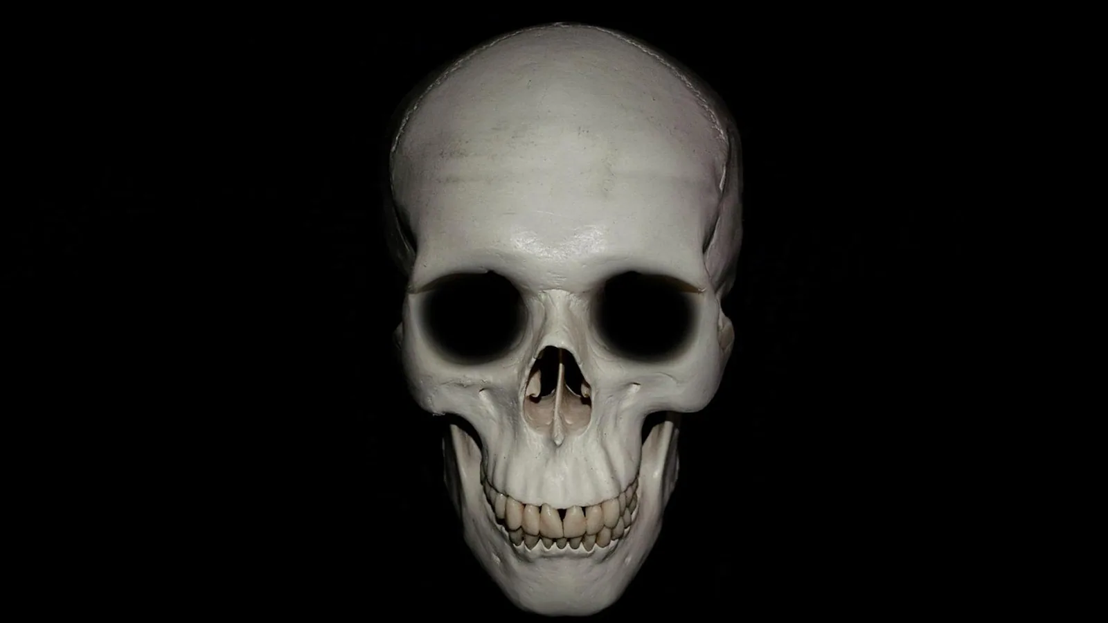
Head anatomy is the study of the skull bones, the muscles that move the jaw and face, and the cranial nerves that supply sensation and movement to the head and neck. In domestic animals, the head is a compact region where bone, muscle, nerve, blood vessel, and special sense organ sit within a few centimeters of one another, which is why a single penetrating wound or a fast-growing mass can affect eating, breathing, vision, and swallowing at the same time.
This overview organizes the region the way a clinician approaches it: skull bones first, then the masticatory muscles that close the jaw, then the facial muscles of expression, then the cranial nerves with the foramina they pass through. A species comparison table follows, along with the clinical relevance of these structures and the mistakes that are most often made when interpreting head anatomy.
This article is educational and is not a substitute for veterinary diagnosis or treatment.
The Skull Bones: A Regional Map
The skull is built from two functional groups of bones. The neurocranium forms the box around the brain. The viscerocranium (the facial skeleton) forms the muzzle, orbits, nasal cavity, and jaws. In the dog, the neurocranium and viscerocranium vary enormously in proportion between breeds, and that variation changes how every other head structure is positioned.
Frontal Bone
The frontal bone forms the forehead and the roof of the cranial cavity. It contributes to the medial wall of the orbit and contains the frontal sinus, which communicates with the nasal cavity. In dogs and cats the frontal sinus is relatively small compared with that of the horse, where it is a large, clinically important cavity that can be involved in sinus disease and is a surgical landmark.
Parietal Bone
The parietal bone forms the caudal part of the cranial roof and the upper part of the cranial wall. It meets the frontal bone rostrally and the occipital bone caudally. In many species a sagittal crest rises along the midline where the left and right parietal and interparietal bones meet, providing attachment for the temporalis muscle. The crest is prominent in carnivores with powerful jaw-closing muscles and is reduced in brachycephalic dogs.
Temporal Bone
The temporal bone is one of the most complex bones of the skull. It contains the external acoustic meatus, the middle ear cavity, and the bony labyrinth of the inner ear. Its squamous part contributes to the wall of the cranial cavity and to the zygomatic arch. The tympanic part surrounds the ear canal and forms the tympanic bulla, a rounded, hollow structure that is well developed in the dog and cat and is a landmark for ear surgery.
Occipital Bone
The occipital bone forms the caudal wall and floor of the cranial cavity and surrounds the foramen magnum, the large opening through which the medulla oblongata continues as the spinal cord. The occipital condyles articulate with the first cervical vertebra. The external occipital protuberance and the nuchal crest are palpable landmarks on the back of the head. In small dogs, the position of the osseous tentorium cerebelli and the transverse sinus canal varies relative to these external landmarks, and a computed tomography study of 23 small-breed dogs found that this deviation differed significantly between breeds, with Chihuahuas showing greater deviation than Toy Poodles and Miniature Dachshunds [1].
Maxilla
The maxilla forms most of the upper jaw, the lateral wall of the nasal cavity, and the floor of the orbit. It carries the upper cheek teeth and, in ruminants, the upper dental pad. The infraorbital foramen opens on its lateral surface and transmits the infraorbital nerve, a branch of the trigeminal nerve. The maxilla and the zygomatic bone are common sites for surface bone tumors in dogs. A retrospective evaluation of 10 dogs with parosteal osteosarcoma of the maxillofacial bones found that the caudal maxilla and/or zygoma was the most common site of origin, accounting for 50 percent of tumors, and that exophthalmos was a prominent clinical sign in 40 percent of cases [2].
Mandible
The mandible is the lower jaw. It consists of a body that carries the lower teeth and a ramus that rises to the temporomandibular joint. The coronoid process provides attachment for the temporalis muscle, the condylar process articulates with the temporal bone, and the angular process gives attachment to the pterygoid muscles. The mandibular canal runs through the body of the mandible and carries the inferior alveolar nerve and vessels. It opens rostrally at the mental foramina. Accessory mental foramina are common and vary by species. In a comparative study of Beetal goats and Kajali sheep, accessory mental foramina were present in 87.5 percent of sheep but only 12.5 percent of goats [3].
Species Differences in Skull Shape
Skull shape is not a cosmetic detail. It changes the geometry of the nasal cavity, the orbit, the palate, and the airway. A study of 852 domestic dogs using archived computed tomography images found that smaller dogs more frequently had rounder crania, meaning cranial morphology does not scale isometrically with body size [4]. The same study found that each 1.0 kg reduction in body weight increased the odds of belonging to a group of breeds predisposed to syringomyelia by 25 percent [4].
Brachycephalic breeds are not interchangeable. A morphometric study of 75 healthy French Bulldogs, Pugs, and Shih Tzus classified all three breeds as extreme brachycephalic, with a craniofacial ratio below 0.2 and a cephalic index above 0.81, yet found significant breed-specific differences. French Bulldogs were the heaviest and had the widest skulls and largest palpebral width, while Pugs had the lowest craniofacial ratio and the shortest muzzles [5].
The hyoid apparatus, which suspends the tongue and larynx, also varies with skull shape. A computed tomography study of 26 dogs found that brachycephalic breeds had a more dorsoventrally positioned and compact hyoid structure, while mesocephalic breeds had a more aligned and elongated configuration of the stylohyoid and thyrohyoid bones [6].
Masticatory Muscles: Closing and Moving the Jaw
The masticatory muscles are the muscles of chewing. They move the mandible against the maxilla and are innervated by the mandibular branch of the trigeminal nerve (cranial nerve V). They are among the strongest muscles in the body relative to their size.
Masseter
The masseter is a thick, powerful muscle that runs from the zygomatic arch to the lateral surface of the mandibular ramus. It elevates the mandible and, because its fibers run obliquely, also helps move the jaw sideways during grinding. In herbivores such as the horse and ruminant, the masseter is enormous and is a major contributor to the lateral jaw movement needed for chewing fibrous feed. In carnivores it is smaller but still prominent.
Temporalis
The temporalis fills the temporal fossa and attaches to the coronoid process of the mandible. It is the principal elevator of the jaw. In carnivores, the temporalis is large and the sagittal crest is correspondingly well developed. In brachycephalic dogs, the temporal fossa is shallower and the muscle is correspondingly reduced in volume.
Pterygoid Muscles
The pterygoid muscles lie medial to the mandibular ramus. The lateral pterygoid protrudes the mandible and moves it from side to side. The medial pterygoid elevates the mandible and works with the masseter to close the jaw. Because they are deep, they are difficult to palpate and are usually assessed indirectly through jaw movement and imaging.
Digastricus
The digastricus is technically a jaw opener rather than a masticatory muscle, but it is grouped with them functionally. It runs from the paracondylar process of the occipital bone to the ventral border of the mandible and opens the jaw. In the dog it is innervated by the mandibular branch of the trigeminal nerve, while in the horse and ruminant it receives innervation from the facial nerve. This difference matters when interpreting jaw function in neurologic examination.
Facial Muscles: Expression, Ear Movement, and Cheek Control
The facial muscles are the muscles of facial expression. They are thin, sheet-like muscles that attach to the skin rather than to bone, and they are innervated by the facial nerve (cranial nerve VII). In domestic animals, they also control the ear, the eyelid, the nostril, and the lips.
The major groups include the muscles of the ear (scutuloauricularis, cervicoauricularis, and related muscles), the muscles of the eyelid (orbicularis oculi, levator palpebrae superioris is innervated by the oculomotor nerve, not the facial nerve), the muscles of the nose and lip (levator nasolabialis, dilator naris, orbicularis oris), and the muscles of the cheek and lip (buccinator, zygomaticus). The platysma, a broad sheet in the neck, is also a facial muscle and is often the first muscle to show signs of facial nerve dysfunction.
Facial muscle function is assessed by watching the animal blink, move its ears, and retract its lips. A drooping ear, a drooping lip, and an inability to blink on one side point to a facial nerve lesion on that side.
Cranial Nerves of the Head and Their Foramina
The cranial nerves emerge from the brainstem and exit the skull through specific openings. Knowing which nerve uses which foramen lets a clinician localize a lesion to a specific point along the nerve.
Trigeminal Nerve (Cranial Nerve V)
The trigeminal nerve is the great sensory nerve of the face and the motor nerve of the masticatory muscles. It has three main divisions.
The ophthalmic division passes through the orbital fissure. It supplies sensation to the cornea, the conjunctiva, the skin of the forehead, and the nasal cavity.
The maxillary division passes through the round foramen (foramen rotundum). It continues as the infraorbital nerve through the infraorbital foramen and supplies sensation to the upper lip, nose, and upper teeth.
The mandibular division passes through the oval foramen (foramen ovale). It supplies sensation to the lower jaw, lower teeth, tongue (via the lingual nerve), and the skin of the temple and cheek. Its motor branches supply the masseter, temporalis, and pterygoid muscles.
Facial Nerve (Cranial Nerve VII)
The facial nerve is the motor nerve of the muscles of facial expression. It also carries taste from the rostral two-thirds of the tongue via the chorda tympani and parasympathetic fibers to the lacrimal, mandibular, and sublingual glands.
The facial nerve exits the skull through the stylomastoid foramen, a small opening just caudal to the tympanic bulla. From there it runs through the parotid salivary gland and divides into branches that fan out across the face. Because it runs superficially, it is vulnerable to trauma, surgical injury during ear canal procedures, and compression by tumors. In dogs, the facial nerve is one of several cranial nerves that can be involved in infiltrative disease. A case report of IgG4-related disease in a human patient described perineural extension involving the optic, infraorbital, facial, and auriculotemporal nerves [7], which illustrates how a single process can track along multiple cranial nerve pathways.
Glossopharyngeal Nerve (Cranial Nerve IX)
The glossopharyngeal nerve exits the skull through the jugular foramen. It supplies sensation to the caudal third of the tongue, the pharynx, and the middle ear. It also carries parasympathetic fibers to the parotid gland and contributes to the gag reflex. In the dog, the gag reflex is tested by stimulating the caudal pharynx and watching for contraction of the pharyngeal muscles.
Vagus Nerve (Cranial Nerve X)
The vagus nerve also exits through the jugular foramen. It is the longest cranial nerve and supplies the pharynx, larynx, and most of the thoracic and abdominal viscera. In the head, its branches include the pharyngeal branches, which contribute to swallowing, and the cranial laryngeal nerve, which supplies the cricothyroid muscle. Damage to the vagus or its laryngeal branches can cause voice change, difficulty swallowing, or aspiration.
Hypoglossal Nerve (Cranial Nerve XII)
The hypoglossal nerve exits through the hypoglossal canal. It is the motor nerve of the tongue. A lesion causes the tongue to deviate toward the affected side when protruded, and in severe cases the tongue cannot be used to lap or to move food to the back of the mouth.
Summary Table of Cranial Nerves and Foramina
| Nerve | Number | Main function | Foramen or exit |
|---|---|---|---|
| Trigeminal, ophthalmic division | V1 | Sensation to cornea, forehead, nasal cavity | Orbital fissure |
| Trigeminal, maxillary division | V2 | Sensation to upper jaw, nose, upper teeth | Round foramen |
| Trigeminal, mandibular division | V3 | Sensation to lower jaw, tongue, motor to masticatory muscles | Oval foramen |
| Facial | VII | Motor to facial muscles, taste rostral tongue, parasympathetic to glands | Stylomastoid foramen |
| Glossopharyngeal | IX | Sensation caudal tongue and pharynx, gag reflex | Jugular foramen |
| Vagus | X | Pharynx, larynx, thoracic and abdominal viscera | Jugular foramen |
| Hypoglossal | XII | Motor to tongue | Hypoglossal canal |
Species Differences in Head Anatomy
| Feature | Dog | Horse | Ruminant |
|---|---|---|---|
| Skull shape | Highly variable, from dolichocephalic to extreme brachycephalic | Long, with a large nasal chamber and extensive sinuses | Variable, with a horn core in horned breeds |
| Nasal chamber | Length varies with breed, short in brachycephalics | Long and narrow, with a large dorsal and ventral conchal system | Moderate, with a well-developed ethmoidal region |
| Horn core | Absent | Absent | Present in horned breeds, a bony core covered by horn |
| Hyoid apparatus | Compact and dorsoventrally positioned in brachycephalics, elongated in mesocephalics [6] | Large and mobile, supports a long tongue | Well developed, supports the tongue and larynx during grazing |
| Mandible | Variable, with a high frequency of accessory mental foramina in some species | Long and robust, with a large diastema | Broad, with a large diastema and a dental pad on the maxilla |
| Occipital region | Variable, with breed differences in tentorium position [1] | Prominent occipital crest | Pentagonal occipital base in Beetal goats, quadrilateral in Kajali sheep [3] |
The horse has one of the longest nasal chambers among domestic species. This length increases the surface area for warming, filtering, and humidifying air, but it also means that a foreign body or mass in the nasal cavity can be difficult to reach and can cause a long-standing unilateral nasal discharge. The horse also has extensive paranasal sinuses that communicate with the nasal cavity and are common sites of infection and neoplasia.
Ruminants have a horn core in horned breeds. The horn core is a bony extension of the frontal bone covered by a layer of horn-producing epidermis. It is hollow in some species and is connected to the frontal sinus, which is why dehorning or horn injury can open the sinus and lead to infection. The anatomy of the horn core and its relationship to the frontal sinus is a key consideration in dehorning procedures and in the management of horn trauma.
The comparative study of Beetal goats and Kajali sheep found distinct species-specific differences in cranial bones. Beetal goat skulls were elongated, narrow, and dolichocephalic, while Kajali sheep skulls were shorter, broader, and ranged from mesaticephalic to brachycephalic. Beetal goats had a pentagonal occipital base, a posteriorly placed facial tuberosity, and a consistently present intercondyloid cleft, while Kajali sheep had a quadrilateral occipital base, a premolar-level facial tuberosity, and no cleft [3].
Clinical Relevance, Limitations and Common Mistakes
Head anatomy matters in clinical practice because so many important structures are packed into a small space. A mass in the caudal maxilla can push the eye outward, as seen in the parosteal osteosarcoma cases where exophthalmos was a prominent sign in 40 percent of dogs [2]. A defect in the cribriform plate can allow brain tissue to herniate into the nasal cavity, as reported in a young American Cocker Spaniel with an encephalocele that was treated surgically [8]. A persistent fontanelle in a toy breed is not by itself a sign of disease, and a study of 41 toy-sized dogs found no evidence that fontanelle status influenced clinical or neurologic presentation [9].
Imaging has changed how head anatomy is assessed. Computed tomography and magnetic resonance imaging allow clinicians to measure structures that cannot be palpated, such as the pituitary gland. A study of 89 dogs with pituitary macrotumors found that 21.3 percent were smaller than 1 cm in height, and the authors proposed that a pituitary gland height greater than 0.60 cm or a volume greater than 0.17 cm3 may represent a macrotumor in brachycephalic dogs [10]. This is a good example of why breed and skull shape must be considered when interpreting measurements.
Common mistakes in head anatomy include assuming that all brachycephalic breeds have the same anatomy, confusing the foramina that transmit the trigeminal divisions, and forgetting that the facial nerve exits through the stylomastoid foramen rather than through the jugular foramen. Another common error is to assume that a drooping ear or lip is always due to a facial nerve lesion, when it can also result from a lesion in the brainstem or the facial nucleus.
Limitations of this overview are that it is a regional summary and does not cover the detailed neuroanatomy of the brain or the full course of each nerve. Individual animals vary, and a veterinarian should evaluate any specific clinical problem in person.
Frequently Asked Questions
What is head anatomy?
Head anatomy is the study of the bones, muscles, nerves, blood vessels, and organs of the head and neck. It includes the skull, the masticatory and facial muscles, and the cranial nerves.
Which foramen does the facial nerve exit through?
The facial nerve exits the skull through the stylomastoid foramen, which is located just caudal to the tympanic bulla.
Which foramina do the trigeminal nerve branches use?
The ophthalmic division passes through the orbital fissure, the maxillary division through the round foramen, and the mandibular division through the oval foramen.
What is the difference between masticatory and facial muscles?
Masticatory muscles move the jaw and are innervated by the trigeminal nerve. Facial muscles control expression, ear movement, and blinking and are innervated by the facial nerve.
How does skull shape differ between dog breeds?
Skull shape ranges from dolichocephalic (long and narrow) to extreme brachycephalic (short and broad). A study of 852 dogs found that smaller dogs more frequently had rounder crania [4].
Do all brachycephalic dogs have the same head anatomy?
No. A study of French Bulldogs, Pugs, and Shih Tzus found significant breed-specific differences in skull width, muzzle length, and craniofacial ratio even though all three were classified as extreme brachycephalic [5].
What is a horn core in ruminants?
A horn core is a bony extension of the frontal bone covered by horn-producing epidermis. It is present in horned ruminant breeds and is connected to the frontal sinus in some species.
Why does head anatomy matter in veterinary practice?
Head anatomy matters because structures are close together, so a small lesion can affect eating, breathing, vision, and swallowing. Knowing the foramina and nerve pathways helps clinicians localize lesions and plan surgery.
Related Articles
- Bovine Neurological Anatomy: Cranial Nerves and Brainstem
- Surgical Approaches to the Head and Neck: Oral and Pharyngeal
- Equine Muscular System: Major Muscle Groups and Function
- Canine Thoracic Limb Nerves: Brachial Plexus and Innervation Patterns
- Raw BARF Diet for Dogs: Muscle Meat Bone and Organ Ratio Guide
- Equine Hindlimb Anatomy: Bones, Joints, and Ligaments
Sources
- Anatomical Variations of the Osseous Tentorium Cerebelli and Transverse Sinus Canal/Groove in Small-Breed Dogs: A Computed Tomographic Study.
- Canine Maxillofacial Parosteal Osteosarcoma: A Retrospective Case Evaluation in 10 Dogs.
- Diagnostic anatomy of the head region of Beetal goat and Kajali sheep with special reference to its importance in regional anaesthesia.
- Miniaturization in Domestic Dogs: Relationships Among Cranial Shape, Head Indexes, and Body Weight.
- Comparative applied craniofacial morphometry in popular brachycephalic breeds: A tool for preventive medicine and risk stratification.
- Comparative Anatomy of the Hyoid Apparatus in Various Dog Breeds: Insights From Clinical Imaging.
- IgG4-related disease involving facial, infraorbital, auriculotemporal and optic nerves: multinervous cranial involvement in an adult patient.
- Surgical Management of a Canine Encephalocele Communicating with the Nasal Cavity.
- The bregmatic fontanelle alone is an unreliable indicator of health in Pomeranian and other toy-sized dogs.
- Defining Variations in the Size of Normal Pituitary Glands and Pituitary Macrotumors Based on Canine Skull Morphology.