Epithelial Cells: Types, Locations, and Functions
By Dr. Zubair Khalid, DVM, MS, PhD ·
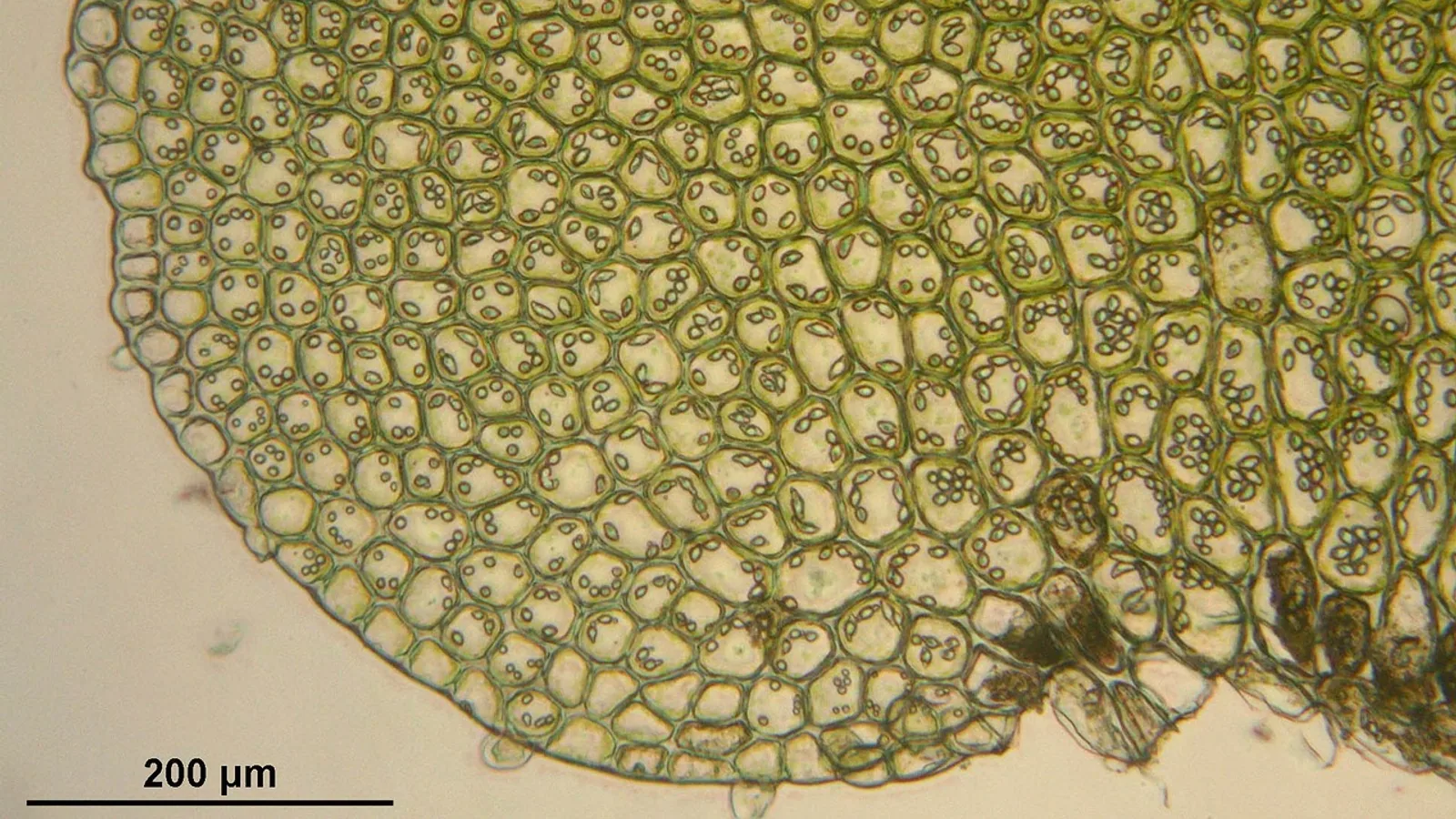
Epithelial cells are polarized cells that sit on a basement membrane, join tightly to their neighbors, and form continuous sheets that cover body surfaces and line every hollow organ, cavity, and gland. They are classified by the number of cell layers (simple versus stratified) and by the shape of the cells in the outermost layer (squamous, cuboidal, or columnar).
Epithelium is the tissue a clinician meets first. Skin, oral mucosa, the cornea, the esophagus, the stomach, the intestine, the airway, the bladder, and the mammary gland are all lined or built by epithelial cells. Because epithelium is the interface between the body and the outside world, its structure tells you what a surface is built to do. A one-cell-thick sheet is optimized for exchange or secretion. A multilayered sheet is optimized for protection. Recognizing that logic turns histology from a memory exercise into a diagnostic tool.
The Two Core Rules of Epithelial Architecture
Two principles organize the entire classification.
The first is the number of layers. Simple epithelium has a single layer of cells resting on the basement membrane. Every cell touches the basement membrane, and every cell reaches the free surface. This design minimizes the distance molecules must travel, so simple epithelia dominate where absorption, secretion, or gas exchange occurs. Stratified epithelium has two or more layers. Only the basal layer contacts the basement membrane. Cells above it are stacked, and only the top layer faces the lumen or the outside air. This design tolerates mechanical wear because lost surface cells are replaced from below.
The second principle is cell shape, judged at the apical (free) surface. Squamous cells are flat and wider than they are tall, like a fried egg seen from the side. Cuboidal cells are about as tall as they are wide, with a round central nucleus. Columnar cells are taller than they are wide, with an oval nucleus usually positioned toward the base.
Combining the two principles produces the standard names. Simple squamous, simple cuboidal, simple columnar, pseudostratified columnar, stratified squamous, stratified cuboidal, and stratified columnar. Pseudostratified epithelium looks layered because nuclei sit at different heights, but every cell contacts the basement membrane, so it is a single layer.
The Basement Membrane and Why Epithelium Has No Blood Vessels
Epithelial tissue is avascular. It contains no blood vessels of its own. Oxygen and nutrients arrive by diffusion from capillaries in the underlying connective tissue, which is why epithelia are thin or folded and why they sit directly on a supporting layer.
That supporting layer is the basement membrane, a thin sheet of specialized extracellular matrix at the base of every epithelium. It anchors the epithelium, provides a substrate for cell migration during repair, and acts as a selective filter. In stratified squamous epithelia, the basal cells form protrusion-like structures that interact with the basement membrane and connect to a mechanosensitive network of microfibrils. Research in mouse epithelium shows that the subcellular positioning of a single mRNA, Net1, at the dermal-epidermal junction controls the cortical accumulation of the Net1 protein and its activity as a RhoA exchange factor, and that disrupting this localization alters junction morphology and keratinocyte-matrix connections, which in turn affects tissue homeostasis [1]. The practical takeaway is that the basement membrane is not passive scaffolding. It is an active signaling interface that epithelial cells continuously read and remodel.
Simple Epithelium: One Layer for Exchange and Secretion
Simple Squamous Epithelium
Simple squamous epithelium is a single layer of flattened cells with flattened nuclei. It lines surfaces where rapid diffusion matters: the pulmonary alveoli, the lining of blood and lymphatic vessels (endothelium), and the serous membranes of the pleural, pericardial, and peritoneal cavities (mesothelium). In the lung, the extreme thinness of the alveolar wall keeps the air-to-blood diffusion distance short.
Simple Cuboidal Epithelium
Simple cuboidal epithelium is a single layer of roughly square cells. It forms the walls of many ducts and the secretory units of many glands, and it lines the tubules of the kidney. Its functions are secretion and absorption, often with active transport proteins concentrated on the apical and basolateral membranes.
Epithelial Tissue Simple Columnar
Simple columnar epithelium is a single layer of tall cells. It lines the stomach, the small intestine, and much of the large intestine, and it is the workhorse of absorption and secretion in the gut. Two features recur. First, the apical surface often carries microvilli, a dense border of finger-like projections that multiplies surface area for absorption. Second, the sheet often contains goblet cells, unicellular glands that release mucus. In the stomach, surface mucous cells secrete a bicarbonate-rich mucus layer that protects the epithelium from acid and pepsin. In the intestine, enterocytes absorb nutrients while goblet cells lubricate and protect the lining.
A ciliated variant lines the uterine tube and parts of the respiratory tract, where coordinated ciliary beating moves fluid, mucus, or the oocyte.
Pseudostratified Columnar Epithelium
Pseudostratified columnar epithelium appears layered but is a single layer. All cells rest on the basement membrane, but their nuclei sit at different levels, and not all cells reach the lumen. The classic example is the respiratory epithelium of the trachea and bronchi, which contains ciliated cells, mucus-secreting goblet cells, and basal cells that act as progenitors. Single-cell profiling of the mouse upper respiratory epithelium has resolved 17 cell types organized into three spatially distinct compartments along the pharyngolaryngeal-to-tracheobronchial axis, with the pharyngolaryngeal epithelium composed of stratified squamous epithelium and the tracheobronchial epithelium composed of pseudostratified epithelium [2]. That study also identified regional keratin gene expression codes within the stratified squamous portion, a reminder that "stratified squamous" is not one uniform tissue along the airway.
Stratified Squamous Epithelium: Multiple Layers for Protection


Stratified squamous epithelium is the protective epithelium of the body. It is built from a proliferative basal compartment, several intermediate layers, and a superficial layer of flattened cells. As cells move from the basal layer toward the surface, they enlarge, then flatten, and their nuclei become smaller and more condensed. Cell shape therefore changes systematically from basal to apical layers, and this gradient is one of the most reliable features used to identify the tissue on a slide [3][4].
The basal layer contains the stem and progenitor cells that renew the sheet. In the corneal epithelium, a constantly self-renewing stratified squamous tissue, basal cell proliferation and centripetal migration maintain the surface. Protein arginine methyltransferase 1 regulates this process. Mice with an inducible knockout of Prmt1 developed significant corneal epithelial thinning, and the defect traced to reduced basal cell proliferation and migration rather than increased apoptosis [5]. This is a clean illustration of a general rule: stratified squamous epithelia are maintained from the bottom up.
Keratinized Stratified Squamous Epithelium
Keratinized stratified squamous epithelium has a superficial layer of dead, flattened cells filled with keratin and lacking nuclei. This is the epidermis of the skin and the stratum corneum at its surface. Keratin makes the surface mechanically tough, water-resistant, and resistant to abrasion. The keratin filaments themselves are molecular markers of epithelial type. A 50,000-dalton and a 58,000-dalton keratin class occur in all stratified epithelia but not in simple epithelia, and a 56,500-dalton and a 65,000 to 67,000-dalton keratin class occur only in keratinized epidermis [6]. In other words, the biochemical identity of the cell matches its morphological identity, and both track the simple-versus-stratified and keratinized-versus-nonkeratinized distinctions.
Non-Keratinized Stratified Squamous Epithelium
Non-keratinized stratified squamous epithelium keeps living, nucleated cells at the surface and is kept moist. It lines the oral cavity, the oropharynx, the esophagus, the vagina, and the anal canal. It resists abrasion but not desiccation, which is why it is found at wet internal surfaces rather than exposed skin.
The oral mucosa is a good teaching example because it does two jobs at once. It is a physical barrier, and it contributes to innate immunity by producing antimicrobial peptides and cytokines. Oral epithelial cells express toll-like receptors, which are pattern recognition receptors that detect pathogen-associated molecular patterns on microorganisms. Receptor binding activates downstream signaling that stimulates innate and adaptive immune responses and helps suppress infection [7].
Stratified Cuboidal and Stratified Columnar Epithelium
These are uncommon and usually limited to short segments of ducts, such as the ducts of some sweat glands and larger salivary gland ducts. Their function is largely protective and secretory, and they are rarely the primary finding in a clinical sample.
Comparison Table: Epithelium Type, Layers, Shape, Keratinization, Locations, and Function
| Epithelium type | Layers | Apical cell shape | Keratinization | Key locations | Main function |
|---|---|---|---|---|---|
| Simple squamous | One | Flat | None | Alveoli, endothelium, mesothelium | Diffusion, filtration, lubrication |
| Simple cuboidal | One | Cuboidal | None | Kidney tubules, small ducts, gland secretory units | Secretion, absorption |
| Simple columnar | One | Columnar | None | Stomach, small and large intestine, uterine tube (ciliated) | Absorption, secretion, transport |
| Pseudostratified columnar | One (appears layered) | Columnar with cilia | None | Trachea, bronchi, parts of upper airway | Mucus transport, barrier, defense |
| Stratified squamous, keratinized | Many | Squamous at surface | Present | Epidermis, hoof, horn, planum nasale | Abrasion and water barrier |
| Stratified squamous, non-keratinized | Many | Squamous at surface | Absent | Oral cavity, esophagus, vagina, anal canal | Abrasion barrier at moist surfaces |
| Stratified cuboidal | Usually two | Cuboidal | None | Ducts of sweat and salivary glands | Protection, secretion |
| Stratified columnar | Many | Columnar | None | Large excretory ducts, male urethra | Protection, secretion |
| Transitional (urothelium) | Many | Dome-shaped when relaxed | None | Renal pelvis, ureter, urinary bladder | Distension barrier against urine |
Species Differences in Keratinization
Keratinization is not a fixed property of a location. It varies by species, by region, and by mechanical demand, and veterinary students are expected to know the classic exceptions.
The equine stomach is the standard example. The equine stomach is divided into a non-glandular (squamous) region and a glandular region, separated by a sharp border called the margo plicatus. The non-glandular portion is lined by stratified squamous epithelium that is keratinized, a continuation of the esophageal lining. This is why equine gastric ulcers are described by location: ulcers in the squamous region and ulcers in the glandular region have different underlying causes and different management. The squamous mucosa has no mucus-bicarbonate barrier of the kind found in the glandular stomach, so it is vulnerable to acid exposure, particularly during exercise and periods of reduced feed intake.
Ruminants show a different arrangement. The rumen, reticulum, and omasum are lined by stratified squamous epithelium, and the ruminal papillae are projections of that epithelium that increase surface area for absorption of volatile fatty acids. The rumen epithelium is keratinized in the parts exposed to the most abrasive ingesta. The esophagus of dogs and cats is non-keratinized stratified squamous throughout, whereas the esophagus of ruminants and horses is heavily keratinized.
The avian crop and the avian esophagus are also lined by stratified squamous epithelium, and the crop is a site of food storage and some microbial fermentation. The avian proventriculus, by contrast, is glandular.
The cornea is a stratified squamous epithelium that is non-keratinized in health. It stays transparent because it is thin, non-keratinized, and relatively dehydrated. Corneal epithelial stem cells reside in the limbal niche, and loss of that niche causes limbal stem cell deficiency, which allows conjunctival epithelium to invade the cornea and produce opacity. Experimental work in a rabbit model of limbal stem cell deficiency showed that mesenchymal stem cells seeded onto amniotic membranes and transplanted to the ocular surface were well tolerated without immunosuppression and were found mainly in the superior limbal stroma eight weeks later, with treated groups showing less corneal opacity and more corneal epithelial layers than untreated controls [8]. This is a research model rather than a clinical protocol, but it shows how dependent corneal clarity is on maintaining the correct stratified squamous phenotype.
How Epithelium Is Identified in Practice
Histology is the primary method. A tissue section stained with hematoxylin and eosin shows the number of layers, the shape of the surface cells, the presence or absence of a keratin layer, and the position of nuclei. The basal-to-apical shape gradient is the key to calling a stratified squamous epithelium correctly [3][4].
Immunohistochemistry adds molecular confirmation. Cytokeratin staining distinguishes epithelial lineages and differentiation states. In esophageal organoid work, cultured esophageal organoids were lined by stratified epithelial structures with cytokeratin 14 positive basal cells and cytokeratin 13 positive luminal cells, resembling native esophageal epithelium. After engraftment into de-epithelialized mouse colon, the graft first formed an immature multilayered epithelium and then a mature differentiated epithelium by day 14, with spatially organized CK14 and CK13 expression and no colon-specific CK20, confirming that the esophageal identity was preserved [9]. The same principle applies in diagnostic pathology: keratin profiles help confirm the origin and differentiation state of an epithelial population.
Single-cell RNA sequencing is increasingly used to resolve epithelial heterogeneity. Paired nasal brushing and nasal tissue samples from the same anatomic site differed in composition. Brushing samples contained a significantly higher proportion of epithelial cells than tissue samples, with enrichment of ciliated and secretory cells, fewer basal cells, and rare glandular basal and secretory cells [10]. For clinicians and researchers, this means the sampling method itself biases which epithelial cell types you see.
Cytology is the practical bedside version. A surface scrape or impression smear of non-keratinized stratified squamous mucosa yields superficial squamous cells with small, dense nuclei and abundant cytoplasm. Keratinized surfaces yield anucleate squames.
Clinical Relevance, Limitations and Common Mistakes
Epithelial structure explains a great deal of clinical behavior. Tissues built for exchange fail differently from tissues built for protection. A simple columnar epithelium in the gut can be damaged by ischemia or infection and recover quickly because it turns over rapidly. A stratified squamous epithelium in the esophagus can be eroded by reflux or by coarse ingesta, and it heals by basal cell proliferation and migration.
Barrier function is not only physical. The oral epithelium produces antimicrobial peptides and cytokines and expresses toll-like receptors that detect microbial patterns and drive innate and adaptive responses [7]. The respiratory epithelium does similar work with a different cell mix, and its regional composition varies along the airway [2].
Mechanical integrity depends on cell-matrix adhesion. Plectin is a cytolinker and a critical component of hemidesmosomes, the adhesion complexes that anchor basal epithelial cells to the basement membrane. In esophageal stratified squamous epithelium, plectin is expressed in all proliferative and differentiating cell types, but its localization is controlled by crosslinking to different macromolecular structures, including hemidesmosomes, desmosomes, and cytoskeletal filaments [11]. When anchoring structures are disrupted, the epithelium separates from its substrate, which is the mechanism behind blistering diseases and certain forms of mucosal erosion.
Common mistakes students make:
- Calling pseudostratified epithelium "stratified." If every cell touches the basement membrane, it is a single layer regardless of how the nuclei are arranged.
- Judging cell shape from the middle of the sheet instead of the apical layer. Shape is defined at the surface.
- Assuming all stratified squamous epithelium is keratinized. Most internal stratified squamous epithelia are non-keratinized.
- Assuming keratinization is fixed by location. The equine stomach and the ruminant forestomach show how much it varies.
- Forgetting that epithelium is avascular. Every epithelial sheet depends on diffusion from underlying connective tissue.
- Treating the basement membrane as inert. It is an active signaling and adhesion interface [1].
This article is educational and is not a substitute for veterinary diagnosis or treatment.
Quick Review
- Epithelium is classified by number of layers and by the shape of the apical cells.
- Simple epithelium is one layer thick and optimized for exchange, absorption, or secretion.
- Stratified epithelium is two or more layers thick and optimized for protection.
- Epithelium is avascular and always rests on a basement membrane.
- In stratified squamous epithelium, cells flatten progressively from the basal layer to the surface.
- Keratinized stratified squamous epithelium covers dry, abrasion-prone surfaces. Non-keratinized covers moist internal surfaces such as the oral cavity, esophagus, and vagina.
- Keratin expression patterns track epithelial type: the 50,000 and 58,000 dalton keratins mark stratified epithelia, and the 56,500 and 65,000 to 67,000 dalton keratins mark keratinized epidermis [6].
Frequently Asked Questions
What is the difference between simple and stratified epithelium?
Simple epithelium is a single layer of cells, all of which contact the basement membrane, and it is built for exchange, absorption, or secretion. Stratified epithelium has multiple layers, only the basal layer contacts the basement membrane, and it is built for protection against abrasion.
Where is stratified squamous epithelium found in animals?
Non-keratinized stratified squamous epithelium lines the oral cavity, oropharynx, esophagus, vagina, and anal canal. Keratinized stratified squamous epithelium forms the epidermis, the equine non-glandular stomach, the ruminant forestomach, and other heavily worn surfaces.
Why is epithelium avascular?
Epithelial cells have no blood vessels of their own. They receive oxygen and nutrients by diffusion from capillaries in the underlying connective tissue, which keeps the epithelial sheet thin and allows it to sit directly on its basement membrane.
What does keratinization mean in a stratified squamous epithelium?
Keratinization means the superficial cells are dead, flattened, and filled with keratin filaments, with no visible nucleus. It produces a tough, water-resistant surface. Non-keratinized stratified squamous epithelium retains living, nucleated surface cells and stays moist.
How do epithelial cells change shape from the basal layer to the surface?
Cells are produced in the basal layer, then enlarge and mature as they are pushed upward. Near the surface they flatten into squamous cells with condensed or absent nuclei. This basal-to-apical shape gradient is a defining feature of stratified squamous epithelium.
Why does the equine stomach matter for epithelial classification?
The equine stomach has a non-glandular region lined by keratinized stratified squamous epithelium and a glandular region lined by columnar epithelium, separated by the margo plicatus. It shows that keratinization depends on local mechanical and environmental demands, not only on the organ.
Related Articles
- RNA Location: Types, Functions, and Cellular Distribution
- Prokaryotic DNA: Location, Structure, and Function
- Z-DNA Found in Biology: Location, Function, and Detection
- Plasmid in Bacteria: Structure, Types, and Functions
- Histone Acetyltransferase: Function, Types, and Mechanism
- Plasmid Definition: Structure, Types, and Functions in Genetics
- Stratified Epithelium: Types, Locations, and Function
- Keratinized Stratified Squamous Epithelium Guide
- Pseudostratified Columnar Tissue: Cells and Locations
Sources
- Control of epithelial tissue organization by mRNA localization.
- A Single-Cell Atlas of the Upper Respiratory Epithelium Reveals Heterogeneity in Cell Types and Patterning Strategies.
- Stratified Squamous Epithelium - Epithelium
- Epithelium - Histology Guide
- Protein arginine methyltransferase 1 stimulates basal cell proliferation and migration to maintain corneal epithelial homeostasis.
- The 50- and 58-kdalton keratin classes as molecular markers for stratified squamous epithelia: cell culture studies.
- Oral Innate Immunity and the Role of Oral Epithelial Cells.
- Bone marrow-versus adipose tissue-derived mesenchymal stem cells for corneal failure in an experimental model of limbal stem cell deficiency.
- Regenerative reconstruction of mature esophageal epithelium by engraftment of esophageal organoids in de-epithelialized mouse colon.
- Single-cell profiling of paired nasal brushing and tissue samples reveals distinct cellular landscapes and immune phenotypes.
- Unraveling the Oncogenic Characteristics of the Cytolinker, Plectin, in Esophageal Squamous Cell Carcinoma.